Endoscopic Treatment of Zenker's Diverticulum: Comparable Treatment Outcomes in Treatment-Naïve and Pretreated Patients
←
→
Page content transcription
If your browser does not render page correctly, please read the page content below
Hindawi
Gastroenterology Research and Practice
Volume 2021, Article ID 9237617, 6 pages
https://doi.org/10.1155/2021/9237617

Research Article
Endoscopic Treatment of Zenker’s Diverticulum: Comparable
Treatment Outcomes in Treatment-Naïve and Pretreated Patients

          Johannes Manzeneder , Christoph Römmele, Carolin Manzeneder, Alanna Ebigbo,
          Helmut Messmann, and Stefan Karl Goelder
          Department of Internal Medicine III at the University Hospital Augsburg, Germany

          Correspondence should be addressed to Johannes Manzeneder; sjmanze@gmail.com

          Received 28 June 2020; Revised 14 February 2021; Accepted 22 February 2021; Published 16 March 2021

          Academic Editor: Agata Mulak

          Copyright © 2021 Johannes Manzeneder et al. This is an open access article distributed under the Creative Commons Attribution
          License, which permits unrestricted use, distribution, and reproduction in any medium, provided the original work is
          properly cited.

          Background and Aims. Flexible endoscopic treatment plays an important role in the treatment of Zenker’s diverticulum (ZD). This
          study analyzes long-term symptom control and the rate of adverse events in treatment-naïve patients and patients with recurrence,
          using the stag beetle knife junior (sb knife jr). Methods. From August 2013 to May 2019, 100 patients with symptomatic ZD were
          treated with flexible endoscopy using the sb knife jr. Before treatment, as well as 1 and 6 months afterwards, symptoms were
          obtained by a nine-point questionnaire, with symptoms weighted from 0 to 4. Results. Overall, 126 interventions were
          performed. The median follow-up period was 41 months (range 7-74). For the three most frequent symptoms, regurgitation,
          dysphagia, and dry cough, a significant reduction of the mean score could be achieved, from 2.85/3.45/2.85 before the initial
          treatment to 0.56/1.09/0.98 6 months later. 17 patients were retreated because of recurrence. Out of these, 12 patients underwent
          a 2nd, 4 patients a 3rd, and 1 patient a 4th session, respectively. The mean dysphagia score for successfully treated patients could
          be reduced from initially 2.34 to 0.49/0.33/0.67 after the 1st/2nd/3rd session, the frequency of dysphagia from 3.45 to
          0.92/1.00/1.33, and the score for regurgitations from 2.85 to 0.35/1.00/0.67. In first-line treatment, as well as in retreatment, no
          severe adverse event occurred. Conclusion. Patients with ZD can be treated safely and effectively with the sb knife jr.
          Retreatment leads to equal symptom relief as compared to a successful first-line treatment and is not associated with a higher
          rate of adverse events.

1. Introduction                                                          been introduced [6]. Currently, flexible endoscopy plays an
                                                                         important role in the treatment of ZD. Basically, endoscopic
Zenker’s diverticulum (ZD) is a rare disease of the laryngo-             approaches have in common the incision of the diverticular
pharynx that appears mainly in elderly people. It emerges                bridge with varying success and recurrence rates. However,
in a weak part of the inferior pharyngeal constrictor muscle             the recurrence rate remains an issue of controversy, especially
called the triangle of Killian which is located superior to the          when comparing endoscopic and surgical techniques [1, 6, 7].
upper esophageal sphincter [1, 2]. Patients with ZD primarily                The aim of this study is to investigate symptom control
suffer from dysphagia and regurgitations. But there are a                 and the rate of adverse events in flexible endoscopic treat-
number of other symptoms such as halitosis, aspiration, dry              ment of ZD with the stag beetle knife junior (sb knife jr) in
cough, and vomiting in varying frequency and intensity asso-             first-line therapy as well as in the therapy of symptomatic
ciated with ZD [1, 3].                                                   recurrence.
    For a long time, the treatment had been either surgical or
peroral with a rigid endoscope mostly done by Ear-Nose-                  2. Methods
Throat (ENT) physicians. In the midnineties, an approach
with flexible endoscopy was presented for the first time [4,               2.1. Patients. Data from patients with symptomatic ZD who
5]. Since then, a huge variety of techniques and devices have            were treated at the Department of Internal Medicine III at
2 Gastroenterology Research and Practice
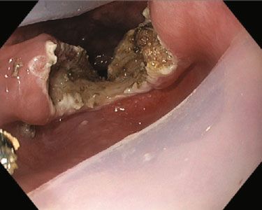
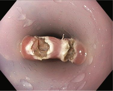
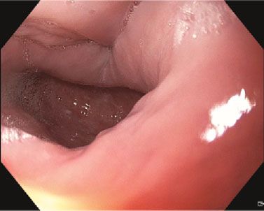
(a) (b) (c) (d) (e)
(f) (g) (h) (i) (j)
Figure 1: (a) Symptomatic ZD of a 73-year-old male patient. (b) Fixation of the diverticulum with an overtube. (c) Incision of the mucosa and
the upper muscular fibers of the diverticular bridge. (d) Cutting down the diverticular bridge with the sb knife jr. (e) Final result after the first
session. (f–j) Second session because of recurrence 16 months later.
the University Hospital Augsburg, Germany, from August Each patient received a single dose of antibiotics (2 g ceftri-
2013 to May 2019 were evaluated. All patients were treated axone) during the intervention. Clipping of the bottom of the
with flexible endoscopy and the sb knife jr as the cutting incision line was not done routinely. Prophylactic clipping to
device. Patients who had had a previous treatment, either prevent delayed bleeding or perforation was performed in
surgical or endoscopic, were excluded. some cases, based on the judgment of the endoscopist.
Recurrence was defined as a relapse of symptoms with
2.2. Endoscopic Procedure and Devices. All interventions were substantial limitations in a patient’s quality of life. Adverse
done by two experienced endoscopists (H.M., S.K.G.). A sin- events were classified according to the American Society for
gle treatment protocol was implemented. Gastrointestinal Endoscopy (ASGE) [9].
On the day prior to the intervention, a gastroscopy was
performed to clean the diverticulum of food remnants, to 2.3. Postprocedural Management and Follow-Up. On the day
measure the size, and to inspect the diverticulum in order after the intervention, a contrast swallow was performed. If
to exclude patients with a candida infection. Diverticulotomy there was no evidence of perforation, the transition to a soft
was performed in deep sedation with midazolam, pethidine, diet for five days was commenced.
and propofol. First, a soft diverticuloscope (ZD overtube, The symptoms of the patients were recorded before treat-
ZDO-22/33 Cook Medical, Limerick, Ireland) was placed to ment as well as one and six months after the intervention by a
stretch the diverticular bridge between the esophagus and questionnaire developed in our clinic [3, 10]. The question-
the diverticular lumen. The endoscope (GIF-HQ190, Olym- naire contains nine points: Dakkak and Bennett’s dysphagia
pus Europa, Hamburg, Germany) was inserted, and the score [11]; the frequency of dysphagia, odynophagia, regurgi-
mucomyotomy was done with the sb knife jr (Sumitomo tation, vomiting, dry cough, halitosis, and nocturnal awaken-
Bakelite, Tokyo, Japan). This is a scissors-shaped device with ing due to Zenker-related symptoms; and the general
an opening width of 3.5 mm that can be rotated by 360 condition of the patient including body weight, duration of
degrees. Additional to electrical cutting, the device is able to symptoms, and weight loss. The symptoms were registered
simultaneously compress the tissue. Due to the mechanical on an ordinal scale with values from zero to four (Table 1).
effect, the directly adjacent tissue is more strongly bonded All patients were informed about the possibility of readmis-
together (Figure 1). The electrosurgical current was gener- sion in case of recurring symptoms. Additionally, patients
ated by the VIO 300 unit (Endocut Q Effect 1, soft coagula- who stated a high point value (>12) in the questionnaire six
tion 40 W, Erbe, Tübingen, Germany). months after the initial intervention were called to evaluate
The technique was modified in the course of the study; whether further treatment was necessary.
initially, only one incision was done in the middle of the 2.4. Statistics. The evaluation was performed using Microsoft
diverticular bridge. Later, two incisions were made and the Excel. Data were stated as mean, median, and standard devi-
part in-between was resected with a snare (double incision ation. For statistical analysis, different t-tests and chi-squared
and snare resection (DISR)) in order to get a broader incision tests were used. Statistical significance was assumed at a p
of the diverticular bridge. The incision was then continued in value of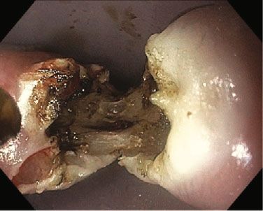
Gastroenterology Research and Practice                                                                                                                           3

                                                          Table 1: Content of the questionnaire.

                                                      0                       1                         2                      3                         4
Recorded symptoms
  Frequency of dysphagia                           Never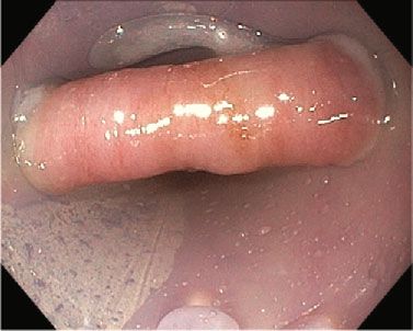
4                                                                                         Gastroenterology Research and Practice

         Initial treatment            Successful treatment          eight (8%) later than six months. Five patients needed a third
                      ⁎
              n = 100                        n = 83                 session and one a fourth.
                                                                        The demographic and clinical characteristics of patients
                                                                    who developed a recurrence did not differ from patients
            Recurrence                Successful treatment          without recurrence (Table 4). Furthermore, the size of the
              n = 17                         n = 12                 diverticulum did not correlate with the risk of recurrence
                                                                    (p = 0:26). The median diverticular size in patients with
                                                                    recurrence was 15 mm (range 5-35 mm). Considering the
          2nd recurrence              Successful treatment
               n=5                           n=4
                                                                    recurrence rate, there was no significant difference between
                                                                    DISR and single incision technique (DISR 17.1%, single inci-
                                                                    sion 16.9%, and p = 0:99). Also, the use of clips during the
          3rd recurrence              Successful treatment          first intervention had no influence on the recurrence rate
               n=1                           n=1                    (clips used 10.5%, no clips 18.5%, and p = 0:40).
                                                                        After retreatment, symptom scores of patients with
Figure 2: Flow chart of patients with recurrence. Recurrence:       recurrence could be reduced to a level comparable to patients
recurring symptoms after a temporary improvement. ∗ 103             who were recurrence-free after one intervention. Dakkak and
sessions in 100 patients due to three two-stage treatments.         Bennett’s dysphagia score was reduced from 2.34 to 0.49
                                                                    (p < 0:001) in patients without recurrence. Those patients
(median score for regurgitations 0.56, p < 0:001; for dyspha-       who developed a recurrence after the initial treatment had
gia 1.09, p < 0:001; and for dry cough 0.98, p < 0:001).            an average score of 1.31 six months after the first interven-
                                                                    tion. Six months after the second treatment, the score was
                                                                    0.33 (p = 0:01) for patients with no further recurrence and
3.2. Adverse Events. Intraprocedural bleeding occurred in 16
                                                                    1.50 for patients with a second recurrence. Besides one
interventions (12.7%). Most cases (15) were stopped by coag-
                                                                    patient, all remaining patients could be treated successfully
ulation with the sb knife jr or by a hemostatic grasper (Coa-
                                                                    in the third session. Their dysphagia score was 0.67 six
grasper Hemostatic Forceps FD-411 QR, Olympus Europa,
                                                                    months after the third intervention which is comparable to
Hamburg, Germany). In four cases, clips (range 1-2, Olym-
                                                                    the values of those patients who did not develop a further
pus Medical Systems Corp., Tokyo, Japan) were used. In
                                                                    recurrence after the first or second treatment. Due to the
three cases, a combination of a hemostatic grasper and
                                                                    small number of patients in this group (available data from
hemoclips was applied. Termination of the procedure due
                                                                    three out of four patients), a reasonable statistical calculation
to intraprocedural bleeding was not necessary since all bleed-
                                                                    is not possible in that case (Figure 3).
ing cases could be treated successfully. There was no case of
                                                                        The same effect was seen for the frequency of dysphagia
delayed bleeding. Procedures performed using the DISR
                                                                    and regurgitation. Regarding the frequency of dysphagia,
technique showed a slightly lower bleeding rate, but this
                                                                    the value declined from 3.45 to 0.92 (p < 0:001)/1.00 (p =
was not significant (DISR 12.2%, single incision 18.6%, and
                                                                    0:02)/1.33 in successfully treated patients (six months after
p = 0:39).
                                                                    the 1st/2nd/3rd session) and for regurgitation from 2.85 to
    In four cases, a perforation was suspected during the
                                                                    0.35 (p < 0:001)/1.00 (p < 0:01)/0.67. Patients with recur-
intervention. Of these, one intervention had to be stopped
                                                                    rence needed in total a mean of 2.35 sessions (range 2-4) to
prematurely. In five other cases, contrast swallow after the
                                                                    achieve symptom control.
treatment showed a small perforation. In case a perforation
                                                                        No bleeding occurred in the treatment of recurrences
was suspected, nil diet and clinical observation were extended
                                                                    which means that the rate of bleeding was significantly lower
and antibiotics were given for several days. But in all these
                                                                    in the retreatment group compared to the initial intervention
cases, the further clinical course was uneventful. Emphysema
                                                                    group (p = 0:04). Also, there was no other severe adverse
was not observed in any of the patients.
                                                                    event in the treatment of recurrences.
    Three patients suffered severe or prolonged postproce-
dural pain. One patient was monitored overnight in the inten-
sive care unit to rule out serious adverse events as a reason for
his pain. All these patients were managed conservatively.
                                                                    4. Discussion
    Another patient developed a hemodynamically relevant
                                                                    The strength of this study is the large cohort of patients and
tachycardia (focal atrial tachycardia) after the intervention,
                                                                    the follow-up over a median of 41 months (range 7-74).
most probably because she had not taken her antiarrhythmic
                                                                    Besides the study of Huberty et al. [13], this is one of the larg-
medication prior to the intervention. She spent one night in
                                                                    est prospectively documented cohort of patients with symp-
the intensive care unit and was treated with amiodarone.
                                                                    tomatic ZD treated by a flexible endoscopic approach. Due
    In all 126 interventions, no severe adverse event was
                                                                    to the long-term follow-up period, late recurrences were also
observed. There was no case of mediastinitis or abscess.
                                                                    included. Of course, there might be patients that are lost to
                                                                    follow-up so that recurrences are not detected. But this prob-
3.3. Recurrence. After initial treatment, 17 patients (17%)         lem is similar to other interventional studies concerning ZD.
developed a recurrence (Figure 2). Nine (9%) recurrences            The recurrence rate of 17% is comparable to the results of
occurred within the first six months after treatment, and            other studies with flexible endoscopy [6].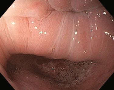
Gastroenterology Research and Practice                                                                                                                5

                                   Table 4: Descriptive characteristics before treatment stratified by no recurrence/recurrence.

                                                                   Nonrecurrence                            Recurrence                       p value
Total number of patients                                                  83                                     17
Median follow-up (months)                                             44 (7-74)                              39 (7-69)
Median age                                                           71 (42-92)                             73 (49-85)                         0.90
Sex (female/male)                                                       32/51                                   4/13                           0.24
Median diverticular size (mm)                                        20 (10-45)                            27.5 (20-40)                        0.26
Median body mass index (kg/m2)                                     25.8 (17.3-38.0)                       27.1 (17.5-34.5)                     0.70
Mean weight loss (kg)                                                 2.1 (0-20)                             2.3 (0-20)                        0.83
Values express absolute numbers with (range).

                               ⁎                                                      events such as mediastinitis or permanent palsy of the re-
                                                                                      current laryngeal nerve. Moreover, surgical meta-analysis
                   ⁎⁎⁎
                                                                                      shows a small number of therapy-related deaths [1, 14].
  4
                                                          n=5

                                                                                      Also, in approaches with rigid endoscopes, some major
                                                                                      adverse events, such as abscesses requiring external drain-
                                   n = 17

  3                                                                                   age, occurred [16].
                                                                                          Although the rate of recurrence in our cohort is slightly
                                                                                      higher, this study has shown that treatment can be easily
                          n = 83

                                                                 n=4

  2
         n = 100

                                                                                      repeated with a high success rate. Repetition of a surgical
                                                 n = 11

                                                                       n=1

                                                                                      treatment might be more difficult and challenging due to
  1                                                                                   scarring tissue. In our cohort, retreatment was technically
                                                                                      feasible, and in one patient, retreatment was performed four
  0                                                                                   times. Retreatment of those patients was not associated with
       Prior to              After                 After           After
      treatment          1st session            2nd session     3rd session           a higher rate of adverse events, and the rate of bleeding was
                                                                                      even significantly lower. Dissection of the scar and remaining
          No recurrence                                                               muscle tissue with flexible endoscopy is technically not more
          Recurrence                                                                  challenging than the initial treatment. Patients with recur-
                                                                                      rence could achieve the same control of their symptoms as
Figure 3: Dysphagia score of patients with and without recurrence                     patients without recurrence. Eventually, even patients who
after each session. Mean dysphagia score prior to initial treatment
                                                                                      needed several sessions (mean 2.35 sessions) could be treated
and six months after each session. Dysphagia score: 0: no
dysphagia, 1: solid food, 2: soft food, 3: fluids, and 4: aphagia. ∗∗∗ t
                                                                                      successfully.
-test significance p < 0:001, ∗ t-test significance p < 0:05.                               Tunneling myotomy has been reported recently; how-
                                                                                      ever, further studies are needed to clarify its long-term effec-
                                                                                      tiveness [17, 18]. It is also unclear if a tunneled myotomy
    In surgical transcervical series, the reported recurrence                         could be repeated in case of recurrence.
rate varies widely, depending on the surgical technique, from                             A limitation of this study is that there is no direct com-
1.9% for open suspension [14] to 21% for invagination of the                          parison to other therapeutic methods, especially a transcervi-
diverticulum [1].                                                                     cal surgical approach. A randomized study with different
    In our study, patients who developed recurrence could                             treatment paths would be able to compare the rate of recur-
be divided into two major groups. In group one (9%), after                            rence and adverse events.
an initial distinct improvement of symptoms, recurrence
occurred early within a few months after treatment. In
the second group (8%), the patients were already in remis-                            5. Conclusion
sion before they developed recurrent symptoms (median 16
                                                                                      Symptomatic ZD can be controlled with endoscopic treat-
months, range 8-26). A reason for the early recurrences
                                                                                      ment using the sb knife jr in a safe and effective way. Patients
might be an incomplete initial dissection of the cricophar-
                                                                                      with recurrence can be retreated without an increased risk of
yngeal muscle in the first session. Probably, the myotomy
                                                                                      adverse events and with a high success rate. Patients with
for those patients was not wide or long enough. An incom-
                                                                                      recurrence can ultimately achieve the same long-term symp-
plete myotomy of the upper esophageal sphincter has
                                                                                      tom control as treatment-naïve patients.
already been discussed in the literature as a possible cause
of recurrence [7, 15]. Why other patients, after an initially
successful treatment with good symptom control, suffer a                               Data Availability
recurrence after a longer period of time cannot be conclu-
sively explained at present.                                                          The data on which this study is based have been deposited in
    There were no severe adverse events. In contrast, surgical                        the study secretariat of the Department of Internal Medicine
approaches have a low but relevant number of severe adverse                           III at the University Hospital Augsburg.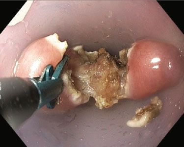
6                                                                                                Gastroenterology Research and Practice

Conflicts of Interest                                                          of Zenker’s diverticulum,” Gastrointestinal Endoscopy, vol. 83,
                                                                               no. 4, pp. 765–773, 2016.
The authors declare that they have no conflicts of interest.               [16] R. Wilken, C. Whited, and R. L. Scher, “Endoscopic staple
                                                                               diverticulostomy for Zenker’s diverticulum: review of experi-
Authors’ Contributions                                                         ence in 337 cases,” The Annals of Otology, Rhinology, and Lar-
                                                                               yngology, vol. 124, no. 1, pp. 21–29, 2015.
Johannes Manzeneder and Christoph Römmele contributed                     [17] J. Yang, X. Zeng, X. Yuan et al., “An international study on the
equally to this work.                                                          use of peroral endoscopic myotomy (POEM) in the manage-
                                                                               ment of esophageal diverticula: the first multicenter D-
                                                                               POEM experience,” Endoscopy, vol. 51, no. 4, pp. 346–349,
References                                                                     2019.
 [1] M. Colombo-Benkmann, V. Unruh, T. Kocher, C. Krieglstein,            [18] Q. L. Li, W. F. Chen, X. C. Zhang et al., “Submucosal tunneling
     and N. Senninger, “Modern treatment options for Zenker’s                  endoscopic septum division: a novel technique for treating
     diverticulum: indications and results,” Zentralblatt für Chirur-          Zenker’s diverticulum,” Gastroenterology, vol. 151, no. 6,
     gie, vol. 128, no. 3, pp. 171–186, 2003.                                  pp. 1071–1074, 2016.
 [2] J. J. van Overbeek, “Pathogenesis and methods of treatment of
     Zenker’s diverticulum,” The Annals of Otology, Rhinology, and
     Laryngology, vol. 112, no. 7, pp. 583–593, 2003.
 [3] J. Brueckner, A. Schneider, H. Messmann, and S. K. Gölder,
     “Long-term symptomatic control of Zenker diverticulum by
     flexible endoscopic mucomyotomy with the hook knife and
     predisposing factors for clinical recurrence,” Scandinavian
     Journal of Gastroenterology, vol. 51, no. 6, pp. 666–671, 2016.
 [4] S. Ishioka, P. Sakai, F. Maluf Filho, and J. M. Melo, “Endo-
     scopic incision of Zenker’s diverticula,” Endoscopy, vol. 27,
     no. 6, pp. 433–437, 1995.
 [5] C. J. Mulder, G. den Hartog, R. J. Robijn, and J. E. Thies, “Flex-
     ible endoscopic treatment of Zenker’s diverticulum: a new
     approach,” Endoscopy, vol. 27, no. 6, pp. 438–442, 1995.
 [6] S. Ishaq, C. Hassan, A. Antonello et al., “Flexible endoscopic
     treatment for Zenker’s diverticulum: a systematic review and
     meta-analysis,” Gastrointestinal Endoscopy, vol. 83, no. 6,
     pp. 1076–1089.e5, 2016.
 [7] H. Feussner, “Zenker’s diverticulum: pro operation,” Chirurg,
     vol. 82, no. 6, pp. 484–489, 2011.
 [8] S. K. Gölder, J. Brueckner, A. Ebigbo, and H. Messmann,
     “Double incision and snare resection in symptomatic Zenker’s
     diverticulum: a modification of the stag beetle knife tech-
     nique,” Endoscopy, vol. 50, no. 2, pp. 137–141, 2018.
 [9] P. B. Cotton, G. M. Eisen, L. Aabakken et al., “A lexicon for
     endoscopic adverse events: report of an ASGE workshop,”
     Gastrointestinal Endoscopy, vol. 71, no. 3, pp. 446–454, 2010.
[10] S. K. Goelder, J. Brueckner, and H. Messmann, “Endoscopic
     treatment of Zenker’s diverticulum with the stag beetle knife
     (sb knife) - feasibility and follow-up,” Scandinavian Journal
     of Gastroenterology, vol. 51, no. 10, pp. 1155–1158, 2016.
[11] M. Dakkak and J. R. Bennett, “A new dysphagia score with
     objective validation,” Journal of Clinical Gastroenterology,
     vol. 14, no. 2, pp. 99-100, 1992.
[12] M. M. Brombart, Radiologie des Verdauungstraktes: Funktio-
     nelle Untersuchung und Diagnostik, Georg Thieme Verlag:
     Stuttgart, New York, 1980.
[13] V. Huberty, S. el Bacha, D. Blero, O. le Moine, S. Hassid, and
     J. Devière, “Endoscopic treatment for Zenker’s diverticulum:
     long-term results (with video),” Gastrointestinal Endoscopy,
     vol. 77, no. 5, pp. 701–707, 2013.
[14] J. Verdonck and R. P. Morton, “Systematic review on treat-
     ment of Zenker’s diverticulum,” European Archives of Oto-
     Rhino-Laryngology, vol. 272, no. 11, pp. 3095–3107, 2015.
[15] G. Costamagna, F. Iacopini, A. Bizzotto et al., “Prognostic var-
     iables for the clinical success of flexible endoscopic septotomy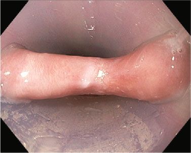
You can also read