Efficacy of krypton laser photodynamic therapy for oral mucosa dysplasia in 9,10 dimethyl 1,2 benzanthracene treated hamsters
←
→
Page content transcription
If your browser does not render page correctly, please read the page content below
1358                                         ONCOLOGY LETTERS 6: 1358-1362, 2013

  Efficacy of krypton laser photodynamic therapy for oral mucosa
       dysplasia in 9,10‑dimethyl‑1,2‑benzanthracene‑treated
                              hamsters
                             LINGYUE SHEN, QING XU, PINGPING LI and GUOYU ZHOU

                            Department of Oral and Maxillofacial Surgery, Ninth People's Hospital,
                           Shanghai Jiao Tong University School of Medicine, Shanghai, P.R. China

                                    Received February 28, 2013; Accepted August 6, 2013

                                                   DOI: 10.3892/ol.2013.1554

Abstract. The present study aimed to evaluate the effi-           Introduction
cacy of krypton laser photodynamic therapy (PDT) with
PsD‑007 for the treatment of oral mucosa dysplasia in             Photodynamic therapy (PDT) is a new technology for the
9,10‑dimethyl‑1,2‑benzanthracene (DMBA)‑treated hamsters.         treatment of cancer that has gained interest within the past two
A DMBA‑induced hamster cheek pouch model of precan-               decades and has been used in the treatment of lung, esopha-
cerous lesions was created and the resultant 25 hamsters were     geal, cervical, bladder and skin cancer and other tumors (1).
divided into five groups. The right side was treated with PDT     Clinically, PDT has been considered to be more applicable to
and the left side was used as the positive control. Following     tumors with a wide range of superficial locations, as it may be
systemic anesthesia, an incision was made in the groin area       combined with fluorescence diagnostic techniques to distin-
to expose the femoral vein. PsD‑007 was administered              guish the range of lesions, selectively kill tumors and reduce
intravenously through the femoral vein. Various doses of          the damage to normal tissues. It is recognized that the efficacy
photosensitizer were used to treat groups A‑E. Subsequent to      of PDT in tumors with a depth of 1.0 cm is extremely high.
closing the incision, the right side of the buccal mucosa was     Precancerous lesions of an oral or maxillofacial location are
irradiated with light using the krypton laser at a wavelength     more superficial, therefore, PDT has a measurable effect on
of 413 nm, a power density of 150 mW/cm2 and an irradiation       these types of lesions (2).
time of 20 min. At six weeks post‑surgery, the response was           The present study used intravenous PDT to treat precan-
analyzed using histological examinations of the buccal pouch      cerous lesions in order to explore the mechanism of the
mucosa. A total of 24 hamsters completed the six‑week obser-      treatment. Through the establishment of a golden hamster
vation period, as one hamster from group C died in the second     leukoplakia model, which was similar to human leuko-
week following the PDT. Of all 24 irradiated sides, 15 formed     plakia, the effect of the hematoporphyrin monomethyl ether
normal mucosal tissues and nine demonstrated mild dysplasia.      (PsD‑007)‑mediated krypton laser PDT on the oral dysplasia
Of the total control sides, six developed moderate dysplasia,     of hamster was observed and the dose‑effect correlation of
five developed severe dysplasia and 13 progressed to carcinoma    PDT was also studied. This provides a basis and reference for
in situ or squamous cell carcinoma (SCC). The results revealed    the treatment of human precancerous lesions in the clinic.
a significant difference between the two sides (P10 mg/kg, there was no statistical difference (P>0.05).
PsD‑007‑mediated krypton laser PDT is effective for the treat-    Experimental animals and establishment of leukoplakia
ment of oral mucosa dysplasia in hamsters.                        model
                                                                  Materials. A total of 25, six to eight‑week‑old female Syrian
                                                                  hamsters (n=25) were purchased from the BK company
                                                                  (Shanghai, China). 9,10‑Dimethyl‑1,2‑benzanthracene
                                                                  (DMBA) was purchased from Sigma (St Louis, MO, USA)
Correspondence to: Dr Guoyu Zhou, Department of Oral
                                                                  and the krypton ion laser was obtained from Coherent (Santa
and Maxillofacial Surgery, Ninth People's Hospital, Shanghai
Jiao Tong University School of Medicine, 639 Zhi Zaoju Road,
                                                                  Clara, CA, USA). The hematoporphyrin monomethyl ether,
Shanghai 200011, P.R. China                                       PsD‑007 (Shanghai Second Military Medical University,
E‑mail: guoyuzhou@hotmail.com                                     Shanghai, China), was prepared prior to performing the
                                                                  study.
Key words: photodynamic therapy, krypton laser, oral dysplasia,       Grouping. The 25 Syrian golden hamsters were randomly
hamster                                                           divided into five groups (A, B, C, D and E; n=5). The concen-
                                                                  tration of the photosensitizer used was 5, 10, 20, 40 and
                                                                  60 mg/kg, respectively in the five groups. In all the animals,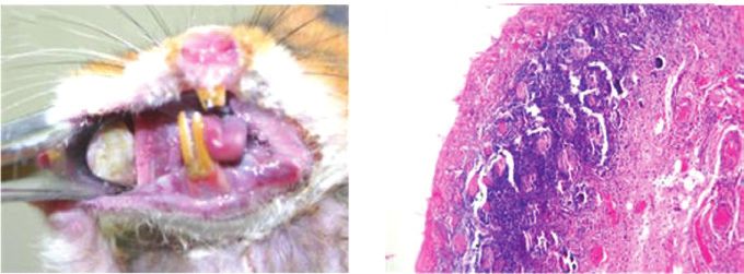
SHEN et al: KRYPTON LASER PDT TREATS ORAL MUCOSA DYSPLASIA                                   1359

the cheek pouch of the right side was set as the PDT group and      were as follows: 2, mild dysplasia; 2‑4, moderate dysplasia;
the left side was set as the control group.                         and >5, severe dysplasia. The typical pathological changes of
    Preparation of leukoplakia model. According to the              the tumor cells were recorded as carcinoma in situ.
methods used by Salley (3), 0.5% DMBA (5 g/l) was dissolved             The efficacy evaluation was based on the post‑operative
in acetone solution and stored in brown bottles. The cheek          cheek pouch mucosal pathology, which was graded as a cure
pouch was everted using tweezers and a number one oil               (normal mucosa) or as effective (mild dysplasia). The lesions
painting pen dipped with 0.5% DMBA acetone solution                 were alleviated compared with the control side.
was used to brush the tissues 10 times within a 1‑cm range
of the cheek pouch vein. The other side of the cheek pouch          Statistics. Values are shown as the mean ± SEM. The differ-
mucosa was prepared using the same method. The hamsters             ences between all the groups were evaluated using one‑way
were made to fast for 2 h following the drug‑smearing. All          ANOVA with a post‑hoc Student‑Newman‑Keuls multiple
the animals were treated with the surface drug on Monday,           comparisons test. The statistical analyses were performed
Wednesday and Friday continuously for six weeks in the same         using SPSS software (v13.0; SPSS, Inc., Chicago, IL, USA)
manner. The study was approved by the ethics committee of           and P
1360 ONCOLOGY LETTERS 6: 1358-1362, 2013
A B
Figure 1. Observations following the treatment. (A) A necrotic tissue covering with white pseudomembrane on the buccal pouch mucosa was observed one to
two weeks subsequent to PDT. (B) One week after PDT (HE staining; magnification, x40). Shedding of the epithelial layer, ulcer formation and inflammatory
cell infiltration in the lamina propri and muscularis is observed. PDT, photodynamic therapy; HE, hematoxylin and eosin.
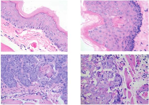
A B
C D
Figure 2. HE results of PDT after six weeks. (A) The PDT‑treated side of the cheek pouch mucosa in group B (HE staining; magnification, x40). (B) The
PDT‑treated side of the cheek pouch mucosa in group C. Mild dysplasia and dyskeratosis (arrow) were observed (HE staining; magnification, x40). (C) Control
side of the cheek pouch mucosa in group C. Severe dysplasia was observed. (HE staining; magnification, x40); (D) Control side of the cheek pouch mucosa in
group E (HE staining; magnification, x40). PDT, photodynamic therapy; HE, hematoxylin and eosin.
process from precancerous lesions to cancer, which may be sion, thereby leading to tumor necrosis (8); iii) PDT may cause
used to observe and evaluate the effect of PDT and obtain an immune response of the tumor cells responding to tumor
reliable results. The golden hamster used in the present study antigens (9); and iv) PDT may affect the gene expression of the
is a small mouse and the tail is extremely short. Therefore, a tumor cells (10). However, due to the shorter photosensitizer
subcutaneous superficial vein or tail vein injection would be absorption lines, the penetration depth of the corresponding
unsuccessful. Consequently, the photodynamic methods of the laser is limited. Therefore, deep and large tumors may not be
present study were established using a femoral vein injection, able to be treated using this method. It has been recognized
which has repeatability as a new method. that the method is only efficient on tumors with a depth of
The mechanism of the destruction of the tumor using ≤1.0 cm (11,12).
PDT may be summarized as follows: i) The hematoporphyrin Precancerous lesions are extremely superficial and only
derivative photosensitizer is combined with a biofilm system, identified in the epithelium, which means that the desired
particularly the cell plasma and nuclear membranes, to destroy treatment results may be obtained with PDT. PDT has gradu-
the mitochondria, lysosomes and liposomes of the tumor ally been focused on the treatment of superficial tumors and
cells. Therefore, the activities of the cells that are necessary precancerous lesions. The treatment of precancerous lesions
for life, including respiration, electron transport and ATP with PDT and 5‑AminoLevulinic Acid (ALA) has become a
synthesis, are affected due to the mitochondrial damage, focus for research (3). ALA is an exogenous photosensitizer,
thereby inducing cell apoptosis (5‑7); ii) PDT causes tumor which is able to indirectly synthesize protoporphyrin IX at a
microvascular dysfunction and structural damage, including 635 nm absorption peak in vivo following absorption through
destruction of the vascular endothelium, platelet adhesion, the skin or mucous (14). ALA‑PDT has been used to treat a
degranulation, siltation of the blood cells and vascular occlu- variety of superficial precancerous lesions, including skin,SHEN et al: KRYPTON LASER PDT TREATS ORAL MUCOSA DYSPLASIA 1361
Table I. Pathological results of the PDT‑treated and control sides of the buccal pouch mucosa in groups A‑E following six weeks
of treatment.
Epithelium dysplasia, n (%)
‑‑‑‑‑‑‑‑‑‑‑‑‑‑‑-------‑‑‑‑‑‑‑‑‑‑‑‑‑‑‑‑‑‑‑‑‑‑‑‑‑‑‑-‑‑‑‑‑‑‑‑‑‑‑‑‑‑‑‑‑‑‑
Group n Normal mucosa, n (%) Mild Moderate Severe Carcinoma in situ, n (%)
A
PDT‑treated side 5 1 (20.0) 4 (80.0) 0 (0.0) 0 (0.0) 0 (0.0)
Control side 5 0 (0.0) 0 (0.0) 1 (25.0) 1 (25.0) 3 (60.0)
B
PDT‑treated side 5 4 (40.0) 1 (60.0) 0 (0.0) 0 (0.0) 0 (0.0)
Control side 5 0 (0.0) 0 (0.0) 2 (40.0) 1 (20.0) 2 (40.0)
C
PDT‑treated side 4 3 (75.0) 1 (25.0) 0 (0.0) 0 (0.0) 0 (0.0)
Control side 4 0 (0.0) 0 (0.0) 0 (0.0) 1 (25.0) 3 (75.0)
D
PDT‑treated side 5 3 (60.0) 2 (40.0) 0 (0.0) 0 (0.0) 0 (0.0)
Control side 5 0 (0.0) 0 (0.0) 2 (40.0) 1 (20.0) 2 (40.0)
E
PDT‑treated side 5 4 (80.0) 1 (20.0) 0 (0.0) 0 (0.0) 0 (0.0)
Control side 5 0 (0.0) 0 (0.0) 1 (20.0) 1 (20.0) 3 (60.0)
Total
PDT‑treated side 24 15 (62.5) 9 (37.5) 0 (0.0) 0 (0.0) 0 (0.0)
Control side 24 0 (0.0) 0 (0.0) 6 (25.0) 5 (20.8) 13 (54.2)
PDT, photodynamic therapy.
Table II. Cure and effective rates of groups A‑E.
The systemic toxicity was observed to be low when partially
Group Dose, mg/kg Cure, n (%) Effective, n (%) n coated with ALA, and light did not have to be avoided following
the surgery due to a rapid metabolism (15,16). However, the
A 5 1 (20.0) 4 (80.0) 5 efficacy of ALA‑PDT was dependent on the level of proto-
B 10 4 (80.0) 1 (20.0) 5 porphyrin that was synthesized in the tumors. The capacity of
C 20 3 (75.0) 1 (25.0) 4 synthesizing protoporphyrin has yet to be elucidated in various
tumor cells and precancerous lesions. Therefore, the method
D 40 3 (60.0) 2 (40.0) 5
lacks a theoretical basis for the treatment of precancerous
E 60 4 (80.0) 1 (20.0) 5
lesions. In the present study, the intravenous PDT method was
Total - 15 (62.5) 9 (37.5) 24 used, in which the mechanism has been confirmed. PsD‑007
was the purified product of the hematoporphyrin derivatives
and the maximum absorption peak was 408 nm, which was
close to the wavelength of the krypton laser (413 nm). The
bladder, lung and gastrointestinal cancer. Tsai et al (15) revealed results revealed that the overall cure rate of dysplasia was
that a combination of 20% ALA and a light-emitting diode 62.5% (15/24) in the PDT group, indicating that PDT had a
(LED) laser (630 nm, 100 J/cm2) was able to effectively treat positive effect on the precancerous lesions.
hamsters with oral dysplasia induced by DMBA. Furthermore The traditional view is that the porphyrin‑based photosen-
the same ALA‑PDT method was used to treat 32 patients sitizer has a selective storage role for tumor tissues. However,
with oral dysplasia. Following a six‑month follow‑up period, there has not been sufficient experimental evidence for this.
the results revealed that a complete response was evident in The mechanism behind the treatment of tumors using PDT
seven cases and a partial response was evident in 13 cases. the was believed to function as the new tumor tissues were rich in
treatment was ineffective in 12 cases. Kübler et al (16) used blood vessels. The hematoporphyrin derivative photosensitizer
20% ALA to treat 12 patients with leukoplakia. After 2 h, the reached a relatively high concentration in the tumor tissues
patients were irradiated using a 630 nm laser (100 J/cm2). The through the blood vessels, thereby selectively destroying the
results revealed that a complete response was evident in five tumors. The objective of the present study was to treat oral
cases and a partial response was evident in four cases. The precancerous lesions, in which the vascular distribution is not
treatment was ineffective in three cases. The lesions disap- clear. Therefore, a confirmation of the differences between
peared following re‑treatment in one case. photosensitizer concentrations in the precancerous lesions1362 ONCOLOGY LETTERS 6: 1358-1362, 2013
and normal tissues was required. In the present study, all References
the animals demonstrated mucosal necrosis and shedding
following the treatment with PDT. The selectivity was also 1. Dougherty TJ, Gomer CJ, Herderson BW, et al: Photodynamic
determined by the range of the irradiation. However, the therapy. J Natl Cancer Inst 90: 889‑905, 1998.
2. Hopper C, Kubler A, Lewis H, et al: mTHPC-mediated photo-
normal tissues were also damaged, which may have been due dynamic therapy for early oral squamous cell carcinoma. Int J
to fact that the vascular distribution of the precancerous lesions Cancer 111: 138-146, 2004.
was not abnormal compared with the tumors. Therefore, the 3. Salley JJ: Experimental carcinogenesis in the cheek pouch of the
Syrian hamster. J Dent Res 33: 253‑262, 1954.
difference in the concentration of the photosensitizer was not 4. Eversole LR: Dysplasia of the upper aerodigestive tract squamous
great enough between the precancerous lesions and the normal epithelium. Head Neck Pathol 3: 63-68, 2009.
tissues in order to obtain selective damage results. 5. Chen HM, Chen CT, Yang H, et al: Successful treatment of
oral verrucous hyperplasia with topical 5‑aminolevulinic
The results of the present study revealed that 15 cases were acid‑mediated photodynamic therapy. Oral Oncol 40: 630‑637,
cured in the PDT group at six weeks post‑treatment, none of 2004.
which appeared carcinogenic. However, in the control group, 6. Gush RJ and King TA: Discrimination of capillary and
arterio‑venular blood flow in skin by laser Doppler flowmetry.
13 cases appeared carcinogenic, indicating that the efficacy Med Biol Eng Comput 29: 387‑392, 1991.
was significantly improved in the PDT group. Furthermore, 7. Tsai JC, Chiang CP, Chen HM, et al: Photodynamic Therapy
although the animals demonstrated mild dysplasia in the PDT of oral dysplasia with topical 5‑aminolevulinic acid and
light‑emitting diode array. Lasers Surg Med 34: 18‑24, 2004.
group, the degree of dysplasia was less than that of the control 8. Biel MA: Photodynamic therapy and the treatment of neoplastic
group. This provides evidence for the efficiency of PDT using diseases of the larynx. Laryngoscope 104: 399‑403, 1994.
a krypton laser and PsD‑007 on the precancerous lesions of 9. Kübler A, Haase T, Rheinwald M, et al: Treatment of oral leuko-
plakia by topical application of 5‑aminolevulinic acid. Int J Oral
the golden hamsters. In addition, there was a significant differ- Maxillofac Surg 27: 466‑469, 1998.
ence between group A (5 mg/kg) and the other four groups 10. Pariser DM, Lowe NJ, Stewart DM, et al: Photodynamic therapy
(>10 mg/kg). However, although the cure rate did not increase with topical methyl aminolevulinate for actinic keratosis: results
of a prospective randomized multicenter trial. J Am Acad
with an increasing concentration, the phototoxicity did. Dermatol 48: 227‑232, 2003.
Therefore, 10 mg/kg was considered a suitable photosensitizer 11. Date M, Sakata I, Fukuchi K, et al: Photodynamic therapy for
concentration. A single hamster in group C died of a severe human oral squamous cell carcinoma and xenografts using a new
photosensitizer, PAD‑S31. Lasers Surg Med 33: 57‑63, 2003.
swelling response and photosensitive reaction at two weeks 12. Nauta JM, van Leengoed HL, Witjes MJ, et al: Photofrin‑mediated
post‑surgery. In groups D and E, the animals also demonstrated photodynamic therapy of chemically‑induced premalignant
photosensitive reactions, including a rash, alopecia and skin lesions and squamous cell carcinoma of the palatal mucosa in
rats. Int J Oral Maxillofac Surg 26: 223‑231, 1997.
ulceration. These reactions may have been due to the larger 13. Collaud S, Juzeniene A, Moan J and Lange N: On the selectivity
dose of photosensitizer and the unintentional exposure to light. of 5‑aminolevulinic acid‑induced protoporphyrin IX formation.
Although PDT exhibited a positive effect on the precan- Curr Med Chem Anticancer Agents 4: 301‑316, 2004.
14. Chang SC, Buonaccorsi G, MacRobert AJ and Bown SG:
cerous lesions, the lesions had characteristics of regional 5‑Aminolevulinic acid (ALA)‑induced protoporphyrin IX
malignancy and multi‑step carcinogenesis. Therefore, the fluorescence and photodynamic effects in the rat bladder: an in
efficiency of the treatment may have been reduced and the vivo study comparing oral and intravesical ALA administration.
Lasers Surg Med 20: 254‑264, 1997.
lesions may have recurred after an extended observation time. 15. Tsai JC, Chiang CP, Chen HM, et al: Potodynamic therapy of oral
PDT requires further study in order to evaluate the long‑term dysplasia with topical 5-aminolebulinic acid and light-emitting
effects of the method on precancerous lesions. diode array. Lasers Surg Med 34: 18-24, 2004.
16. Kubler A, Haase T, Rheinwald M, et al: Treatment of oral leuko-
plakia by topical application of 5-aminolevulinic acid. Int J Oral
Acknowledgements Maxillofac Surg 27: 466-469, 1998.
This study was supported by the Research Fund of Science
and Technology Commission of Shanghai Municipality (grant
no. 08DZ2271100).You can also read