Recurring myositis ossificans traumatica of temporal muscle: A case report
←
→
Page content transcription
If your browser does not render page correctly, please read the page content below
Vojnosanit Pregl 2021; 78(2): 255–260.            VOJNOSANITETSKI PREGLED                                                    Page 255

CASE REPORT                                                                                         UDC: 616.314-089:616.742-089
                                                                                      DOI: https://doi.org/10.2298/VSP180806048J

Recurring myositis ossificans traumatica of temporal muscle: A case
report
                         Recidiv traumatskog osificirajućeg miozitisa temporalnog mišića

                          Saša Jović*, Denis Brajković†, Milena Borilović*, Uroš Marjanović*,
                                 Marko Brkić*, Ružica Kozomara*‡, Srboljub Stošić*‡

                    Military Medical Academy, *Clinic for Maxillofacial Surgery, Belgrade, Serbia; Clinical
                      Center of Vojvodina, †Clinic for Maxillofacial and Oral Surgery, Novi Sad, Serbia;
                    University of Defence, ‡Faculty of Medicine of the Military Medical Academy, Belgrade,
                                                             Serbia

Abstract                                                             Apstrakt

Introduction. Myositis ossificans traumatica (MOT) refers to a       Uvod. Traumatski osificirajući miozitis (Myositis ossificans
benign, localized ectopic bone formation within skeletal             traumatica ‒ MOT) se odnosi na benigno, lokalizovano ek-
muscle bundles related to a traumatic injury. MOT rarely af-         topično formiranje koštanog tkiva unutar skeletnih mišića
fects masticatory muscles, and it represents a major diagnos-        nakon trauma. MOT retko zahvata mastikatorne mišiće i
tic and therapeutic problem for clinicians. Currently, the           tada predstavlja ozbiljan dijagnostički i terapijski problem za
treatment of choice is complete excision of the calcified            hirurge. Trenutno je tretman izbora potpuna ekscizija kalci-
mass after bone maturation and resection of the affected             ficifikovane mase i resekcija zahvaćene kosti. Prikaz
bone. Case report. A 47-year-old male presented with a               bolesnika. Bolesnik, star 47 godina, javio se na pregled
month-long severe restriction of mouth opening that was              zbog otežanog otvaranja usta mesec dana unazad, nakon
followed by extraction of the right lower third molar tooth          ekstrakcije desnog donjeg umnjaka pod lokalnom anestezi-
under local anesthesia. A computed tomography (CT) scan              jom. Nalaz kompjuterizovane tomografije (CT) glave uka-
revealed ectopic bone formation in the right temporal mus-           zao je na stvaranje ektopične kosti u desnom temporalnom
cle extending to the right coronoid process. Surgical exci-          mišiću i koronoidnom nastavku donje vilice. Učinjena je hi-
sion of the calcified mass was performed. Six years after the        rurška ekscizija kalcificirane mase i resekcija zahvaćene kos-
surgery, the patient reported the same symptoms. The CT              ti. Šest godina nakon operacije, bolesnik se žalio na iste
scan revealed a calcified mass of the right temporal muscle          simptome. Nalaz CT glave je pokazao ponovnu poja-
extending to the medial pterygoid muscle. The patient was            vu kalcifikovanog tkiva unutar desnog temporalnog mišića
reoperated, and sent for the postoperative physical treat-           koje se protezalo na medijalni pterigoidni mišić. Bolesnik je
ment. Conclusion. MOT represents a major diagnostic and              reoperisan i upućen na postoperativni fizikalni tretman.
therapeutic challenge for surgeons due to unclear etiology           Zaključak. Zbog nejasne etiologije i čestih recidiva nakon
and frequent recurrences after surgical treatment. Further           hirurškog lečenja MOT predstavlja dijagnostički i terapijski
research is needed to clarify the mechanisms of ossification         izazov za hirurge. Dalja istraživanja su potrebna kako bi se
in MOT in order to develop conservative treatment ap-                razjasnili mehanizmi osicifikacije kod MOT u cilju razvoja
proaches.                                                            konzervativnog tretmana.

Key words:                                                           Ključne reči:
myositis ossificans; temporal muscle; oral surgical                  miozitis osifikans; temporalni mišić; hirurgija, oralna,
procedures; recurrence; treatment outcome.                           procedure; recidiv; lečenje, ishod.

     Introduction                                                    and myositis ossificans traumatica (MOT) 1. MOP is an
                                                                     autosomal dominant disease characterized by systemic
     Myositis ossificans is a rare disease in which ectopic          ossification of muscles and soft tissues with poor
benign bone formation occurs in muscle tissue. It has                prognosis 1. MOT refers to a benign, localized ectopic
been divided into myositis ossificans progressiva (MOP)              bone formation and ossification of fibrous connective
Correspondence to: Denis Brajković, Military Medical Academy, Resident at the Clinic for Maxillofacial Surgery, Crnotravska 17, 11 000
Belgrade, Serbia; E-mail: denis.brajkovic@gmail.com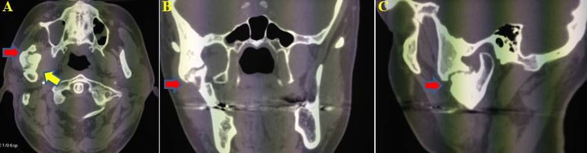
Page 2                                      VOJNOSANITETSKI PREGLED                                             Vol. 78, No. 2

tissue within skeletal muscle bundles related to traumatic   injury 1–3. The pathological mechanism of MOT is not
completely understood, and proposed pathogenesis             and extending superiorly to the right coronoid process. It
theories suggest inflammatory response in muscle tissue      also depicted the insertion of the right temporalis muscle.
related to trauma followed by displacement of                The lesion appeared as a central radiolucency surrounded
osteoprogenitor cells and overexpression of bone             by the circumscribed ossified periphery. Calcification in
morphogenetic proteins, leading to ectopic bone              the right temporal muscle was approximately 43 × 15 mm
formation 1–3.                                               in size when measured from the coronoid processus to the
     MOT is most commonly seen in young patients due         upper part of the lesion. It extended to the temporal fossa
to bone metabolism, renewing periosteum, and richness in     and was well-defined from the surrounding structures.
mesenchymal cells 4. MOT rarely affects masticatory          There were no signs of bone destruction and infiltration of
muscles, and, so far, a limited number of reports have       other masticatory muscles (Figure 1).
been published. MOT of masticatory muscles represents a            At this point, the differential diagnosis of
major clinical problem since there is no unified algorithm   extraskeletal bone formation included myositis ossificans,
for diagnosis and treatment. The main clinical sign of       fibrodysplasia          ossificans,        osteochondroma,
MOT of masticatory muscles is progressive trismus            chondrosarcoma, osteosarcoma, osteoma, and vascular
followed by head and neck trauma. The only treatment         malformation with phlebolithis.
modality widely accepted is complete excision of the               We decided to perform a right mandibular
calcified mass after bone maturation and resection of the    coronoidectomy and extirpation of the osseous tissue. By
affected bone.                                               intraoral approach and elevation of the mucoperiosteal
     In this case report, we presented a case of recurring   flap, a calcified mass extending to the temporalis muscle
MOT affecting the right temporal muscle after extraction     from the coronoid process, 4 × 3 cm in size, was
of the right lower third molar under local anesthesia.       visualized after striping the temporalis attachment from
                                                             the coronoid process. Due to the size and localization of
    Case report                                              the lesion, an extraoral temporal approach was performed
                                                             in order to gain access to the mass in the right temporal
     A 47-year-old male presented to the Department of       fossa, which was excised along with the coronoid
Maxillofacial Surgery in 2011, complaining of a month-       processus. The resected coronoid process and the calcified
long jaw motion restriction. The patient's previous          mass were a normal-appearing bone with no evidence of a
medical history was uneventful. The patient reported that    surrounding bony or soft tissue destruction or infiltration.
he underwent extraction of the right lower third molar due   After resection, there was a 2 cm gap between the
to the pericoronal infection under local mandibular          mandibular ramus and the temporalis muscle. The
anesthesia in a private dental clinic approximately 2        immediate intraoperative maximal incisal opening,
months before the symptoms appeared. There was no            measured from the maxillary to the mandibular incisal
history of other trauma to the head and neck.                edges, was 40 mm, compared to the 2 mm preoperatively.
     Head and neck examinations revealed severe trismus,           A histological finding revealed a zonal pattern of the
with a maximum incisal opening of 2 mm.                      lesion. The innermost zone consisted of an immature
     There was no tenderness of the right masseter and       vascularized fibroblastic zone with a mild degree of
temporalis muscles. Intraoral examination was incomplete     pleomorphism, sparse inflammatory cells, and rare
due to the trismus. Computed tomography (CT) scan of         multinucleated giant cells. The intermediate zone
the facial bones depicted radiopaque entity attached to      consisted of an irregular bone trabecula, and the

    Fig. 1 – Preoperative computed tomography (CT) demonstrating heterotopic calcification in the right temporal
                      muscle (red arrows): A) Sagittal view; B) Axial view; C) 3D reconstruction.

                                                                          Jović S, et al. Vojnosanit Pregl 2021; 78(2): 255–260.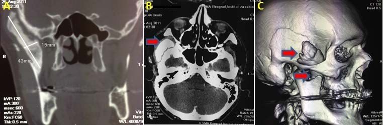
Vol. 78, No. 2                                      VOJNOSANITETSKI PREGLED                                          Page 257

peripheral zone revealed ossification and mature lamellar       uneventful. The patient’s maximum incisal opening was 30
bone (Figure 2). The histological finding was consistent        mm without assistance or pain. However, the CT scan
with myositis ossificans.                                       revealed an osseous lesion in the right temporal fossa, in the
     Postoperatively, the patient was able to open his mouth    vicinity, but not attached to the resected coronoid processus,
passively up to 25 mm without assistance.                       extending to the temporal muscle. The radiological features
     The patient presented to our Clinic for a follow-up        of the lesion were similar to the previous findings (Figure 3).
evaluation two years after undergoing the right mandibular           Since there were no functional problems, the patient
coronoidectomy, partial resection of the right temporal         was scheduled for a follow-up examination after 6 months.
muscle tendon, and extirpation of the osseous tissue from the   Two years later, the CT scan was repeated (2015), and the
temporal fossa. He reported difficulties in mouth opening in    revealed condition was unchanged; however, the ossification
the morning but no restriction or pain in mouth opening         extended towards the mandibular attachment of the right
during the day. Extraoral and intraoral examinations were       medial pterygoid muscle (Figure 4).

     Fig. 2 – Photomicrograph of an excised extraosseous bony lesion: A) Section of specimen showing central loose
    connective tissue with immature bone containing osteocytes and mature bone at the periphery (hematoxylin and
    eosin – H&E stain, ×100); B) Photomicrograph showing central connective tissue zone surrounded by immature
           bone containing osteocytes in the lacunae (H&E stain, ×200); C) Periodic acid-Schiff (PAS) staining.

             Fig. 3 – Postoperative computed tomography (CT) 2 years after surgical therapy demonstrating
               recurrence of heterotopic calcification in the right temporal muscle (red arrows). Coronoid
                                 processus is resected. A) Coronal view; B) Sagittal view.

     Fig. 4 – Postoperative computed tomography (CT) 4 years after surgical therapy demonstrating recurrence of
    heterotopic calcification in the right temporal muscle (red arrows) extending towards the right medial pterygoid
               muscle (yellow arrow). Note demarcated ectopic bone formation in the right temporal fossa.
                                      A) Axial view; B) Sagittal view; C) Coronal view.

Jović S, et al. Vojnosanit Pregl 2021; 78(2): 255–260.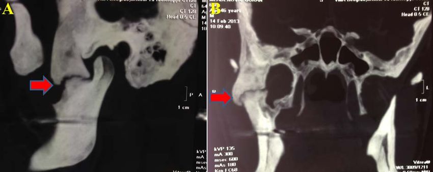
Page 258 VOJNOSANITETSKI PREGLED Vol. 78, No. 2
The patient was referred again to our Clinic in April visualized and partially extirpated. Due to poor visibility and
2017 because of a two-week-long inability to open his intraoperative bleeding, an extraoral submandibular incision
mouth. The CT scan revealed enlargement of the ossified was performed. With the preservation of major vascular and
mass in the right temporal muscle extending from the nerve structures, the right temporal fossa was approached
temporal fossa to the muscular space affecting the medial and an osseous lesion was identified in the temporal muscle
pterygoid muscle (Figure 5). extending towards the medial pterygoid muscle tendon. The
The decision on the surgical treatment was made. An osseous mass was extirpated. The maximal incisal opening
intraoral incision was made along the external oblique ridge was 40 mm postoperatively. Macroscopically, the tissue
of the mandible, and a calcified mass extending from the resembled the previously extirpated tumor, and the
resected mandibular coronoid to the temporal muscle was histological finding was similar (Figure 6).
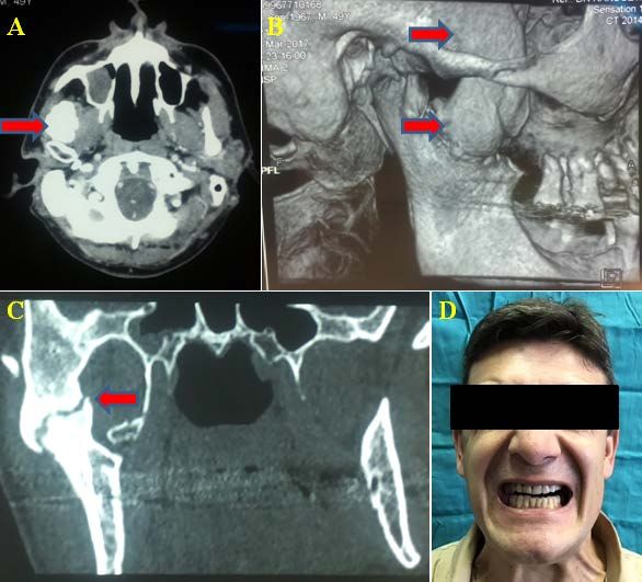
Fig. 5 – Postoperative computed tomography (CT) 6 years after surgical therapy
demonstrating recurrence of heterotopic calcification in the right temporal muscle (arrow).
Note fusion of calcification in temporal muscle to resected coronoid processus.
A) Axial view; B) 3D reconstruction; C) Coronal view; C) Sagittal view; D) Preoperative
photograph of the patient showing minimal mouth opening.
Fig. 6 – Photomicrograph of an excised extraosseous bony lesion: A) Section of specimen showing central loose
connective tissue with immature bone containing osteocytes and mature bone at the periphery (hematoxylin and
eosin – H&E stain, ×100); B) and C) Photomicrograph showing central connective tissue zone surrounded by
immature bone containing osteocytes in the lacunae (H&E stain, ×200).
Jović S, et al. Vojnosanit Pregl 2021; 78(2): 255–260.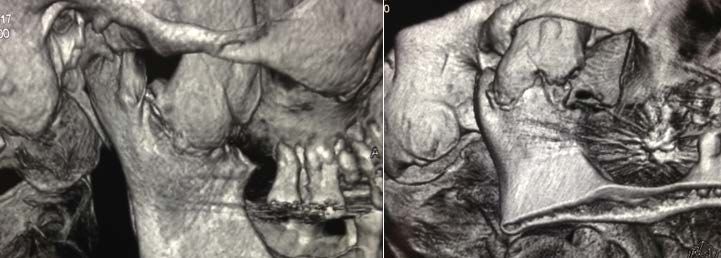
Vol. 78, No. 2                                      VOJNOSANITETSKI PREGLED                                           Page 259

     Fig. 7 – Postoperative computed tomography (CT) 3D reconstruction 6 months after second surgical treatment
       showing resected coronoid processus, and ectopic calcified mass in temporal and medial pterygoid muscle.

       After the patient was released from the Clinic, he was    most interesting clinical feature for discussion were
able to open his mouth passively up to 10 mm without any         difficulties in diagnosis and differential diagnosis. The
assistance, and he was provided with physical treatment          diagnosis of MOT was based on the clinical picture,
consisting of aggressive mandibular range-of-motion              radiological examination, and histological findings. Bone
exercises. At the regular check-ups, the mouth opening was       formation was confined to the tendon of the temporal
still reduced to 10 mm, and the patient was sent to perform      muscle and mandibular coronoid processus with no other
the proposed physical therapy. On the CT scan follow-up 6        areas of ectopic bone formation. Laboratory tests revealed
months after the surgery, the ectopic calcified mass affecting   no specific abnormalities of bone metabolism. However,
the right temporal and medial pterygoid muscles was still        even after mandibular coronoidectomy and resection of the
present (Figure 7).                                              temporal muscle tendon, the recurrence of the disease was
                                                                 observed after two years. Furthermore, seven years
     Discussion                                                  following the surgical treatment, the patient had limited
                                                                 mouth opening because of the enlargement of the ectopic
      Although MOT of masticatory muscles presents a             bone and the affection of the nearby medial pterygoid
benign reparative ectopic bone formation in muscles,             muscle by the ossification process. The following surgical
accompanying trismus is a major functional problem to the        treatment included extirpation of the osseous lesion in the
patients. Initial trauma causes an inflammatory response in      temporal muscle and the medial pterygoid muscle tendon.
the muscle and periosteum with subsequent displacement of        The uneventful postoperative course and relapse of the
bone fragments and osteoprogenitor cells into muscle             disease could be linked to the ongoing inflammatory
bundles, which induce ectopic bone formation 3–6. However,       process and activity of osteoprogenitor cells, leading to a
in about 25% of cases, a history of trauma is not found 1.       continuous process of ectopic bone formation, as proposed
      Previous reports found that the most commonly              in other trials 1–3.
affected masticatory muscles were the masseter and medial              MOT represents a significant diagnostic challenge for
pterygoid muscle, but in several reports, more than one          clinicians. The main criteria for diagnosing MOT include a
muscle was affected 1–3. Reports of affection of more than       history of local injury, clinical and radiological evidence of
one muscle indicate that the inflammatory response               ossification within two months following the injury, and
following trauma is not localized on one individual              localization of the ossification in the muscle tissue 8. The
muscle 7. Most MOT lesions were caused by direct trauma          differential diagnosis for MOT includes other benign bone-
to the masseter muscle or trauma to the medial pterygoid         forming lesions, such as fibro-osseous dysplasia progressive,
muscle after local anesthetic injection 1. Temporal muscle       calcified fibromatosis, phleboliths, osteoma, osteoblastoma,
is not commonly affected with MOT. In the present case,          but     also     malignancies,    such      as   osteosarcoma,
MOT could not be linked to any apparent trauma to the            chondrosarcoma, and rhabdomyosarcoma 9.
head except the mandibular anesthesia and extraction of the            The radiographic appearance of MOT depends on the
third molar, which occurred two months before the initial        maturity of the lesion 10. Radiologically, Shirkoda et al. 10
symptoms began. However, this type of surgical trauma            described 4 phases of MOT. The initial phase is
would not cause direct trauma to the temporal muscle.            characterized by inflammation and mesenchymal stem cell
There are indices that chronic subclinical infection, which      proliferation, without calcification. Initial bone formation is
often accompanies third molars due to the pericoronitis,         seen 1–2 weeks after trauma. The intermediate phase with
could lead to the inflammation and periosteal reaction and       peripheral ossification is seen after 4 weeks. The mature
subsequently cause bone formation 1–3. In the present case,      phase is seen after 6 weeks, and the lesion appears as a
the trigger of ectopic ossification is still questionable. The   central radiolucency surrounded by peripheral mature bone.

Jović S, et al. Vojnosanit Pregl 2021; 78(2): 255–260.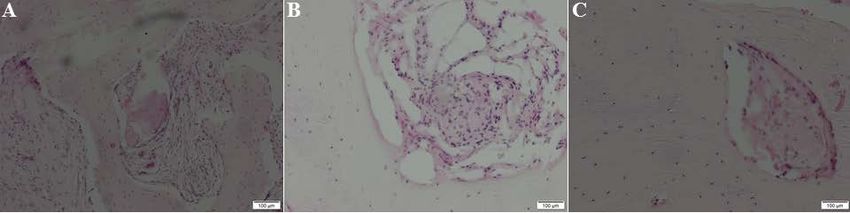
Page 260                                            VOJNOSANITETSKI PREGLED                                                  Vol. 78, No. 2

During this phase, the lesion is well delineated from the              functional impairment. Several trials reported the use of anti-
surrounding tissue, and surgical treatment could be                    inflammatory drugs, radiotherapy, and drugs affecting bone
performed with minimal adverse events. CT scan is sensitive            metabolism as means of controlling postoperative
for identifying ossification. The radiological appearance of           inflammation and the ossifying potential of the tissue 6.
MOT is consistent with the zonal histological pattern of the           Other treatment modalities include physical therapy, acetic
lesion with a well-circumscribed ossified periphery and a              acid iontophoresis, magnesium therapy, and bisphosphonate
low attenuating central portion. Early lesions appear as               therapy 1. However, these reports are confined to single case
amorphous calcifications within the soft tissue, while mature          studies, and the development of new treatment methods is
lesions are well separated from the surrounding bone by a              needed.
thin radiolucent area; however, older lesions can appear
attached to the adjacent bone 11.                                           Conclusion
      Histologically, the hallmark of MOT is the zonal
pattern 12, 13. The central or cellular zone represents the                  MOT represents a major diagnostic and therapeutic
innermost region of the lesion, showing mitotic activity,              challenge for surgeons. It is fundamental that patients with
undifferentiated cells, necrotic muscular tissue, giant                an unspecific clinical history leading to trismus are referred
multinucleated cells, and loose fibrovascular tissue. The              to specialized centers for diagnosis. When MOT of
middle or intermediate zone contains active osteoblasts and            masticatory muscles is suspected, a CT scan could be both a
immature osteoid. The peripheral or outer zone of the lesion           diagnostic and prognostic radiological tool. Surgical
shows mature bone with active osteoclasts and collagenous              treatment remains the treatment of choice and should involve
fibrous stroma. The microscopic and radiographic zone                  excision of osseous lesion and osteotomy of muscle
pattern is strongly suggestive of a reactive lesion and helps          attachment region of the bone. In cases when several muscles
rule out a diagnosis of sarcoma 11, 14.                                are affected, surgical treatment may resolve a major
      Standard treatment of MOT is surgical excision of the            masticatory disfunction. Thus, further research is needed to
ossification along with osteotomy. In the mature phase, the            clarify the mechanisms of ossification in order to develop
ectopic bone is well demarcated from the surrounding tissue,           conservative treatment approaches.
and it is easiest to excise 9. Several authors proposed
interposition of soft tissue graft between resected bone and                Acknowledgements
muscle to prevent the bone and hematoma formation 3–6.
Although wide surgical excision of the lesion is performed,                 This study was supported by the Serbian Ministry of
relapses are often reported. When several muscles are                  Education, Science and Technological Development, grant
affected, surgical treatment may resolve significant                   no. 175021.

                                                      R E F E R E N C E S
1. Aoki T, Naito H, Ota Y, Shiiki K. Myositis ossificans traumatic
   of the masticatory muscles: Review of the literature and report     9. Jayade B, Adirajaiah S, Vadera H, Kundalaswamy G, Sattur AP,
   of a case. J Oral Maxillofac Surg 2002; 60(9): 1083‒8.                   Kalkur C. Myositis ossificans in medial, lateral pterygoid, and
2. Kim DD, Lazow SK, Har-El G, Berger JR. Myositis ossificans               contralateral temporalis muscles: A rare case report. Oral
   traumatica of masticatory musculature: A case report and liter-          Surg Oral Med Oral Pathol Oral Radiol 2013; 116(4): e261‒6.
   ature review. J Oral Maxillofac Surg 2002; 60(9): 1072‒6.           10. Wang SY, Lomasney LM, Demos TC, Hopkinson WJ. Radiologic
3. Schiff MJ, Meara DJ. Myositis ossificans of the temporalis mus-          case study. Traumatic myositis ossificans. Orthopedics 1999;
   cle: Case report and review of the literature. J Oral Maxillofac         22(1000): 991‒5.
   Surg 2013; 71(11): 1893‒8.                                          11. Shirkoda A, Armin AR, Bis KG, Makris J, Irwin RB, Shetty AN.
4. Torres AM, Nardis AC, Da Silva RA, Savioli C. Myositis ossifi-           MR imaging of myositis ossificans: Variable patterns at dif-
   cans traumatica of the medial pterygoid muscle following a               ferent stages. J Magn Reson Imaging 1995; 5(3): 287‒92.
   third molar extraction. Int J Oral Maxillofac Surg 2015; 44(4):     12. Radunović A, Košutić M, Vulović M, Milev B, Janjušević N, Ivošević
   488‒90.                                                                  A, et al. Ilizarov method as limb salvage in treatment of mas-
5. Guarda-Nardini L, Piccotti F, Ferronato G, Manfredini D. Myositis        sive femoral defect after unsuccessful tumor arthroplasty.
   ossificans traumatica of the temporalis muscle: A case report            Vojnosanit Pregl 2016; 73(8): 779‒82.
   and diagnostic considerations. Oral Maxillofac Surg 2012;           13. Geist JR, Bhatti P, Plezia RA, Wesley RK. Fibrodysplasia ossifi-
   16(2): 221‒5.                                                           cans circumscripta of the masseter muscle. Dentomaxillofac
6. Thangavelu A, Vaidhyanathan A, Narendar R. Myositis ossificans          Radiol 1998; 27(3): 182‒5.
   traumatica of the medial pterygoid. Int J Oral Maxillofac Surg      14. Both DW, Westers BM. The management of athletes with myo-
   2011; 40(5): 545‒9.                                                     sitis ossificans traumatic. Can J Sport Sci 1989; 14(1): 10‒6.
7. Arima R, Shiba R, Hayashi T. Traumatic myositis ossificans in
   masseter muscle. J Oral Maxillofac Surg 1984; 42(8): 521‒6.                                                 Received on August 6, 2018.
8. Reddy SPD, Prakash AP, Keerthi M, Rao BJ. Myositis ossificans                                                Revised on April 19, 2019.
   traumatica of temporalis and medial pterygoid muscle. J Oral                                                   Accepted April 23, 2019.
   Maxillofac Pathol 2014; 18(2): 271‒5.                                                                          Online First April, 2019.

                                                                                      Jović S, et al. Vojnosanit Pregl 2021; 78(2): 255–260.
You can also read