Computational modelling of cervix radiation procedure using a virtual anthropomorphic phantom and the MCNPX code
←
→
Page content transcription
If your browser does not render page correctly, please read the page content below
Journal of Physics: Conference Series

PAPER • OPEN ACCESS

Computational modelling of cervix radiation procedure using a virtual
anthropomorphic phantom and the MCNPX code
To cite this article: William S. Santos et al 2021 J. Phys.: Conf. Ser. 1826 012040

View the article online for updates and enhancements.

 This content was downloaded from IP address 46.4.80.155 on 25/05/2021 at 19:26

10th Brazilian Congress on Metrology (Metrologia 2019) IOP Publishing
Journal of Physics: Conference Series 1826 (2021) 012040 doi:10.1088/1742-6596/1826/1/012040

Computational modelling of cervix radiation procedure using
a virtual anthropomorphic phantom and the MCNPX code

 William S. Santos a,b,*, Lucio P. Neves a,c, Ana P. Perini a,c, Carla J. Santos c,
 Walmir Belinato d, Rogerio M. V. Silva e, Maria R. Soares f, Caio C. Valeriano g,
 Linda V. E. Caldas b
 a
 Instituto de Física, Universidade Federal de Uberlândia (INFIS/UFU), Uberlândia,
 MG, Brazil.
 b
 Instituto de Pesquisas Energéticas e Nucleares, Comissão Nacional de Energia
 Nuclear (IPEN-CNEN/SP), São Paulo, SP, Brazil.
 c
 Programa de Pós-Graduação em Engenharia Biomédica, Faculdade de Engenharia
 Elétrica, Universidade Federal de Uberlândia, Uberlândia, MG, Brazil.
 d
 Instituto Federal da Bahia (IFBA), Vitória da Conquista, BA, Brazil.
 e
 Hospital Haroldo Juçaba, Fortaleza, CE, Brazil.
 f
 Fundação Universidade Federal de Rondônia (UNIR), Porto Velho, RO, Brazil.
 g
 Programa de Pós-Graduação em Educação, Universidade Tiradentes, Aracaju, SE,
 Brazil.

 *william@ufu.br

 Abstract. There is a serious, and growing concern about the increased risk of the emergence of
 a secondary cancer, radio-induced, associated with radiotherapy treatments. To assess the radiation
 doses to organs outside the target volume, in this work, several computational exposure scenarios
 were modelled, based on Monte Carlo simulation (MCNPX code). A Varian 2100c accelerator,
 and a female virtual anthropomorphic phantom were used, in a simulated treatment of cervical
 cancer. The determination of the dispersed dose would be important for assessing the risk in
 different organs or tissues. Four treatment fields were applied, varying the gantry angle. It was
 possible to observe that the conversion factors for equivalent dose were higher for the AP
 projection. For the RLAT and LLAT projections, the results were similar, fact that may be
 attributed to the symmetrical distributions of the organs in relation to the radiation source. The
 results presented in this work showed that the computational exposure scenario provides a versatile
 and accurate tool to estimate in a ready way the absorbed doses during a cervical treatment.
 Keywords: Dosimetry, radiotherapy, Monte Carlo simulation, MCNPX, anthropomorphic
 phantom

 Content from this work may be used under the terms of the Creative Commons Attribution 3.0 licence. Any further distribution
 of this work must maintain attribution to the author(s) and the title of the work, journal citation and DOI.
Published under licence by IOP Publishing Ltd 1
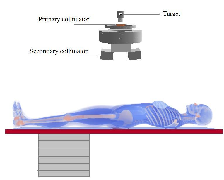
10th Brazilian Congress on Metrology (Metrologia 2019) IOP Publishing
Journal of Physics: Conference Series 1826 (2021) 012040 doi:10.1088/1742-6596/1826/1/012040

1. Introduction
According to Bray et al. [1] the cervix cancer is the sixth most common cancer type in women
worldwide. This type of tumor is the third most frequent among the women population, behind the breast
and colorectal cancer. It occupies the fourth place in the cause of women death [2]. With the diagnosis
of cervical cancer, treatment options vary, and depending on the stage external radiotherapy may be
indicated. Despite the treatment efficiency, radiotherapy may cause injuries at patients, since healthy
tissues located closer, or inside, the treated area are exposed to the photon beam. These effects are
characterized as acute effects, such as erythema or a late effect such as a secondary tumor. As presented
in several works in the literature [3 - 8], the determination of the dispersed dose would be important for
assessing the risk and biological consequences in different organs or tissues. This subject is of great
concern, mainly when new treatment techniques may increase the leakage radiation to patients.
 The absorbed doses to organs and tissues outside the radiation planning field are unavoidable during
the treatment, and may contribute to the induction of a secondary cancer. These doses are derived from
the scattered radiation throughout the patient's body, walls, floor and ceiling of the radiotherapy room,
head, collimators and other structures of the LINAC [9].
 Most studies found in the literature are focused on the determination of absorbed dose distributions
inside the treatment planned area [10,11]. These works aimed the modelling of the main components of
the beam line, such as the target, primary collimator, jaws, and the flattening filter. The presence of the
patient, which is more important to scattered radiation, was not considered.
 The Monte Carlo (MC) simulation has been widely used to model primary treatment fields of
various LINAC models, such as Varian, Siemens and Elekta [4]. To determine the doses in different
organs and tissues, the Monte Carlo simulation is used coupled with virtual anthropomorphic phantoms.
The aim of this study is to estimate the absorbed doses in tissues and organs located outside the volume
of the radiotherapeutic planning. In this study the 2D planning mode was considered for cervix cancer.
For this, a computational model was developed using Monte Carlo simulation coupled with the
computational anthropomorphic phantom (FASH3), representing the patient, and a Varian 2100c
LINAC. The results are presented in the format of conversion factors (CF).

2. Material and Methods

2.1. Computational model description
In this work the MCNPX software (version 2.7.0) [12] was used to model the scenario with a medical
Varian 2100c LINAC. The photon and electron transport were carried out based on the ENDF/B-VI8
cross section libraries, with a cutoff energy of 0.01MeV for photons, and 0.1MeV for electrons. The
photon transport was considered including photoelectric absorption with the possibility of fluorescent
emission and Auger electron, coherent and incoherent scattering. The Varian 2100c LINAC, using a
6 MV photon beam, was modelled with the target, wedge filter, jaws and a detailed 80 leaf multi-leaf
collimator (MLC) [5,6]. The primary and secondary collimators, as well as other structures that may
affect the leakage and scattering radiation were also considered. The validation was obtained comparing
the simulated results with those obtained experimentally.
 An 8×8×3 m³ radiotherapy room with walls and concrete ceiling (2.43 g/cm³ density) with 1 m
thickness was modeled, as well as the extra slabs for primary radiation shielding. In addition, the floor
has 20 cm thick concrete. Besides the equipment, the room was filled with atmospheric air
(0.001205 g/cm³ density). Figure 1 presents the computational scenario with the adult phantom on the
positioning table, the gantry of the LINAC, the positioning table made of carbon fiber, with 10 cm
thickness, 17 cm wide and 200 cm length, supported by a metallic base. The adult patient was
represented by a female virtual anthropomorphic phantom, developed at the Federal University of
Pernambuco (UFPE – Brazil) [13]. This phantom has 60 kg of body mass and 1.63 m of height and is
composed of more than a hundred organs and tissues with dosimetric importance. It was formed with
voxels of dimensions 2.4×2.4×2.4 cm³, each one. To avoid unnecessary exposure of the patient's arms

 2
10th Brazilian Congress on Metrology (Metrologia 2019) IOP Publishing
Journal of Physics: Conference Series 1826 (2021) 012040 doi:10.1088/1742-6596/1826/1/012040

and, consequently, higher doses in the red bone marrow (RBM) and bone surface (BS), as performed in
a real treatment, the arms of this phantom were removed.

Figure 1. Computational model employed in this work for the radiotherapy procedure.

 During the treatment, four treatment fields were applied. The radiation field was directed to the
cervix. The treatment fields, and gantry angles, were: 16×16 cm2 (projections antero-posterior (AP/0°)
and postero-anterior (PA/180°) and 16×12 cm2 (projections right lateral (RLAT/90°) and left lateral
(LLAT/270º). The MCNP mesh tally was used to determine the photon fluence profile and to test the
beam collimation, with the LINAC operating at 6 MV with the MLC.

2.2. Scenario validation
The validation of the beam properties was done by means of comparisons of the percentage depth dose
(PDD) and dose profiles (Off-axis), calculated with the results of experimental measurements. The PDD
results were simulated at various depths within a water phantom, using the tally *F8 (MeV/particle-
source) to evaluate the energy deposited in the water, along the central axis of the field.
 To determine the PDD and the Off-axis, it was necessary to model a set of voxels to represent the
radiation detectors. The first set consists of 400 voxels distributed along the central axis and the second
set with 100 voxels along the axial axis. The energy deposited in each voxel of the central and the axial
axes were used to determine the PDD and Off-axis, respectively. For the determination of the PDD,
voxels with dimensions 2.5×2.5×0.1 cm3 were used, and for Off-axis calculations 1.0×1.0×0.4 cm3
voxels were used. The PDD calculations were made with the detectors spaced 1 mm apart, and during
experimental measurements, they were spaced 0.5 cm apart.
 A Gaussian spatial distribution was chosen for the electron source, along the xy axis, with a half
height width (FWHM) of 0.13 cm. The PDD and Off-axis simulations were performed using a PMMA
phantom of dimensions 40×40×40 cm³ filled with water. The phantom has an open top and 1 cm thick
PMMA on its sides.
 The Varian 2100c LINAC was operated (experimentally) with a voltage of 6 MV and the photon
beam irradiating a field of 10×10 cm² with a source-surface distance (SSD) of 100 cm. Figure 2 shows
the computational model used to determine the dose profile (Off-axis) and PDD.

 3
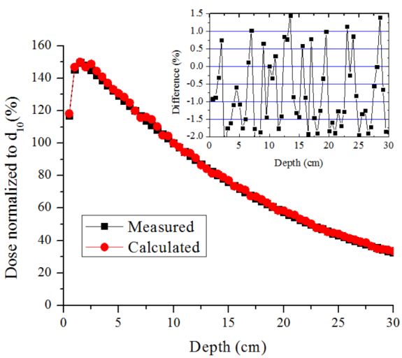
10th Brazilian Congress on Metrology (Metrologia 2019) IOP Publishing
Journal of Physics: Conference Series 1826 (2021) 012040 doi:10.1088/1742-6596/1826/1/012040

Figure 2: Computational model used in determination of the PDD and Off-axis ratio.
 The PDD and Off-axis measurements were performed using a Farmer ionization chamber (model
FC65/P – IBA dosimetry) with a sensitive volume of 0.6 cm³. The PDD was measured with the
ionization chamber positioned along the central axis with depths ranging from 0.5 to 30 cm in 0.5 cm
steps. The Off-axis was measured along the axial axis with 0.025 cm steps. PDD and Off-axis were
normalized by the absorbed dose at 10 cm depth on the central axis.

2.3. Dosimetric calculations
A set of absorbed doses in organs and tissue located inside, or in the proximity, of the target area during
cervix radiotherapy, for an adult patient, were determined. The dosimetric results were due to the
scattered primary photons and secondary electrons. The absolute dose of a set of organs and tissues with
dosimetric importance, recommended by the International Commission on Radiological Protection [14],
were calculated using Equation 1. The assumed prescribed treatment delivered 45 Gy to the target area
of the cervix. This amount was divided into 28 fractions, where each day 1.8 fractions were delivered.
The treatment took 25 days, divided in 5 days groups in a row, as normally carried out in these
treatments.

 ( ) = (1)

where is the estimated absorbed dose in each organ or tissue T, is the dose in the cervix volume

and is the total dose prescribed for all radiotherapy treatment. In this study, the ratio is determined

as the conversion factor (CF).
 The dose deposition was determined using the tally F6:p (MeV/g/source-particle), which could be
converted into Gy, for all organs and tissues, except for the red bone marrow (RBM), where the tally
∗F4 (MeV/cm2/particle) was used. For all simulations, to determine the organ doses 5.0E8 particles
histories were used. To assess the beam properties (PDD, Off-axis and photon fluence) 1.0E9 particles
histories were used, providing results with less than 2% uncertainty.

3. Material and Methods

3.1. Dosimetric properties of the simulated radiation beam
Figure 3 shows the comparison between the PDD curves calculated with the MCNPX and those
measured with the ionization chamber, at various depths in the water phantom, for a field size of

 4
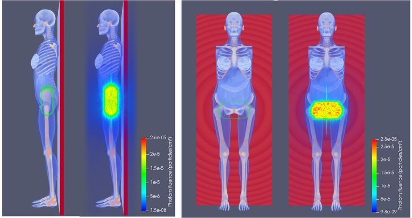
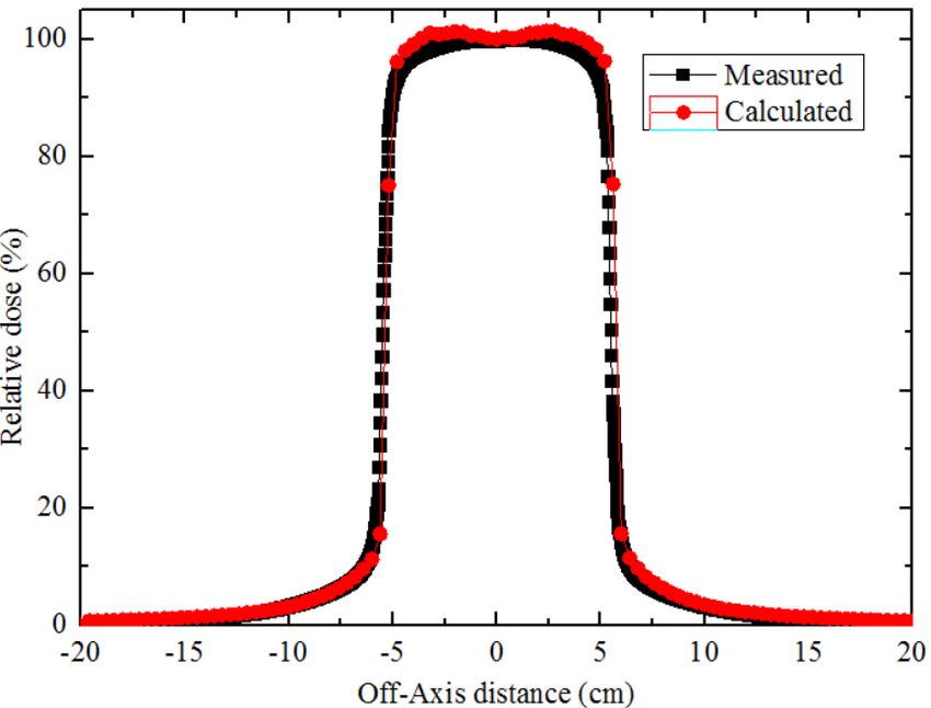
10th Brazilian Congress on Metrology (Metrologia 2019) IOP Publishing
Journal of Physics: Conference Series 1826 (2021) 012040 doi:10.1088/1742-6596/1826/1/012040

10×10 cm². The results were normalized to a dose corresponding to a depth of 10 cm (d 10), a region of
more stability than dmax. The difference between the experimental and simulated results is within ±2%.

Figure 3. Measured and calculated (MCNPX) PDD for a Varian 2100c with 6 MV beam.

 The lateral profiles, measured and calculated, at a depth of 10 cm, are shown in Figure 4. It is noted
that the distances of the detectors decrease with respect to the center of the beam. The simulated Off-
axis is slightly overestimated in the region between -5 cm and -2.5 cm and 2.5 cm and 5 cm. These
differences (< 4%) between the simulated and experimental results are attributed to the small volume of
the detectors, immersed in the water phantom. However, this does not affect the quality of the results.
In the overall set of results, the Off-axis ratio is practically uniform in the field of 10×10 cm2, showing
that the geometry of the flattening filter and the modelled collimators are correct. In the regions near the
edge, the calculated and experimental results presented differences of less than 2%.

Figure 4. Measured and calculated (MCNPX) lateral profile – Varian 2100c LINAC with 6 MV beam
and 10×10 cm² field.

 Besides the dose profile, the beam collimation, for the 16×16 cm2 (AP/PA) and 16×12 cm2
(RLAT/LLAT) reference fields with the MLC were also evaluated. In this part of the work, the mesh
tally MCNPX command was utilized. This command allowed the creation of the photon fluence maps

 5
10th Brazilian Congress on Metrology (Metrologia 2019) IOP Publishing
Journal of Physics: Conference Series 1826 (2021) 012040 doi:10.1088/1742-6596/1826/1/012040

for the four projections studied in this work. In Figure 5, it is possible to view that the radiation field
margins with MLC are properly delimited, confirming the correct collimation.

 (a) (b)
Figure 5. Photon fluence maps (particles/cm2): View of the beam collimation for right
lateral projection RLAT (a) and antero-posterior AP projection (b) of the treated region of
the FASH3 anthropomorphic phantom.

3.2. Dosimetric results
The CF values were calculated, as a function of the gantry angle, for absorbed doses of a set of organs
and tissues located inside and outside the treatment region. The results of these factors by projection,
and the absorbed dose of the complete treatment, are shown in Table 1. Using the mean CF value, by
projection of each organ, the absolute absorbed dose was determined for the major organs/tissues, as
suggested by the ICRP 103 [14].
 It is possible to observe that the highest dose values were obtained for organs located near, or within,
the irradiated volume (cervix), such as large intestine (colon), small intestine, gonads and bladder. The
doses calculated for the bladder (44.3 Gy) and gonads (45.6 Gy), which are within the irradiated area,
received a dose similar to the prescribed dose of the cervix (45 Gy).
 Another important fact is that for the same field size (AP/PA), the results of the CF values for AP/0°
projection, were mostly higher than for the PA/180° projection. In these cases, the gantry is under the
table, which may shield the low energy photons. The spine of the patient may also shield these photons,
reducing the dose for the organs located in this region. For the RLAT/90º and LLAT/270º projections,
the CF values were mostly similar, fact that can be attributed to the symmetrical distributions of the
organs in relation to the radiation source. For the same irradiation conditions, the skin and the bone
surface, which are evenly distributed throughout the human body, the differences in CF values were
small, i.e., they do not depend on the irradiation position.
 In addition to these differences, it is important to note that the breasts, organs located superficially
and distant from the target organ, obtained an average absorbed dose of 0.30 Gy, which is a value close
to the mean value calculated for the heart (0.29 Gy). This fact can be associated with the contribution of
scattered radiation from the collimators and the leakage radiation of the LINAC head. The organs

 6
10th Brazilian Congress on Metrology (Metrologia 2019) IOP Publishing
Journal of Physics: Conference Series 1826 (2021) 012040 doi:10.1088/1742-6596/1826/1/012040

relatively distant from the non-targeted ones presented the lowest CF values, since they received just
the scattered radiation.
 The percentage relative uncertainty of each organ and tissue estimated by the MCNPX code is
presented between parentheses. The eye and eyes lenses have relatively small volumes and, therefore,
the statistical uncertainties of these organs were the largest, and they varied between 7% and 27%. For
all other organs and tissues, in all evaluated situations, the uncertainties in the CF values were below
4%.

Table 1. CF values for absorbed doses per beam projection for a treatment using a 2D planning system.
The relative uncertainties of the simulations are shown in parentheses.
 Conversion Factor (CF) Average
Organs and tissues AP LLAT PA RLAT dose (Gy)
 Bone marrow 2.55E-02 (0.3%) 5.26E-02 (0.4%) 2.67E-02 (0.3%) 5.94E-02 (0.4%) 1.85E+00
 Colon 2.52E-01 (0.1%) 2.93E-01 (0.2%) 2.74E-01 (0.1%) 2.89E-01 (0.2%) 1.25E+01
 Lung 6.46E-03 (0.4%) 5.63E-03 (0.6%) 6.76E-03 (0.4%) 5.53E-03 (0.6%) 2.74E-01
 Stomach 2.36E-02 (0.5%) 1.63E-02 (0.8%) 1.92E-02 (0.5%) 1.66E-02 (0.8%) 8.51E-01
 Breast 9.05E-03 (0.6%) 6.03E-03 (1.0%) 5.69E-03 (0.7%) 6.14E-03 (1.0%) 3.03E-01
 Remainder tissues 1.80E-02 (0.0%) 2.37E-02 (0.1%) 1.60E-02 (0.1%) 2.29E-02 (0.1%) 9.07E-01
 Gonads 1.08E+00 (0.3%) 1.03E+00 (0.5%) 8.92E-01 (0.3%) 1.05E+00 (0.4%) 4.56E+01
 Bladder 1.14E+00 (0.2%) 9.12E-01 (0.3%) 9.07E-01 (0.2%) 9.85E-01 (0.3%) 4.43E+01
 Oesophagus 5.84E-03 (1.5%) 5.44E-03 (2.1%) 5.62E-03 (1.3%) 5.48E-03 (2.3%) 2.52E-01
 Liver 2.09E-02 (0.4%) 1.16E-02 (0.6%) 1.73E-02 (0.4%) 1.17E-02 (0.6%) 6.93E-01
 Thyroid 4.81E-03 (2.6%) 2.87E-03 (4.1%) 2.54E-03 (3.3%) 2.53E-03 (4.0%) 1.43E-01
 Bone surface 1.26E-01 (0.1%) 1.89E-01 (0.1%) 1.03E-01 (0.1%) 1.91E-01 (0.1%) 6.85E+00
 Brain 1.66E-03 (1.1%) 1.87E-03 (1.3%) 1.23E-03 (1.3%) 1.89E-03 (1.3%) 7.48E-02
 Salivary glands 3.72E-03 (1.7%) 2.63E-03 (2.2%) 1.68E-03 (2.3%) 2.63E-03 (2.1%) 1.20E-01
 Skin 9.01E-02 (0.1%) 7.55E-02 (0.1%) 6.94E-02 (0.1%) 7.77E-02 (0.1%) 3.52E+00
 Uterus 1.00E+00 (0.2%) 1.00E+00 (0.2%) 1.00E+00 (0.2%) 1.00E+00 (0.2%) 4.50E+01
 Heart 7.26E-03 (0.9%) 5.99E-03 (1.2%) 6.44E-03 (0.8%) 5.88E-03 (1.2%) 2.88E-01
 Thymus 6.71E-03 (2.3%) 4.59E-03 (3.3%) 4.40E-03 (1.8%) 4.67E-03 (3.4%) 2.29E-01
 Pancreas 5.33E-02 (0.5%) 2.32E-02 (1.0%) 4.25E-02 (0.5%) 2.35E-02 (1.0%) 1.60E+00
 Spleen 1.49E-02 (1.0%) 1.40E-02 (1.1%) 1.36E-02 (1.0%) 1.36E-02 (1.1%) 6.30E-01
 Lymphatic nodes 1.65E-01 (0.1%) 1.90E-01 (0.2%) 1.19E-01 (0.1%) 2.33E-01 (0.2%) 7.95E+00
 Adrenals 1.60E-02 (1.8%) 1.20E-02 (2.3%) 1.57E-02 (1.6%) 1.19E-02 (2.3%) 6.25E-01
 Gall bladder 4.57E-02 (1.0%) 1.74E-02 (1.5%) 3.25E-02 (1.0%) 1.79E-02 (1.6%) 1.28E+00
Extra-thoracic region 3.69E-03 (2.5%) 2.20E-03 (4.2%) 1.68E-03 (3.5%) 2.28E-03 (4.0%) 1.11E-01
 Kidneys 2.31E-02 (0.5%) 1.74E-02 (0.8%) 2.35E-02 (0.4%) 1.67E-02 (0.9%) 9.09E-01
 Oral mucosa 3.27E-03 (2.2%) 2.56E-03 (2.8%) 1.35E-03 (2.8%) 2.68E-03 (2.9%) 1.11E-01
 Small intestine 2.91E-01 (0.1%) 2.39E-01 (0.2%) 1.97E-01 (0.1%) 3.12E-01 (0.2%) 1.17E+01
 Muscle 1.62E-02 (0.0%) 2.21E-02 (0.1%) 1.47E-02 (0.1%) 2.09E-02 (0.1%) 8.31E-01
 Eyes 3.03E-03 (3.3%) 2.05E-03 (4.8%) 8.79E-04 (6.5%) 2.15E-03 (6.0%) 9.12E-02
 Eye lens 3.08E-03 (8.8%) 2.09E-03 (16%) 1.09E-03 (27%) 1.79E-03 (14%) 9.05E-02

4. Conclusions
The gantry angles considerably affected the results. The majority of the organs near or inside the target
volume were those that received the highest radiation doses. Regarding the influence of the gantry angle,
the higher CF values were obtained for the AP/0º projection. Usually, the absorbed dose of the organs
depends on the location, distribution of the organs in the body and the distance of the treated volume.
Comparing the dose values with the prescribed value for the cervix, the organs and tissues outside the
treated region that received the highest doses were the large intestine (28%), small intestine (26%), the
lymph nodes and the bone surface (15%).

 7
10th Brazilian Congress on Metrology (Metrologia 2019) IOP Publishing
Journal of Physics: Conference Series 1826 (2021) 012040 doi:10.1088/1742-6596/1826/1/012040

 During the treatment, it is impossible to measure experimentally the doses due to the scattered
radiation within the patient's body. Therefore, the CF values calculated in this study will be useful to
readily estimate the absorbed doses during a cervix treatment. Besides the traditional analytical planning
systems, the computational modelling presented in this study may be an alternative tool for dosimetry
calculations.

Acknowledgements
The authors would like to thank Dr. Richard Kramer for kindly providing the virtual anthropomorphic
phantoms used in this work. This work was partially supported by the Brazilian agencies: Fundação de
Amparo à Pesquisa do Estado de Minas Gerais (FAPEMIG, Grants No. APQ-03049-15 and APQ-
02934-15), Conselho Nacional de Desenvolvimento Científico e Tecnológico (CNPq Grants No.
421603/2016-0, 420699/2016-3 and 301335/2026-8) and Fundação de Amparo à Pesquisa do Estado de
São Paulo (FAPESP, Grant No. 2018/05982-0).

References
[1] Bray F, Soerjomataram I, Siegel RL, Torre LA, Jemal A. Global cancer statistics 2018:
 GLOBOCAN estimates of incidence and mortality worldwide for 36 cancers in 185 countries.
 CA Cancer J Clin. 68, p.394–424, 2018.
[2] INCA. Instituto Nacional do Câncer. Estimativa de casos de câncer em homens e mulheres entre
 2010 e 2014. (Estimates of cancer cases in men and women between 2010 and 2014), 2018.
 Accessed in 26/06/2018: http:www.inca.gov.br.
[3] Xu XG, Bednarz B, Paganetti H. A review of clinical data and radiation dosimetry methods on
 secondary cancers from external beam radiation treatments. Phys. Med. Biol. 53, p. 193- 241,
 2008.
[4] Bednarz B, Xu XG. A feasibility study to calculate unshielded fetal doses to pregnant patients in
 6-MV photon treatments using Monte Carlo methods and anatomically realistic phantoms.
 Med Phys. 35, p. 3054 - 61, 2008.
[5] Brenner DJ, Curtis RE, Hall EJ, Ron E. Second malignancies in prostate carcinoma patients after
 radiotherapy compared with surgery. Cancer, 88, p. 398 - 406, 2000.
[6] Hall E, Wu CS. Radiation-induced second cancers: the impact of 3D-CRT and IMRT. Int. J.
 Radiat. Oncol. Biol. Phys. 56, p. 83-88, 2003.
[7] Mesbahi A, Seyednejad F, Gasemi-Jangjoo A. Estimation of organs doses and radiation-induced
 secondary cancer risk from scattered photons for conventional radiation therapy of
 nasopharynx: a Monte Carlo study. Jpn. J. Radiol. 28, p. 398-403, 2010.
[8] Kry SF, Salehpour M, Followill DS, Stovall M, Kuban DA, White RA, et al. Out-of-field photon
 and neutron dose equivalents from step-and-shoot intensity-modulated radiation therapy. Int.
 J. Radiat. Oncol. Biol. Phys. 62, p.1204-1216, 2005.
[9] Taylor ML, Kron T. Consideration of the radiation dose delivered away from the treatment field
 to patients in radiotherapy. J. Med. Phys. 36, 59-71, 2011.
[10] Verhaegen F, Seuntjens J. Monte Carlo modelling of external radiotherapy photon beams. Phys.
 Med. Biol. 48, p. 107-164, 2003.
[11] Rijkee AG, Zoeteliefl J, Raaijmakers CPJ, Van DMSC and Van DZW. Assessment of induction
 of secondary tumours due to various radiotherapy modalities. Radiat. Prot. Dosim. 118, p. 219-
 226, 2006.
[12] Pelowitz D B. MCNPX User`s Manual, Version 2.7.0. Report LA-CP-11-00438. Los Alamos
 National Laboratory, 2008.
[13] Cassola V F, de Lima V J, Kramer R, Khoury H J. FASH and MASH: Female and male adult
 human phantoms based on polygon mesh surfaces. Part II. Dosimetric calculations. Phy. Med.
 Biol. 55, p. 163-189, 2010.
[14] ICRP 103. International Commission on Radiological Protection. Recommendations of the ICRP
 publication 103. Elsevier, 37, p. 61-62, 2007.

 8
You can also read