Clavicle Fracture Management: An Updated Overview for Recent Options of Fixation
←
→
Page content transcription
If your browser does not render page correctly, please read the page content below
European Journal of Molecular & Clinical Medicine
ISSN 2515-8260 Volume 08, Issue 03, 2021
Clavicle Fracture Management: An Updated Overview for
Recent Options of Fixation
Ali Abdallah Ali Alghazzawi1,ElsayedAbdelmoty Mohammed2,Mohammed Mansour
Elzohairy3,and Mohamed Ismael Abdelrhman4
1
M.B; B.Ch.; Faculty of MedicineAzzawyah University- Libya.
2
Professor of Orthopedic Surgery,Faculty of MedicineZagazigUniversity.
3
Ass. Professor of Orthopedic Surgery,Faculty of Medicine Zagazig University.
4
Lecturer of Orthopedic Surgery, Faculty of Medicine Zagazig University.
Correspondingauthor:Ali Abdallah Ali Alghazzawi
Email :Doctormax35@gmail.com
Abstract
Background:Clavicle fractures are common fractures, comprising 5% to 10% of all fractures
seen in emergency departments. Fractures of the middle third, or midshaft, are the most
common, accounting for up to 80% of all clavicle fractures. They occur due to falls on the
lateral aspect of the shoulder, the outstretched hand or due to high-energy direct impact over
the bone. The incidence of clavicle fractures has increased in recent years and the operative
treatment of these fractures has increased disproportionately. Clavicle fractures are most
commonly classified according to the Allman classification and the Robinson classification. The
location and type of fracture are important in decision-making as it influences management
strategies. The clavicle acts as the only osseous link between the upper extremity skeleton and
the thorax. In animals that do not bear weight on their forelimbs, it is absent. In such animals,
the scapula is stabilized to the thorax by numerous powerful muscles. The absence of a clavicle
improves running and agility on four limbs. In brachiating animals however, including man, it
serves as a solid strut to position the upper limb away from the trunk and enhance more global
positioning and use of the limb. Intramedullary fixation is often preferred over plate fixation
because it is a simpler procedure. Intramedullary fixation has a smaller surgical incision, less
invasive, easier hardware removal, and shorter hospital stay. Various devices can be used with
this surgical option including Knowles pins, Hagie pins, Rockwood pins, and minimally
invasive titanium nails. Devices need to be very flexible; the implant needs to be very stable, and
it must be small enough to pass through the medullary space at its most narrow point in the
midclavicle.
Keywords:Midshaft Fracture Clavicle,Intra Medullary Fixation
Anatomy of The Clavicle:
Gross Anatomy:
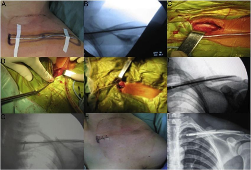
The clavicle is an elongated, S-shaped bone that rests horizontally at the sternum across the upper
part of the ribcage, and the acromial end of the scapula[1].
It is made up of a medial two-thirds which is circular in section and convex anteriorly, and a
lateral one third which is flattened in section and convex posteriorly, it has a role as main stability
between the axial skeleton medially (through the sternoclavicular joint) and the upper limb
laterally (through the acromioclavicular joint), in addition to its superficial location, explains why
it is liable to injury[2].
This bone is an important part of the skeletal system since it plays an essential role in everyday
3070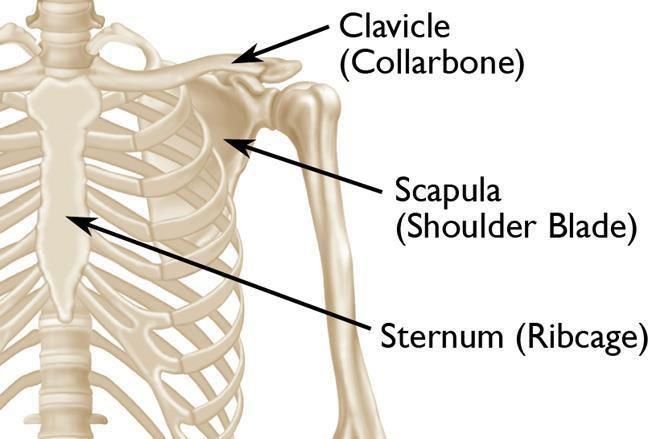
European Journal of Molecular & Clinical Medicine
ISSN 2515-8260 Volume 08, Issue 03, 2021
functional movement, serving as the connection between the axial skeleton and the pectoral
girdle[1] (Figure1).
Figure (1): The clavicle and its location in the shoulder girdle [92]
As a result, the clavicle is able to act as a brace for the shoulder, allowing weight to be transferred
from the upper limbs to the axial skeleton. Injuries of the clavicle seriously compromise everyday
activities [1].
Articulations:
Due to the clavicle’s structure, there are only two planar diarthrosis articulations that can be found.
This type of articulation is also known as a ‘double plane joint’ where two joint cavities are
separated by a layer of articular cartilage [1].
Acromioclavicular joint:
Which is formed by the acromial end of the clavicle and the acromion of the scapula respectively.
It enables slight gliding movement about the shoulder region. The synovial joint is surrounded by
a capsule of articular cartilage filled with intra-articular synovium [1].
From infancy, the articular cartilage starts as hyaline cartilage but soon develops into
fibrocartilage (at the scapula acromion and the clavicle acromial end at ages 17 and 24,
respectively). The acromioclavicular ligament forms a strong connection between the clavicle and
the scapula acromion, which restricts movement about the clavicle at its acromial end [1].
The coracoclavicular (conoid and trapezoid) ligaments pass from the inferior surface of the
clavicle to the base of the coracoid process of the scapula, these strong ligaments provide vertical
stability to the acromioclavicular joint, they constitute the primary support by which the scapula is
suspended from the clavicle; complete division of these ligaments is necessary for a complete
superior dislocation to occur [1] (Fig 2).
Sternoclavicular joint:
Which is formed by the sternal end of the clavicle and the manubrium of the sternum. This synovial
joint is important as it anchors the clavicle and scapula to the axial skeleton. However, the joint enables
a variety of limited movements of the arm, including:
Protraction and retraction.
Depression and elevation.
Slight rotation.
Like the acromioclavicular joint, the sternoclavicular joint is surrounded by an articular cartilage
3071
European Journal of Molecular & Clinical Medicine
ISSN 2515-8260 Volume 08, Issue 03, 2021
capsule, but with a fibrocartilage articular disk inside that creates a clavicular and a sternal synovial
cavity. Sternoclavicular joint ligaments stabilize the joint on its anterior and posterior surfaces [1].
The joint is also reinforced by two accessory ligaments:
The anterior interclavicular ligament, which covers the superior surface of the joint. This ligament is
responsible for preventing dislocation of the clavicle upon shoulder depression [1].
The posterior costoclavicular ligament, which runs from the clavicle costal tuberosity to the superior
and medial surface of the first rib. In contrast, this ligament prevents clavicle dislocation upon
shoulder elevation [1] (Fig 2).
Figure (2): Articulation of the clavicle; acromioclavicular(ac)joint, sternoclavicular (sc) joint, with
related ligaments [3].
Treatment of Fractures Clavicle
Traditionally, midshaft clavicular fractures have been managed non-operatively, even when
substantially displaced [4,5] with good to excellent results [6,7]. Recent evidence, however, reveals
that the final result of non-surgically midshaft clavicular fractures, particularly those with quite
large displacements or shortening, is not like that which was previously thought, demonstrating
higher rates of delayed union, non-union, shoulder weakness and residual pain [8,9].
Conservative (Non-operative) treatment:
Non-operative treatment is warranted for fractures in children, simple fractures with minimal
displacement, and fractures in patients with low compliance without indication for surgical
treatment [10].
Figure (3): Simple midshaft fracture with minimal displacement.[11]
Non-operative treatment is focused on pain management and either a sling or figure-of-eight
3072European Journal of Molecular & Clinical Medicine
ISSN 2515-8260 Volume 08, Issue 03, 2021
bandage. The figure-of-eight bandage allows for an extension by pulling the shoulders back in an
attempt to prevent clavicular shortening and malunion. For the figure-of-eight bandage to work
appropriately, it must maintain its tension and often needs to be tightened daily.
The use of a sling has also shown to be beneficial and is often more comfortable for the patient
than the figure-of-eight bandage [11].
The sling assists in immobilization of the affected arm as well as allows for comfort by supporting
the weight of the arm.
Figure (4): Arm sling (above), and Figure-of-eight bandage (below) as a conservative treatment
of fracture clavicle.[77]
Adults who sustain a midshaft clavicle fracture do heal non-operatively. When treating non-
operative, the initial treatment is immobilization (sling or figure-of-eight bandage) until the pain is
resolved, usually 2 to 4 weeks. At the time of pain resolution, patients are allowed to work on a
range of motion and then light resistance to be added at six weeks. Athletic could be returned to
play after non-operative treatment is on the average three months.
The limits associated with non-operative treatment are, in fact, the risk of nonunion, malunion,
altered biomechanics of the upper girdle, deformity with unsatisfactory cosmetic results, and
upper extremity weakness [13,14].
Surgical (operative) treatment:
Absolute indications are the presence of open fractures, high commination and/or displacement of
the fragments, high risk for in–out skin wounds, a shortening superior to 20 mm, floating shoulder
and neurovascular lesions. Relative indications are polytraumas, painful malunions or
nonunions[14,15]
Operative treatment of displaced MSCFs can be achieved successfully using plates or
intramedullary (IM) implants like Rush pins, Kirschner wires, or titanium elastic nails [14].
Plate fixation:
Operative measures of internal fixation have included dynamic compression plates, tubular plates,
or reconstruction plates. Plate fixation is commonly used to surgically treat displaced midshaft
clavicle fractures. 3.5mm Plate fixation was been a procedure of choice since it allows for
immediate stability and rapid postoperative mobilization [15,16]
There have been many reports of successful plate implants, however, complications have also
been reported. 3.5mm Plate fixation is also associated with higher neurovascular risks [65]. Due to
the local anatomical structures, it is recommended option the plate be removed because of undue
pain or discomfort. Plate fixation complications have included implant failure, nonunion,
3073European Journal of Molecular & Clinical Medicine
ISSN 2515-8260 Volume 08, Issue 03, 2021
infection, and refracture after plate removal, hypertrophic or dysesthetic scars, implant loosening,
intraoperative vascular injury [65,69,71]. Reconstruction plates are prone to leading to deformation at
the site of the fracture, decreasing the chance of union [16].
Figure (5): Fracture midshaft clavicle with plate fixation; pre & post operation x-ray.[16]
A majority of the infections reported from plate fixations were found to be wound infections that
were treated successfully with oral antibiotics. furthermore, 3.5mm plates and screws require
significant soft tissue stripping, which may compromise the blood supply to the clavicle and
interfere with subsequent healing. The bicortical screws on the clavicle may act as multiple stress
raisers leading to fractures [17]
Figure (6): Fracture clavicle with broken plate fixation due to stress.[20]
3074European Journal of Molecular & Clinical Medicine
ISSN 2515-8260 Volume 08, Issue 03, 2021
Intramedullary fixation:
Intramedullary fixation is often preferred over plate fixation because it is a simpler procedure.
Intramedullary fixation has a smaller surgical incision, less invasive, easier hardware removal, and
shorter hospital stay [17]. Various devices can be used with this surgical option including Knowles
pins, Hagie pins, Rockwood pins, and minimally invasive titanium nails [16]
Devices need to be very flexible, the implant needs to be very stable, and it must be small enough
to pass through the medullary space at its most narrow point in the midclavicle[7].
Additionally, the implant must be stable enough to neutralize the potentially disruptive forces.
Though intramedullary fixation is often preferred, it does not allow for as much rotational stability
as plate fixation [18].
Figure (7): Fracture clavicle with different devices of intramedullary fixation; at Lt Titanium nail,
at Rt Rockwood pin.[19]
In addition, pin migration has also been reported as a concern. Intramedullary fixations was
concluded the procedure was minimally invasive with excellent functional and cosmetic results.
It has been suggested intramedullary fixation should not be the treatment of choice if plate fixation
will maintain the clavicular length [20].
In addition, complications include implant shortening, implant breakage, temporary brachial
plexus palsy, and skin break down where the hardware is inserted [65].
However, besides the great response in healing rates, intramedullary fixation has been associated
with complications.
The majority of reported complications have been superficial infections, however, delayed union,
non-union and refractures have also been reported [20].
Biomechanically, intramedullary bracing using elastic titanium nails allows the optimal position of
the implant to maintain tension adequate position under loaded stress [20]. It is minimally invasive
and provides pain relief. Intramedullary nailing allows for a stable fixation for early function and
restitution of clavicle symmetry. The disadvantages are the cost of the implant and the need for
implant removal after the union.
All hardware was removed once the union was complete on an average of 6 months postoperative
[21]
.
Surgical procedures of intramedullary fixation:
Intramedullary fixation (Elastic nailing) was done using the technique described first by Jubel et
al[22]. A small skin incision was made approximately 1 cm opened using an awl pointed laterally
and angled at about 30 degrees to the coronal plane in line with the clavicle. Care was taken not to
perforate the dorsal cortex in order to avoid major complications. Single elastic nails of different
3075European Journal of Molecular & Clinical Medicine
ISSN 2515-8260 Volume 08, Issue 03, 2021
diameters varying from 2 to 3.5 mm, were used, depending on the width of the bone. To obtain the
exact position of the titanium elastic nail (TEN), fluoroscopy with true perpendicular views was
used. Closed reduction was done under an image intensifier and provisionally fixed with two
percutaneously pointed reduction clamps. In some cases, close reduction of the fracture site could
not be done, so an additional small incision was made above the fracture site for direct
manipulation of the main fragments before the nail was introduced into the lateral fragment and
the fracture was compressed. Care was taken to avoid perforation of the dorsolateral cortex of the
lateral clavicle. The TEN was cut as short as possible at the medial end[22]
Figure (8): Surgical procedures of intramedullary nailing using TEN. (A) external location with
Kirschner wire; (B) the length of TEN measured under the fluoroscopic control of C-arm machine;
(C) the medullary cavity opening with a small size incision in the inner end of clavicle; (D) the
insertion of TEN from the inner end of clavicle; (E) the outlook after TEN is completely inserted
into the medullary cavity; (F) a good position of TEN is seen using C-arm machine; (G) locking
the screw at the tail part of TEN; (H) the postoperative outlook of small size incision; (I) a
radiograph obtained just after surgery shows a good position of TEN and satisfactory reduction of
fracture.[24]
ConflictofInterest: Noconflictofinterest.
References
1. Jeremy M, Burnham MD, Daniel C, Kim MD, Srinath KamineniMD (2016): Midshaft
Clavicle Fractures: A Critical Review. Orthopaedic journal; 39 (5): e814-e821.
doi.org/10.3928/01477447-20160517-06.
3076European Journal of Molecular & Clinical Medicine
ISSN 2515-8260 Volume 08, Issue 03, 2021
2. Craig EV. (1998): Fractures of the clavicle. Rockwood CA Jr, and Matsen FA 2nd edition. 1:
428-482.
3. Robert L Hatch, James R Clugston, Jonathan Taffe, et al. (2020):ClavicleFracture. In
Uptodate marketing professional. ISBN-1105 - 154.179.59.41 - 90D55FA593.
4. Xu JJ, Xu L, Xu W, Gu Y, Xu JJ. (014): Operative versus nonoperative treatment in the
management of midshaft clavicular fractures: ameta-analysis of randomized controlled trials. J
Shoulder Elb Surg; 23:173–181.
5. Hu¨bner EJ, Hausschild O, Su¨dkamp NP, StrohmPC. (2011) Clavicle fractures—is there a
standard treatment? Acta ChirOrthopTraumatol Cech; 78:288–296.
6. NeerCS.(1960): Nonunion of the clavicle. J Am Med Assoc; 172(10):1006–1011.
7. Rowe CR. (1968): An atlas of anatomy and treatment of midclavicular fractures. Clinical
Orthopedics and Related Research; 58:29–42
8. Simon P. (2009): Fractures and non-unions of the clavicle. European instructional
lectures.9:75–80.
9. Narsaria N, Singh AK, Arun GR, Seth RRS.(2014): Surgical fixation of displaced midshaft
clavicle fractures: elastic intramedullary nailing versus pre contoured plating. J
OrthopTraumatol; 15:165–171.
10. Smekal V, Oberladstaetter J, Struve P, Krappinger D. Shaft fractures of the clavicle: current
concepts. Arch Orthop Trauma Surg 2009; 129(60: 807-15.
11. Matthew Pecci, , Jeffrey B. Kreher.(2008): Clavicle fracture. In American Academy of
Family Physicians ;77(1):65-71. PMID: 18236824.
12. Anderson K, Jensen PO, Lauritzen J.(1987): Treatment of clavicular fractures. Figure-of-
eight bandage versus a simple sling. Acta Orthop Scand;58(1):71-4.
13. Erik N, Kublak, Kenneth J, Koval Joseph, D Zuckerman.(2016): Clavicle Fracture;
shoulder fracture practice guide to management. 10:180-87.
14. Ledger M, Leeks N, Ackland T, Wang A.(2005): Short malunions of the clavicle: an
anatomic and functional study. J Shoulder Elb Surg; 14:349–354.
15. Hill J, McGuire M, Crosby L.(1997): Closed treatment of displaced middle-third fractures of
the clavicle gives poor results. J Bone Joint Surg Br; 79(4):537–538.
16. Kabak S, Halici M, Tuncel M, Avsarogullari L, KaraogluS.(2004): Treatment of mid-
clavicular non-union: comparison of dynamic compression plating and low contact dynamic
compression plating techniques. J Shoulder Elbow Surg;13(4):296-403.
17. Thyagarajan DS, Day M, Dent C, Williams R & Evans R.(2009): Treatment of mid-shaft
clavicle fractures: a comparative study. International journal of shoulder surgery, 3(2), 23.
18. Bachoura A, Deane AS, KamineniS.(2012): Clavicle anatomy and the applicability of
intramedullary midshaft fracture fixation. Journal of Shoulder and Elbow Surgery;21(10):1383-
90.
19. Qing-Hua Sang, Zhi-Gang Gou, Hua-Yong Zheng, et al.(2015). The Treatment of Mid-shaft
Clavicle Fractures. Chinese Medical Journal, 128(21): 2946–2951.
20. Preston CF, EgolKA.(2009): Midshaft clavicle fractures in adults. Bull NYU Host
JtDis;67:52-57. PMID: 19302058.
21. Rehm KE, Andermahr J, JubelA.(2005): Intramedullary nailing of midclavicular fractures
with an elastic titanium nail. Euro J Trauma. 2005; 31(4): 409-416.
22. Jubel A, Andermahr J, Schiffer G, Tsironis K, Rehm KE.(2003): Elastic stable
intramedullary nailing of midclavicular fractures with a titanium nail. Clinical Orthopedics and
Related Research; 408:279–285.
3077European Journal of Molecular & Clinical Medicine
ISSN 2515-8260 Volume 08, Issue 03, 2021
23. BeigangFu.(2016): Minimally invasive intramedullary nailing of clavicular fractures by a new
titanium elastic nail. Acta Orthopaedica et Traumatologica Turcica; 50:494- 500.
24. Robert L Hatch, , James R Clugston, Jonathan Taffe, et al.(2020):ClavicleFracture. In
Uptodate marketing professional. ISBN-1105 - 154.179.59.41 - 90D55FA593.
3078You can also read