Computer aided manufacturing and design of fixed bridges restoring the lost dentition, soft tissue and the bone
←
→
Page content transcription
If your browser does not render page correctly, please read the page content below
Volume 81                      International Scientific Journal
                                                           Issue 2                        published monthly by the
                                                           October 2016                   World Academy of Materials
                                                           Pages 68-75                    and Manufacturing Engineering

     Computer aided manufacturing and
     design of fixed bridges restoring the lost
     dentition, soft tissue and the bone
          P. Malara a,*, L.B. Dobrzański        b
          a Institute of Engineering Materials and Biomaterials, Silesian University of Technology,
          ul. Konarskiego 18a, 44-100 Gliwice, Poland
          b Centre of Medicine and Dentistry SOBIESKI, ul. Sobieskiego 12, 44-100 Gliwice, Poland

          * Corresponding e-mail address: piotr.malara@pols.pl

          ABSTRACT

          Purpose: The aim of the paper is to present the methodology of computer aided designing
          and manufacturing of an all-ceramic multi-unit bridge restoring missing teeth and the lost
          soft and hard tissues of the oral cavity as a result of surgical treatment of oral tumor.
          Design/methodology/approach: The methodology of computer aided designing and
          manufacturing of the multi-unit all-ceramic bridge was presented on the basis of an actual
          clinical case of a patient who underwent the surgical treatment of myxoma of the oral cavity.
          All the steps of clinical and technical production of the bridge were described and illustrated.
          Findings: It is possible to use the CAD/CAM technology to design and manufacture
          all-ceramic multi-unit bridges restoring missing teeth and the lost soft and hard tissues of
          the oral cavity. The design of the bridge must be clinically validated using mock-ups and only
          then can be implemented for the CAM software.
          Practical implications: Thanks to the method of designing and manufacturing of multi-
          unit all-ceramic bridges for patients with significant lost of the soft and hard tissues of the
          mouth it is possible to carry out a prosthetic rehabilitation of patients after trauma and tumor
          surgery.
          Originality/value: : The execution of extensive bridges with the maximum available height
          of about 25 mm requires a high technological rigor at the design and manufacture stage. To
          ensure longevity of the reconstruction, it is necessary to plan all the work while maintaining
          the maximum thickness of the substructure. It is desirable to provide minimum of 2 mm
          thick substructure and the surface of at least 20 mm2 or more in the cross-sections. At the
          same time, the structure of the bridge must be supported on the alveolar ridge to provide
          aesthetics and endurance.
          Keywords: Zirconia dioxide; Prosthetic bridge; All-ceramic denture; CAD/CAM; PMMA
          Reference to this paper should be given in the following way:
          P. Malara, L.B. Dobrzański, Computer aided manufacturing and design of fixed bridges
          restoring the lost dentition, soft tissue and the bone, Archives of Materials Science and
          Engineering 81/2 (2016) 68-75.

          METHODOLOGY OF RESEARCH, ANALYSIS AND MODELLING

     68                                         © Copyright by International OCSCO World Press. All rights reserved. 2016
68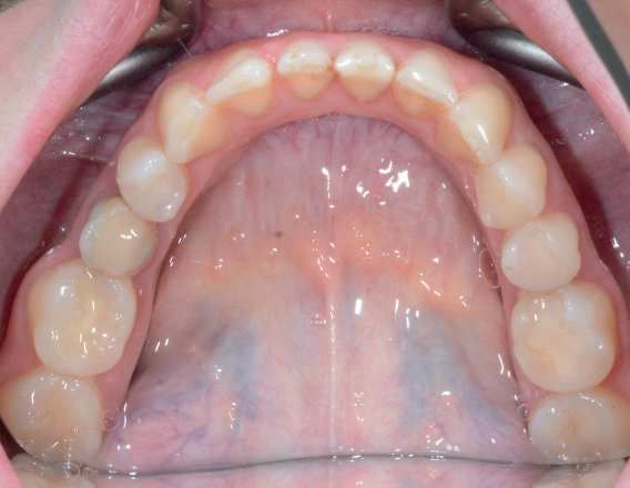
Introduction
1. Introduction                                                        It must be noted that prostheses restore the lost teeth but
                                                                   also soft and hard tissues that are lost as a result of the loss
                                                                   of teeth. Restoration of the soft and hard tissues of the oral
    Dental prosthetics uses many technical solutions for
                                                                   cavity that were lost due to trauma or tumor is one of the
reconstruction of the lost teeth. Biomechanics of these
                                                                   most difficult clinical problems in contemporary dental
restorations is significantly different from the biomechanics
                                                                   prosthetics. A significant problem is not limited only to the
of the natural patient's dentition [1,2]. During designing
                                                                   missing teeth. Designed prosthesis must then restore the
process of a prosthetic restoration it is necessary to take into
                                                                   lack of soft and hard tissues of the oral cavity [9]. A
account the configuration of residual dentition, the state of
the hard tissues and the pulp of the remaining teeth, their        restricted area of the oral cavity that is able to transfer the
anatomical structure and fixation in their sockets. These          chewing forces becomes a real problem as well as in many
                                                                   instances disadvantageous configuration of the prosthetic
factors determine the possibility of using residual dentition
                                                                   area and a considerable volume and mass of the produced
as a possible support for the planned restorations [3-5].
    Taking into account the way of transmission of chewing         restoration. The need to obtain adequate aesthetics of the
forces from the artificial teeth onto the patient's own            prosthesis also requires combining multiple materials with
tissues, dentures are divided into periodontally supported,        different color and translucency to imitate pink mucosa and
mucosal supported or mixed. Mucosal supported prostheses           white teeth. All the aspects pointed above make the desi-
transmit the chewing force onto the oral mucosa. Under             gning and execution of the posttraumatic and postoperative
physiological conditions, these tissues are not adapted to         dental restorations quite challenging from both clinical and
receive a significant occlusal load. Therefore, oral mucosa        technical point of view [10-12].
charged with such loads can react with hypertrophy,                    The aim of the paper is to present the methodology of
atrophy, inflammation or a combination of these patholo-           computer aided designing and manufacturing of an all-
gical conditions. In addition, the chewing forces transmit-        ceramic multi-unit bridge restoring missing teeth and the
ted to the oral mucosa cause considerable discomfort               lost soft and hard tissues of the oral cavity as a result of
during use. The instability of the prosthesis results in           surgical treatment of oral tumor.
discomfort for the patient who can feel the movement of
the prosthesis on the mucosal surface [6]. The movements
of the prosthesis on the mucosa may also cause abrasion of
the oral mucosa. The use of these types of prostheses
                                                                   2. Clinical
                                                                   2.          case presentation
                                                                      Clinical case presentation and
                                                                                                 and
significantly impairs the pronunciation [6,7].                        methodology
                                                                      methodology
    Another group of prosthetic solutions are the peri-
odontally supported prostheses. This group consists of
                                                                   2.1. Clinical case presentation
                                                                        Clinical case presentation
prostheses which transmit the chewing forces on the
patient's own teeth, still remaining in the oral cavity. These
prostheses are very well tolerated by the patients, because            The patient was a 38-year-old woman. She underwent a
during chewing, they do not exert pressure on the oral             surgery of tumor removal from the oral cavity in the region
mucosa. The feelings of patients related to the use of these       13-15 10 years ago. The tumor was confirmed histo-
prostheses are virtually the same as when chewing with             pathologically to be a myxoma. Myxoma is a locally
their own teeth. It should be remembered that, during the          malignant tumor. This means that it grows aggressively
use of the periodontally supported prostheses the suppor-          locally in an uncontrolled way causing infiltration and
ting teeth receive greater loads than physiologically accepted     destroying the surrounding anatomical structures.
chewing forces. It may result in adverse consequences to the       However, it does not give metastases to distant tissues.
suspension apparatus of the teeth [6-8].                           A surgical procedure to treat myxoma assumes radical
    The last group of prostheses consists of dentures with         removal of the tumor with a margin of healthy tissues.
mixed support. Biomechanics of these solutions assume              Such a surgical procedure was used in the patient. The
that a part of the chewing forces is transmitted through the       teeth 13, 14, 15 lying in the tumor mass were also removed
natural teeth of the patient and the other part through the        during the operation. To this day, there have not been the
oral mucosa. These prostheses form a very diverse group in         recurrence of the tumor.
terms of construction. Depending on the ability to remove              A removable prosthetic restoration was fabricated for
the denture from the oral cavity by the patient they are           the patient 5 years ago. It was a periodontally and
divided into fixed or removable prostheses [6].                    mucosally supported denture (Figs. 1-5).

READING DIRECT: www.archivesmse.org                                                                                          69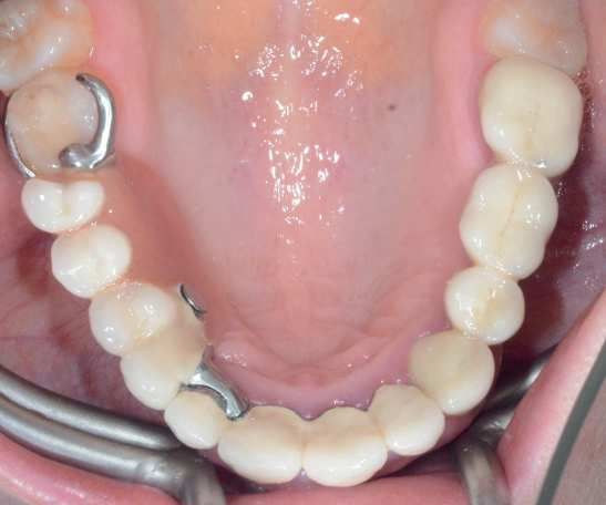
P. Malara, L.B. Dobrzański

     Fig. 1. Intraoral view of the region 13-15 with significant
     lost of the soft and hard tissues as a result of surgical
     treatment of the tumor and the fixed portion of the           Fig. 4. Intraoral view of the prosthetic restoration of the
     prosthetic restoration                                        upper arch; the removable portion of the restoration
                                                                   snapped on the fixed portion

     Fig. 2. The removable portion of the prosthetic restoration

                                                                          Fig. 5. Intraoral view of the opposite dentition

                                                                       The patient was not satisfied with the restoration,
                                                                   because it was not stable enough and caused irritation to
                                                                   the soft tissues of the oral cavity. This was the reason for
                                                                   recurrent inflammation of the oral mucosa. It was
                                                                   particularly dangerous due to the neoplastic process in the
                                                                   patient's medical history. All irritants could be in fact be
                                                                   the reason for a local recurrence of the tumor.
                                                                       Therefore, the patient was qualified for the treatment
                                                                   with an all-ceramic multi-unit bridge restoring missing
     Fig. 3. Intaglio view of the removable portion of the         teeth, as well as the lost soft and hard tissues as a result of
     prosthetic restoration                                        the surgical treatment of the oral tumor.

       70                                                                 Archives of Materials Science and Engineering
70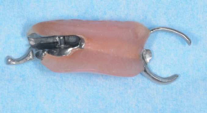
Computer aided manufacturing and design of fixed bridges restoring the lost dentition, soft tissue and the bone
2.2.
2.2. Clinical and technical
Clinical and technicalmethodology
methodology
Firstly, the old prosthetic restoration was removed and
preparation of all the remaining teeth in the upper arch was
performed. Impressions were taken in a traditional way
with polyether precision impression material. Impressions
of the opposite dentition were taken with alginate dental
impression material. Due to the reduced vertical height of
the occlusion, it was necessary to reconstruct also the
height of the occlusion.
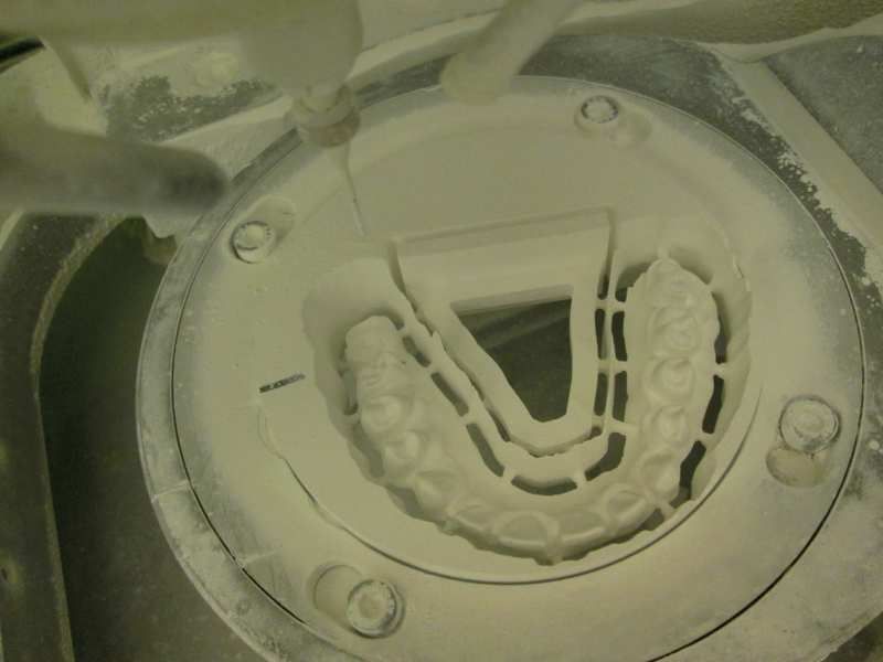
The impressions were scanned using a laboratory
scanner (Fig. 6). Using the computer, the final prosthetic
restoration was designed in a virtual environment (Figs. 7-9).
The bridge was designed to reconstruct the lost teeth as Fig. 8. Lateral view of the designed bridge with the portion
well as the lost soft and hard tissues of the oral cavity. restoring the lost soft and hard tissues marked in red.
Fig. 9. The virtual prototype of the bridge superimposed on
the virtual model of the scanned maxilla.
Fig. 6. The virtual model of the prepared upper dentition
To verify the correctness of the designing process, the
first prototype of the bridge was manufactured in wax
(Fig. 10). The use of wax makes it possible to try-in the
prototype intraorally and to make necessary adjustments of
the shape of the teeth.
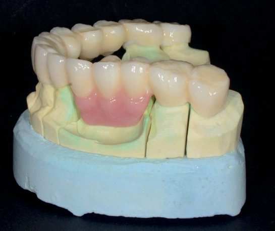
Fig. 7. Designed model of the final bridge; the part of the
prostheses restoring the lost soft and hard tissues is marked
in red Fig. 10. A wax prototype of the designed bridge
Volume 81 Issue 2 October 2016 71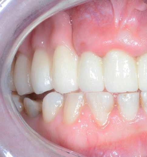
P. Malara, L.B. Dobrzański

         In case of such an extensive prosthetic work, prior to
     the final manufacturing of expensive and time-consuming
     prosthetic solution, it was necessary to perform a prototype
     of a cheaper material. For prototyping polymethyl metha-
     crylate (PMMA) was selected (Fig. 11).

                                                                     Fig. 12. Intraoral verification of the PMMA prototype
                                                                     bridge

         Fig. 11. Prototype of the bridge made from PMMA

         The PMMA bridge was also verified intraorally.
     Verification of the prototype bridge included an asses-
     sment of:
     • the proper settlement of the bridge on the abutment
         teeth,
     • the correct run of subgingival margin of the prosthetic
         bridge on the necks of the abutment teeth,                  Fig. 13. Milling the bridge substructure in a CNC milling
     • the correctness of the design of the shape of the dental      machine
         arch,
     • the correct selection of the size and shape of the teeth,
     • correct occlusal relationship with the opposing teeth,
     • the correctness of the design of the part of the bridge
         restoring the lost soft and hard tissues,
     • no irritation of the soft tissues in the static and dynamic
         states of the oral mucosa,
     • lack of compression of the oral mucosa in the vicinity
         of the pontics.
         The prototype was verified intraorally (Fig. 12). The
     patient accepted the new height of occlusion, as well as the
     shape and size of the teeth.
         Having positively verified the PMMA prototype of the
     bridge, the ceramic substructure was milled from a block of        Fig. 14. The ceramic substructure tried-in intraorally
     zirconium dioxide using a CNC milling machine (Fig. 13).
         The milled ceramic substructure was then subjected to           Over the zirconium dioxide layer the veneering ceramic
     high-temperature sintering process. The substructure was        was applied giving the final shape of the bridge. At this
     tried-in again intraorally to verify the precision of           stage the bridge was verified intraorally for the last time
     designing and manufacturing process (Fig. 14).                  (Fig. 15).

       72                                                                   Archives of Materials Science and Engineering
72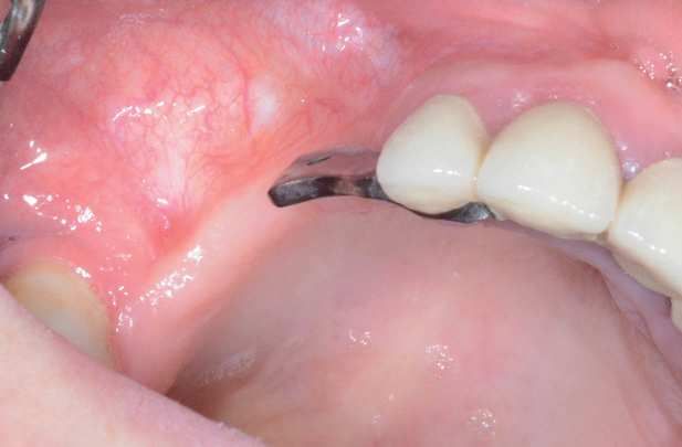
Computer aided manufacturing and design of fixed bridges restoring the lost dentition, soft tissue and the bone

Fig. 15. The bridge covered with veneering ceramic tried-in
intraorally
                                                                   Fig. 18. Intraoral anterior view of the final all-ceramic
    To obtain adequate aesthetics of the final prosthetic          bridge
bridge the ceramic was glazed (Figs. 16,17). The final
bridge was cemented to the abutment teeth with a com-
posite light-curing cement (Figs. 18,19).

Fig. 16. Lateral view of the final all-ceramic bridge on the
plaster model; the lost soft and hard tissues restored with
pink veneering ceramics

                                                                   Fig. 19. Intraoral lateral view of the final all-ceramic
                                                                   bridge; the lost soft and hard tissues restored with pink
                                                                   ceramics

                                                                   3. Discussion
                                                                      Discussion

                                                                     Multi-unit bridges restoring a whole tooth arch must
                                                                   carry the greatest forces arising in the mouth at the area of
                                                                   pontics. If it is planned to achieve a high aesthetic effect it
                                                                   is necessary to use zirconium dioxide to ensure
                                                                   transparency of incisal edges, especially at the front.
Fig. 17. Anterior view of the final all-ceramic bridge on the      Additionally, if, as in the case described above, it is
plaster model; the lost soft and hard tissues restored with        necessary to restore the lost soft and hard tissues of the
pink veneering ceramics                                            mouth, the designing and manufacturing process must be

Volume 81    Issue 2 October 2016                                                                                           73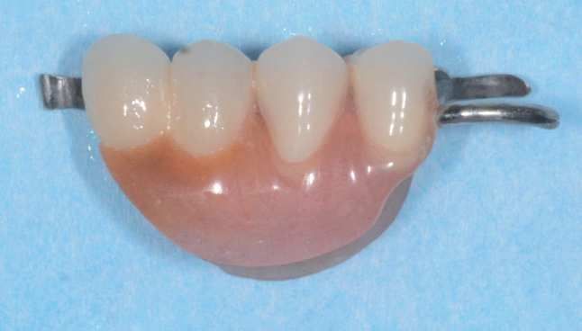
P. Malara, L.B. Dobrzański

     adjusted for the purposes of aesthetics and function                 The substructure of the bridge can be verified again
     [13-15].                                                         intraorally and only then the outermost layer of the
         To ensure the greatest endurance of a full-ceramic           veneering ceramics can be applied.
     multi-unit bridge made of zirconia dioxide it is necessary to
     design the proper thickness of the construction at the points
     of connectors and pontics [16,17]. In our case it was            4. Conclusions
                                                                      4. Conclusions
     decided to manufacture ¾ crowns on all points of the
     bridge paying particular attention to ensure the maximum             It is possible to use the CAD/CAM technology to
     available cross-sectional area of connectors between all the     design and manufacture all-ceramic multi-unit bridges
     points. For this purpose it was decided to prepare a mock-       restoring missing teeth and the lost soft and hard tissues of
     up, which in addition to the verification of occlusion and       the oral cavity. However, it should be noted that the
     the introduction of adjustments intraorally allowed the          execution of such an extensive bridge with the maximum
     patient to see the planned appearance of the final prosthetic    available height of about 25 mm requires a high techno-
     reconstruction and to accept the aesthetic effect of the         logical rigor at the design and manufacture stage. To ensure
     proposed work. The mock-up was then scanned again on             longevity of the reconstruction, it is necessary to plan all
     the model and its shape, particularly in the area of the         the work while maintaining the maximum thickness of the
     occlusal surface, was copied in the final reconstruction.        substructure. It is desirable to provide minimum of 2 mm
     This step is particularly important for two reasons. The first   thick substructure and the surface of at least 20 mm2 or
     is the elimination of the need to make adjustments on the        more in the cross-sections. At the same time, the structure
     occlusal surfaces of the finished prosthetic reconstruction.     of the bridge must be supported on the alveolar ridge to
     The second of these is the possibility of designing the          provide aesthetics and endurance.
     reconstruction having the maximum available thickness of
     the substructure, in particular in sensitive sites such as
     connectors between the abutment teeth and the pontics,           References
                                                                      References
     especially on the chewing surfaces where the transmitted
     forces are the greatest. At the same time, to ensure the         [1]   R.C. Silveira Rodrigues, A.C. Lapria Faria, A.P.
     endurance of the entire structure, it was also extremely               Macedo, M.G. Chiarello de Mattos, R.F. Ribeiro,
     important to provide a stable support for the pontics. For             Retention and stress distribution in distal extension
     this purpose, the sectional bridge shape was designed to be            removable partial dentures with and without implant
     oval. To ensure the proper support at the regions of                   association, Journal of Prosthodontic Research 57
     edentulous maxillary ridge, the anatomical shape of the                (2013) 24-29.
     pontics was designed to extend approximately 0.3 mm              [2]   D.M. Bohnenkamp, Removable Partial Dentures
     towards the gum line.                                                  Clinical Concepts, Dental Clinics of North America
         To design the reconstruction of the alveolar area it is            58/1 (2014) 69-89.
     necessary to determine the extent of the framework in            [3]   G. McKenna, P.F. Allen, D. O’Mahony, M. Cronin, C.
     consultation with the dentist and the patient. For this                DaMata, N. Wood, The impact of rehabilitation using
     purpose, it is also extremely useful to prepare mock-ups               removable partial dentures and functionally orientated
     made of PMMA. It is necessary to take into account the                 treatment on oral health-related quality of life:
     anatomical shape of the teeth and pontics and their exact              A randomised controlled clinical trial, Journal of
     reproduction while ensuring the correct line of gingival shape         Dentistry 43/1 (2015) 66-71.
     in close correlation with the adjacent teeth. The thickness of   [4]   D.R. Nelson, J.F. Palik, Duplicate casts in fabrication
     such an element should be at least 2.5 mm [18].                        of temporary removable partial dentures, The Journal
         The design of the bridge must be clinically validated              of Prosthetic Dentistry 53/3 (1985) 441-442.
     using mock-ups and only then can be implemented for the          [5]   S.K. Hummel, M.A. Wilson, V.A. Marker, M.E.
     CAM software. It must be emphasised that in cases of such              Nunn, Quality of removable partial dentures worn by
     extensive and high bridges (24.5 mm before the sintering               the adult U.S. population, The Journal of Prosthetic
     process), the reconstruction must be machined with the                 Dentistry 88/1 (2002) 37-43.
     supporting structure enabling the execution of sintering         [6]   K. Kono, D. Kurihara, Y. Suzuki, C. Ohkubo, Pres-
     maintaining the originally designed shape. In the absence              sure distribution of implant-supported removable
     of such a support, the bridge could get distortion. For                partial dentures with stress-breaking attachments.
     smaller structures it is not a required procedure [19,20].             Journal of Prosthodontic Research 58/2 (2014) 115-120.

       74                                                                     Archives of Materials Science and Engineering
74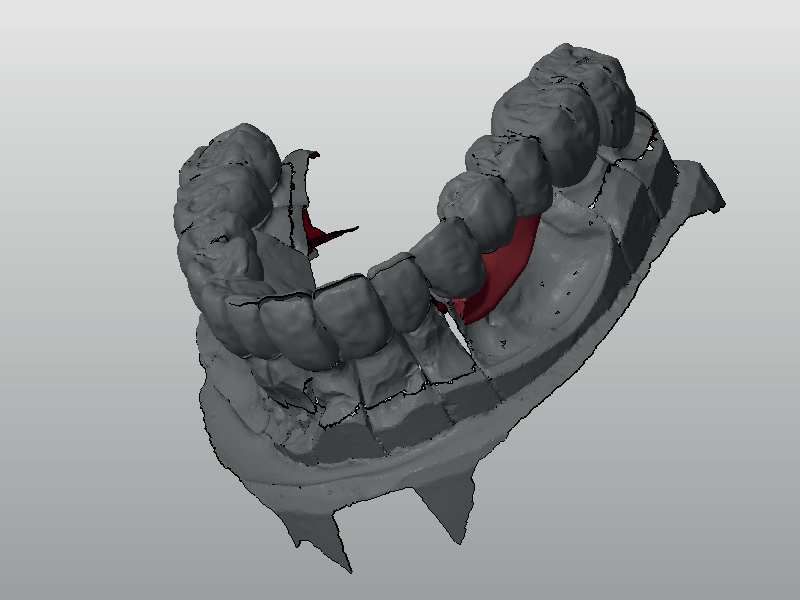
Computer aided manufacturing and design of fixed bridges restoring the lost dentition, soft tissue and the bone

[7]   Y. Goto, J.S. Brudvik, Custom precision                      [14] P. Malara, L.B. Dobrzański, Computer-aided design and
      attachment housings for removable partial dentures,               manufacturing of dental surgical guides based on cone
      The Journal of Prosthetic Dentistry 88/1 (2002)                   beam computed tomography, Archives of Materials
      100-102.                                                          Science and Engineering 76/2 (2015) 140-149.
[8] C.H. Wang, H.E. Lee, J.K. Du, Y Igarashi,                      [15] P. Malara, L.B. Dobrzański, Designing and manufa-
      Connecting rigidities of various precision attachments            cturing of implantoprosthetic fixed suprastructures in
      compared with the conical crown retained telescope,               edentulous patients on the basis of digital impressions,
      The Kaohsiung Journal of Medical Sciences 21/1                    Archives of Materials Science and Engineering 76/2
      (2005) 22-28.                                                     (2015) 163-171.
[9] A.A.A. Mahmoud, N. Wakabayashi, H. Takahashi,                  [16] P. Malara, L.B. Dobrzański, J. Dobrzańska, Computer-
      Prediction of permanent deformation in cast clasps                aided designing and manufacturing of partial
      for denture prostheses using a validated nonlinear                removable dentures. Journal of Achievements of
      finite element model, Dental Materials 23/3 (2007)                Materials and Manufacturing Engineering 73/2 (2015)
      317-324.                                                          157-164.
[10] N. Olvera, J.D. Jones, Alternatives to traditional            [17] J. Żmudzki, G. Chladek, P. Malara, L. Dobrzański,
      complete dentures, Dental Clinics of North America                The simulation of a mastication efficiency of the
      58/1 (2014) 91-102.                                               mucous-borne complete dentures. Archives of Mate-
[11] M. Hundal, R. Madan, Comparative clinical evalu-                   rials Science and Engineering 63/2 (2013) 75-86.
      ation of removable partial dentures made of two              [18] S.C. Gutiérrez-Rubert, M.D. Meseguer-Calas, A.
      different materials in Kennedy Applegate class II                  Gandía-Barberá, Analysis of the feeding system in the
      partially edentulous situation, Medical Journal Armed              injection process of peek in fixed partial dentures,
      Forces India 71/2 (2015) S306-S312.                                Procedia Engineering 132 (2015) 1021-1028.
[12] B. Orzechowska-Wylęgała, M. Kajor, M. Mazur, A.               [19] G. Chladek, J. Żmudzki, P. Malara, L.A Dobrzański,
     Wodołażski, P. Malara, J. Drugacz: Malignant                       C. Krawczyk, Influence of introducing silver
     lymphoma or inflammation? Difficulties diagnostic                  nanoparticles on tribological characteristics of soft
     and therapeutic – two cases, Polish Merkuriusz                     liner, Archives of Materials Science and Engineering
     Medicine 20/118 (2006) 433-436.                                    62/1 (2013) 5-14.
[13] J. Żmudzki, P. Malara, G. Chladek, Full contoured             [20] M. Hannig, B. Wöstmann, M. Balkenhol, Fracture
     tooth-implant supported 3-pointic all-ceramic denture               strength of temporary fixed partial dentures:
     during occlusal load transfer in lateral region, Archives           CAD/CAM versus directly fabricated restorations,
     of Metallurgy and Materials 61/2A (2016) 843-846.                   Dental Materials 27/4 (2011) 339-347.

Volume 81    Issue 2 October 2016                                                                                         75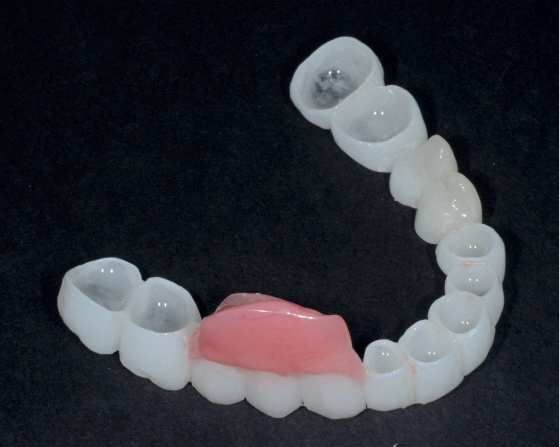
You can also read