Endodontic Management of Mandibular Lateral Incisor with Talon Cusp: A Case Report
←
→
Page content transcription
If your browser does not render page correctly, please read the page content below
Asian Journal of Dental Sciences

                              3(4): 1-7, 2020; Article no.AJDS.59365

            Endodontic Management of Mandibular Lateral
                  Incisor with Talon Cusp: A Case Report
                  N. D. L. Luqman1, A. M. Azmimurad1, S. Zaharudin1, S. H. Hamzah2,
                                                 N. A. Ramlan3 and A. S. Hussein2*
                          1
                         Faculty of Dentistry, Universiti Teknologi MARA, Sg Buloh Campus, Malaysia.
                         2
                          Centre of Paediatric Dentistry and Orthodontics Studies, Faculty of Dentistry,
                                               Universiti Teknologi MARA, Sg Buloh Campus, Malaysia.
             3
              Centre of Comprehensive Care Studies, Faculty of Dentistry, Universiti Teknologi MARA,
                                                                          Sg Buloh Campus, Malaysia.

                                                                                              Authors’ contributions

   This work was carried out in collaboration among all authors. Authors NDLL and AMA designed the
   study and wrote the first draft of the manuscript. Authors SZ, SHH and NAR managed the literature
    searches. Author ASH revised the manuscript. All authors read and approved the final manuscript.

                                                                                                    Article Information

                                                                                                                    Editor(s):
                                                                         (1) Dr. Ketij Mehulić, University of Zagreb, Croatia.
                                                                                                                  Reviewers:
                                       (1) Praveen Kumar Neela, Kaloji Narayana Rao University of Health Sciences, India.
                                                       (2) S. Arbiya Anjum, Ramaiah University of Applied Sciences, India.
                                             Complete Peer review History: http://www.sdiarticle4.com/review-history/59365

                                                                                            Received 24 May 2020
                                                                                            Accepted 31 July 2020
  Case Study
                                                                                         Published 10 August 2020

ABSTRACT

 Introduction: Talon cusp is an uncommon developmental anomaly which is characterized by the
 presence of an extra cusp-like structure projecting from the cingulum area of maxillary or
 mandibular incisors. It consists of enamel, dentine with or without pulp tissue. Its aetiology is still
 unknown; however, its formation is attributed by the hyperactivity of the enamel organs during the
 morpho-differentiation stage.
 Case Report: A healthy 15-year-old Malay female came to the paediatric dental clinic with the
 chief complaint of teeth sensitivity. An intra-oral examination revealed presence of a prominent
 cusp on the lingual surface as well as a deep fissure on the labial surface of mandibular right
 lateral incisor with a slight degree of mobility due to periapical abscess. An intraoral periapical
 radiograph revealed a radiopaque projection. The treatment was a root canal.
 Conclusions: This case highlights the endodontic treatment of periapical lesions on the
 mandibular lateral incisor with talon cusp in an adolescent patient. Diagnosis and management of a
 rare anomaly are essential in the dental practice.
_____________________________________________________________________________________________________

*Corresponding author: Email: dr_alaasabah@yahoo.com, alaa@uitm.edu.my;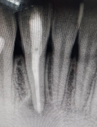
Luqman et al.; AJDS, 3(4): 1-7, 2020; Article no.AJDS.59365

Keywords: Mandibular lateral incisor; talon cusp; root canal treatment.

1. INTRODUCTION                                           incisal edge to create a T-shaped contour or, if
                                                          more cervical, a Y-shaped crown contour [6].
Talon cusp is a rare odontogenic dental anomaly           Moreover, it seems to be a V-shaped radiopaque
projected from the cementoenamel junction                 structure overlapping the affected crown with its
(CEJ) or on the cingulum region of anterior teeth.        apex directed incisally in the radiograph [11].
It is composed of enamel, dentine, and pulpal
tissue with varying extensions [1,2]. It is also          Hattab in 2014 stated that the prevalence of talon
commonly known as a supernumerary cusp,                   cusps varies according to the population from
hyperplastic    cingulum,    dens    evaginatus,          less than 0.06 to 7.7% with racial differences.
evaginated     odontome,      accessory      cusp,        This anomaly was reported to be higher among
supernumerary lingual tubercle, and cusped                the Arab population compared to Caucasians
cingulum [3]. This particular cusp looks like an          and Negroes. Specifically, the prevalence of
eagle’s talon and hence is named after it [4].            talon cusps among the Malaysian population is
                                                          5.2% [4]. Maxillary incisors are the most
Although the aetiology is unclear for this                frequently affected teeth where 55% occur in the
particular anomaly; however, it is suggested that         permanent maxillary central incisor and 33% in
there is multifactorial aetiology that proposed the       the permanent maxillary lateral incisor [12],
formation of talon cusp [5]. It incorporates              followed by mandibular incisors 6%, and
both genetics and environmental influences.               maxillary canines 4% [13]. The affected teeth are
Mutations in the human genes EDA1, EDAR, and              reported to have appeared more in males than
EDARADD frequently lead to more serious                   females in both primary and permanent
phenotypes which would likely lead to tooth loss          dentitions.
and malformations [6]. It occurs during the bell
stage and is characterized by an irregular                The occurrence of talon cusps on mandibular
proliferation of the inner enamel epithelium into         anterior teeth is an extremely rare entity. In
the stellate reticulum of the enamel organ [6].           addition to it, the present case had talon cusps in
Transient focal hyperplasia of the mesenchymal            the permanent mandibular lateral incisor which
dental papilla and altered endocrine functions            was associated with a periapical abscess as late
could also be contributing factors to this                detection and further complication of this
predicament [7,8].                                        anomaly.

According to Hattab et al., talon cusps are               2. CASE REPORT
classified into three types based on the degree of
cusp formation as well as the extension. Type I           A 15-year-old Malay female came to the
(Talon): An additional cusp that projects from the        Paediatric Dental Clinic in Faculty of Dentistry,
anterior tooth palatal surface and reaches at             with the chief complaint of tooth sensitivity on her
least half the length of the cementoenamel                lower anterior region. Her medical and family
junction to the edge of the incisors. Type II             histories were unremarkable whereas the dental
(Semi-talon): An additional cusp of 1 mm or more          history revealed that she had a previous
in length that extends towards the incisor edge           restoration done on her maxillary posterior tooth.
less than half of the length of the cementoenamel
junction. Type III (Trace-talon): Amplified               Extraoral examination disclosed no significant
appearance, the cingulum protrudes with                   findings. Intra-oral examination revealed a
variations such as conical, tubercle-like, or bifid       presence of a cusp-like projection on the lingual
[9,10].                                                   surface of the permanent mandibular right lateral
                                                          incisor including a deep fissure on the labial
The clinical features of talon cusps vary in size,        surface which can be seen in Fig. 1. Additionally,
form, length, and mode of attachment to the               the affected tooth also had mobility grade II. An
crown, ranging from an extended cingulum to a             intraoral periapical radiograph was taken and the
broad, well-linear cusp extending beyond the              presence of radiopacity was noted on the crown
tooth's incisal edge. It may also attach to the           of the tooth indicating a talon cusp. Furthermore,

                                                      2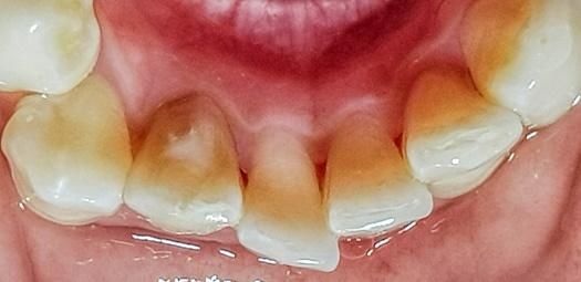
Luqman et al.; AJDS, 3(4): 1-7, 2020;; Article no.AJDS.59365
                                                                                                           no.

   Fig. 1. Intra-oral
                 oral photographs showing deep fissure on the labial surface of permanent
 mandibular right lateral incisor, extra-cusp
                                   extra cusp on the lingual surface and presence of crowding
                                          and cross bite

a radiolucent lesion was seen at the crown and              root canal treatment of affected tooth
the periapical area of the particular tooth which           with talon cusp were performed. The parents
indicates caries and a periapical lesion,                   and the patient were informed about the
respectively. A panoramic radiograph was taken              condition.
to detect possible additional
                         dditional dental anomalies
and for orthodontic assessment (Fig. 2a–b).                 Endodontic treatment was performed in 3 visits.
Sensibility tests including percussion, cold test           During the first visit, after moisture ccontrol
and electrical pulp test were performed to                  protocol was established, the access cavity was
investigate the vitality of the affected tooth. The         done on the palatal surface by removing the
controlled measures were taken with the                     talon cusp. Apex locator was used to determine
permanent mandibular left incisor as a reference.           the working length. Cleaning and shaping of the
The results revealed that the affected tooth was            canal with constant irrigation of 2.5% Sodium
non-vital.
     vital. According to these results, the                 Hypochlorite was done during the second visit.
diagnosis of permanent mandibular right lateral             Intracanal medicament was also done with
incisor with talon cusp type I and pulp necrosis            calcium hydroxide for two weeks. After two
as well as asymptomatic
                    omatic apical periodontitis was         weeks, the patient returned to the clinic
made. Hence, non-surgical
                     surgical endodontic treatment          symptom-free
                                                                       free and the root canal was obturated
of the tooth was planned. Dental examination of             with Gutta-percha
                                                                         percha and the permanent restoration
the other teeth disclosed no noticeable                     was done with composite (Fig. 3a--b). One-month
abnormalities. All the permanent first molars had           post-operative
                                                                   operative visit revealed that the patient was
caries (ICDAS 02 and 03) and mobility on           o        asymptomatic and had no new complaints
maxillary primary right canine was noted.                   pertaining to the endodontic treatment. A 6-      6
Furthermore, she had Class I malocclusion with              month follow-up  up visit including clinical and
skeletal Class II pattern associated with                   radiographic assessment was perform performed. The
moderate crowding on the anterior region of the             tooth was a symptomatic clinically and showing a
upper       and      lower        arch.     Crossbite       reduction in the radiolucency indicating a
between maxillary permanent      nent right lateral         resolution of the periapical lesion (Fig. 4).
incisors and mandibular permanent right canine              Currently the patient is under a 3   3-month review
was present (Fig. 1).. Oral hygiene was                     since the patient is at high caries risk. Moreover,
fair with mild gingivitis. Therefore, routine scaling       the patientt was referred to the orthodontist for
and oral prophylaxis, tooth coloured restorations           initiating orthodontic treatment to correct her
of carious teeth, exodontia of primary
                                     mary canine and        malocclusion.

                                                        3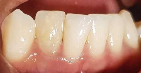
Luqman et al.; AJDS, 3(4): 1-7, 2020;; Article no.AJDS.59365
no.
Fig. 2a. Preoperative periapical radiograph of the permanent mandibular right lateral incisor
showing the presence of talon cusp
Fig. 2b. Panoramic view of the dentition showing the periapical lesion on the permeant
mandibular right lateral incisor
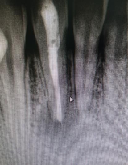
Fig. 3a. An immediate postoperative periapical radiograph of the permanent mandibular right
lateral incisor with talon cusp after root canal treatment
4Luqman et al.; AJDS, 3(4): 1-7, 2020; Article no.AJDS.59365
Fig. 3b. Intra-oral photographs showing the composite restoration on the labial and lingual
surfaces of permanent mandibular right lateral incisor
Upon review of the literature, the treatment
modalities of talon cusp vary according to the
associated diagnosis. Treatment and
management of talon cusp are usually based on
patient presentation and symptoms which should
be as conservative as possible [15]. The
treatment options ranged from simple
prophylactic measures such as fissure sealants
or tooth coloured restorations to the invasive
approach that include total cusp reduction
followed by pulpotomy, root canal treatment, or
tooth extraction followed by orthodontic treatment
and prosthetic rehabilitation [11].
Since talon cusps are plaque retentive,
preventive treatments such as oral prophylaxis
and placement of sealant were performed and
reported for small talon cusps that are
asymptomatic [16]. Moreover, Nuvvula et al.
(2014) reported a different approach including
reduction of the entire thickness of enamel at the
Fig. 4. A 6-month follow-up periapical bulk of the talon cusp by merging the borders
radiograph of the permanent mandibular right with the tooth surface in two planes. Then,
lateral incisor with talon cusp showing preventive resin restoration was performed that
reduction in the radiolucency included placement of fluoride varnish to reduce
sensitivity and stimulate reparative dentine
3. DISCUSSION formation for pulp protection [17]. Both
treatments were performed for paediatric patients
There are many clinical problems noted with and the issue was recognized and addressed
talon cusp cases including plaque retention and immediately. However, the treatment option
caries susceptibility in the developmental differs when there are cases of pulpal
grooves, traumatic occlusion, and aesthetic involvement associated with the talon cusps. In
concerns in cases of facial talon cusp with the present case, the root canal treatment
displacement of the affected and opposing teeth, performed is similar to Gürhan et al. (2017),
attrition of the opposing teeth, periodontal which had reported that two visits involving root
problems, hypersensitivity, pulpal necrosis and canal treatment was done for an adult patient
periapical pathosis due to excessive attrition due to symptomatic apical periodontitis. Similarly,
accidental cusp fracture, irritation of the tongue Lakshman et al. (2013) referred an adult patient
during speech and mastication, and for root canal procedure due to the presence of
temporomandibular joint pain due to excessive bilateral talon cusp on maxillary incisors which
occlusal forces [12,14]. Therefore, it is critical for were associated with calcification of the pulp
dental practitioners to provide early diagnosis, canal. However, in our case, a paediatric patient
intervention, and definitive treatment for the was involved and was too early for the child to
patients with such anomaly. have endodontic treatment at this age. No other
5Luqman et al.; AJDS, 3(4): 1-7, 2020; Article no.AJDS.59365
treatment option was available to avoid such periodontitis: A case report. J Dent Oral
invasive approaches due to late presentation. A Biol. 2017;2(13):1082.
strong evidence of genetic background of talon 4. Lakshman AR, Kanneppady SK, Kalkur C.
cusp was reported by Elmubarak. Therefore, the Bilateral talon cusp on maxillary incisors: A
patients’ siblings were called for dental unique case report. Inter J Oral Health Sci.
examination to provide early preventive 2013;3:109-12.
measures in case they have a similar anomaly. 5. Gupta R, Thaku N, Thakur S, Gupta B,
Gupta M. Talon cusp: A case report
4. CONCLUSION with management guidelines for practicing
dentist. Dent Hypotheses. 2013;4(2):
Despite talon cusp being a rare entity in daily 67-69.
dental practices, proper diagnosis and 6. Shrestha, Ashish, Marla, Vinay, Shrestha,
appropriate management are required. Sushmita, Maharjan, Iccha. Developmental
Dental practitioners should have good knowledge anomalies affecting the morphology of
and thorough understanding of dental teeth - A review. RSBO. 2014;12(11):68-
developmental anomalies, their variations as well 78.
as the clinical implications for early detection and 7. Balcioglu Huseyin, Keklikoglu Nurullah,
avoiding any further complications that may Kökten, Gülseren. Talon cusp: A
happen. Furthermore, the detection and morphological dental anomaly. Roman J
evaluation of the various symptoms are Morphol Embryo. 2011;52:179-81.
necessary to diagnose the condition and to 8. Ramar K, Hariharavel VP, Annamalai S,
identify the required care. Samuel AV. Bilateral fusion of permanent
mandibular incisors with talon's cusp.
CONSENT SRM J Res Dent Sci. 2017;8(3):144-
147.
Written consent was obtained from the parents 9. Hattab FN. Double talon cusps on
for the agreed dental treatment and the use of supernumerary tooth fused to maxillary
her records or photographs for publication central incisor: Review of literature and
purposes. report of case. J Clin Exp Dent. 2014;6(4):
e400–e407.
10. Hattab FN, Yassin OM, Al-Nimri KS. Talon
ETHICAL APPROVAL cusp in perm dentition associated with
other dental anomalies: Review of
It is not applicable. literature and reports of seven cases.
ASDC J Dent Child. 1996;63:368-76.
COMPETING INTERESTS 11. Elmubarak N. Genetic risk of talon cusp:
Talon cusp in five siblings. Case report in
Authors have declared that no competing dentistry; 2019.
interests exist. Available:https://doi.org/10.1155/2019/308
0769
REFERENCES 12. Kumar Rao P, Ram Shetty S, Prabhu VR,
Veena KM, Chatra L, Shenai P. Talon
cusps in mandibular incisors: An unusual
1. Segura-Egea JJ, Jiménez-Rubio A, presentation in a child patient. J. Dent Res
Velasce-Ortega E, Ríos-Santos JV. Talon Dent Clin Dent Prospects. 2011;5(1):
cusp causing occlusal trauma and acute 37–39.
apical periodontitis: Report of a case. Dent 13. Angela C. Chi, Douglas D. Damm, Brad W.
Trau. 2003;19(1):55–59. Neville, Carl M. Allen, Jerry Bouquot. Oral
2. Kulkarni VK, Choudhary P, Bansal AV, and maxillofacial pathology - E-Book.
Deshmukh J, Duddu MK, Shashikiran ND. Elsevier Health Sciences; 2008.
Facial talon cusp: A rarity, report of a case 14. Deshpande SK, Deshpande N. Unusual
with one year follow up and flashback on case report of mandibular talons cusp.
reported cases. Contemp. Clin. Dent. Acta Scientific Dental Sciences. 2018;
2012;3(1):S125–S129. 2(12):40-42.
3. Gürhan C, Şener E. Endodontic 15. Ozcelik B, Atila B. Bilateral palatal talon
management of talon cusp causing apical cusps on permanent maxillary lateral
6Luqman et al.; AJDS, 3(4): 1-7, 2020; Article no.AJDS.59365
incisors: A case report. Euro J Dent. 2011; 17. Nuvvula S, Gaddam KR, Jayachandra B,
5(1):113–116. Mallineni SK. A rare report of
16. Oredugba FA. Mandibular facial talon mandibular facial talon cusp and its
cusp: Case report. BMC Oral Health. 2005; management. J Conserv Dent.
5:9. 2014;17(5):499–502.
© 2020 Luqman et al.; This is an Open Access article distributed under the terms of the Creative Commons Attribution License
(http://creativecommons.org/licenses/by/4.0), which permits unrestricted use, distribution, and reproduction in any medium,
provided the original work is properly cited.
Peer-review history:
The peer review history for this paper can be accessed here:
http://www.sdiarticle4.com/review-history/59365
7You can also read