Clinical Benef|ts of the Immediate Implant Socket Shield Technique
←
→
Page content transcription
If your browser does not render page correctly, please read the page content below

CLINICAL ARTICLE

Clinical Benef|ts of the Immediate Implant Socket
Shield Technique
                                                                 €
REZA SAEIDI POUR, DR MED DENT*, OTTO ZUHR, DR MED DENT†, MARKUS HURZELER, PROF DR MED DENT‡,
                                                                                            {
OTTO PRANDTNER, MDT§, CAROLINE FREITAS RAFAEL                                                   , DANIEL EDELHOFF, PROF DR MED DENT**,
ANJA LIEBERMANN, DR MED DENT*

     ABSTRACT
     Objective: Extraction-socket resorptionis considered a major problemthatcan limit implantologicalrehabilitation options
     and compromise the esthetic outcome.Surgical techniques to reduce remodeling are of restricted predictability and
     commonly require several surgicalinterventions and grafting.Thisincreases the treatmentcost and places a physical and
     psychological strain onthe patient.This clinical case report presents a replacement of an upper canine using the socket-
     shield technique (SST) with a CAD/CAM surgical guide, resulting in a predictable, high esthetic, and functionalresult.
     Clinical considerations: The SST is an alternative approach to curbing remodeling and resorption by retaining the facial
     part of the root during tooth extraction. An immediately placed implant supports the facial root fragment,
     preventing the collapse of the buccal wall.The SSTwith digital precision planning in combination with a CAD/CAM
     surgical guide benef|ts patients by preserving their tissue architecture and causing only insignif|canttrauma.
     Furthermore, the SSTreduces the number of surgical and prosthetic interventions required to one each for
     pre-operative planning, surgical procedures, and prosthetic rehabilitation.
     Conclusions: The socket shield technique is a minimally invasive implantological approach offers patients and clinicians
     multiple benef|ts.

     CLINICAL SIGNIFICANCE
     The socket-shield technique (SST) represents an alternative approach to intervene remodeling and resorption
     processes by the maintenance of the facial part of the root during tooth extraction.The immediate placement of an
     implant supports the facial root fragment and thereby prevents a collapse of the buccal wall.The SSTassociated with
     a CAD/CAM fabricated surgical guide, can reduce the amount of appointments, due to the immediate fabrication of
     the def|nitive restoration with the existing model.Therefore, no further necessary appointments are required apart
     from f|rst pre-operative planning, second for surgical treatment, and third for prosthetic rehabilitation.

INTRODUCTION                                                                            alterations varies and it can result in the loss of ridge
                                                                                        volume and changes in ridge shape, with up to 3.8 mm
Socket healing after tooth loss results in altered                                      horizontal and 1.24 mm vertical reduction.4 Moreover,
dimensions of the alveolar ridge1,2 due to remodeling3                                  the greatest losses occur on the buccal aspect, which is
and tooth-dependent alveolar process.1 The degree of                                    related to a thinner bone wall2 composed of large

*Assistant Professor, Department of Prosthodontics, Ludwig-Maximilians-Universit€
                                                                                at, Goethestrasse 70 Munich, 80336, Germany

 Dentist, Private Practice H€
                            urzeler/Zuhr and Department of Periodontology, Centre for Dental, Oral, and Maxillofacial Medicine (Carolinum), JohannWolfgang
Goethe-University Frankfurt/Main, Frankfurt, Germany
`
    Dentist, Private Practice H€
                               urzeler/Zuhr, Munich, Germany and Department of Operative Dentistry and Periodontology, University of Freiburg, Freiburg, Germany
‰
    Master Dental Technician, Plattform Laboratory, Goethestrasse 47 Munich, 80336, Germany

    PhD Student, Department of Dentistry, Federal University of Santa Catarina, Floriano polis, Brazil and Guest Dentist, Department of Prosthodontics,
Ludwig-Maximilians-Universit€
                            at, Goethestrasse 70, 80336 Munich, Germany
**Director and Chair, Department of Prosthodontics, Ludwig-Maximilians-Universit€
                                                                                at, Goethestrasse 70 Munich, 80336, Germany

C 2017 Wiley Periodicals, Inc.
V                                DOI 10.1111/jerd.12291                                    Journal of Esthetic and Restorative Dentistry Vol 00  No 00  00^00  2017   1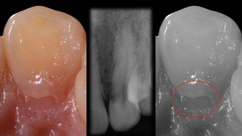
MINIMALLY INVASIVE IMMEDIATE IMPLANT TREATMENT FOR SINGLE-TOOTH REPLACEMENT Saeidi Pour et al

    amounts of bundle bone2 primarily vascularized by the                         extraction, preserving periodontal vascularization,
    periodontal tooth membrane3 and particularly                                  cementum bundle bone16 and the buccal bone wall.17
    susceptible to surgical trauma and resorption.5–7 Other                       Furthermore, the technique has additional advantages:
    important reasons to maintain the bone wall while                             there is no added cost for materials, comorbidity is
    teeth are present include maintenance of the                                  reduced, and it can be applied in the presence of
    periodontal ligament and the provision of nutritional                         endodontic apical pathology, and reduced surgical
    and functional stimuli.8                                                      intervention.16

    Most dimensional changes that compromise socket                               There are suggestions in the literature that a root can
    healing occur during the first to third months.8 A                             be retained to preserve alveolar ridge volume
    reorganization of the alveolar ridge can be observed for                      underneath removable complete prostheses without
    up to 1 year, but with a less pronounced influence on                          complications such as infection.2,18–20 This additionally
    the hard and soft tissues.9 In most situations, these                         allows vertical bone growth coronal to the decoronated
    changes adversely affect with the esthetic outcome,                           root.19,20 No further resorption and no interference
    treatment planning, implant positioning, material                             with implant osseointegration was observed.3,10,11,17
    selection, and osseointegration.1 This is even more                           Enamel matrix derivate (Emdogain; Straumann, Basel,
    critical in the anterior regions10 where these changes                        Switzerland) can be co-administered with the
    directly influence red and white esthetics.11,12 Soft-tissue                   technique (applied on the internal aspect of the
    augmentations immediate or posterior to implant                               fragment) to prevent epithelial proliferation, in
    placement are successful to control the tissue alterations.                   addition to its antimicrobial effect.10,11
    However, it means more surgical interventions.13
                                                                                  Meticulous presurgical planning is mandatory, including
    Several approaches have been described for contouring                         the fabrication of a surgical guide to allow optimal
    the socket alterations caused by tooth extraction10–12:                       implant placement and to ensure esthetic and functional
    implant placement directly after extraction4;                                 restorative success. The use of computer-aided design/
    positioning of the implant on the palatal/lingual wall                        computer-aided manufacturing (CAD/CAM)
    (“palatal approach”), preserving the buccal wall                              stereolithographic (SLA) surgical guides associated with
    contact1; performing the surgery using the flapless                            cone-beam computed tomography (CBCT) facilitates
    technique to maintain vascularization1; and using soft-                       optimal positioning of the implant with more precision
    tissue or bone grafts to maintain the dimension of the                        than with conventional templates. A digital image of the
    ridge by socket augmentation.10 Recent studies                                situation makes it possible to fabricate an individual
    concentrated either on immediate implants or on the                           healing abutment prior to tooth extraction.
    use of grafts, but they also stated that remodeling
    cannot be avoided with these techniques but can                               The aim of this article was to describe the use of the
    continue even after 3 to 6 months of healing.1,14                             innovative SST in the upper canine region, combined
    Moreover, any surgical intervention can result in an                          with a surgical CAD/CAM guide that allows the
    anxiety response on the part of the patient. Anxiety is                       insertion of the final restoration at the second
    a state of discomfort and stress as well as tension, both                     appointment, maintaining the tissue architecture. In
    before and after surgery, according to a definition by                         addition, the SST reduces postoperative patient
    the American Psychiatric Association.15                                       morbidity in terms of swelling and pain.

    The socket-shield technique (SST) may reduce the
    extent of treatment and decrease patient stress and                           CLINICAL CASE PRESENTATION
    pain.10 Additionally, the SST might reduce socket
    resorption and help avoid soft-tissue or hard-tissue                          A 38-year patient presented to the Department of
    grafting. The technique retains the buccal root after                         Prosthodontics at the Dental School in Munich in

2   Vol 00  No 00  00^00  2017 Journal of Esthetic and Restorative Dentistry                         DOI 10.1111/jerd.12291 V
                                                                                                                               C 2017 Wiley Periodicals, Inc.
MINIMALLY INVASIVE IMMEDIATE IMPLANT TREATMENT FOR SINGLE-TOOTH REPLACEMENT Saeidi Pour et al

2014 in connection with pain at the upper left canine.              of martial arts (Figure 1). Clinically, the canine showed
Intraoral examination showed no sensitivity, probably               external resorption; periapical radiographic
as a result of a previously trauma during the practice              examinations showed a related radiolucent area (Figure
                                                                    2). A CBCT scan was therefore indicated to evaluate
                                                                    the depth of the resorption and the possibility to
                                                                    restore the compromised tooth (Figure 3). The patient
                                                                    was referred to an endodontic specialist for a root
                                                                    canal treatment (Figure 4). However, at the 6-month
                                                                    follow-up, a renewed radiographic examination showed
                                                                    progressive resorption, compromising the chance of
                                                                    tooth preservation.

                                                                    This unexpected outcome caused the dental team, in
FIGURE 1. Preoperative frontal view of the upper jaw.               consultation with the patient, to embark on planning

FIGURE 2. Preoperative intraoral/palatal photograph and periapical radiograph of the internal resorption.

FIGURE 3. The 3D scan (CBCT) of the internal palatal resorption at the canine site.

C 2017 Wiley Periodicals, Inc.
V                                DOI 10.1111/jerd.12291               Journal of Esthetic and Restorative Dentistry Vol 00  No 00  00^00  2017   3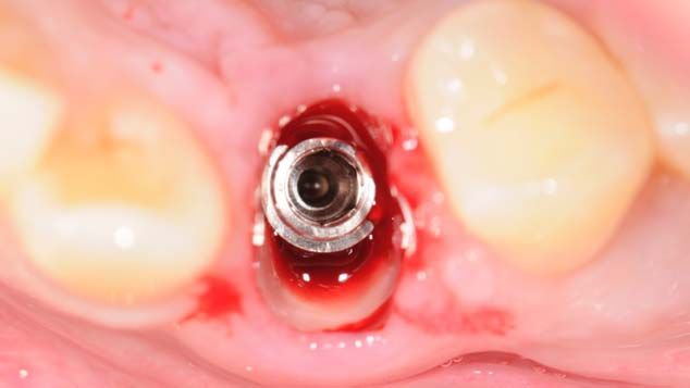
MINIMALLY INVASIVE IMMEDIATE IMPLANT TREATMENT FOR SINGLE-TOOTH REPLACEMENT Saeidi Pour et al

    FIGURE 4. Periapical radiographs, pre- and postendodontic.

    FIGURE 5. Study cast of the first situation, surgical guide fabricated over the cast, installation of the implant on the model, and
    fabrication of a custom healing abutment.

    FIGURE 6. Custom healing abutment with a special contour.                     FIGURE 7. Preoperative lateral photograph of the upper left
                                                                                  canine.
    for the extraction of the afflicted tooth and its
    replacement with an implant. The prominent canine
    contour combined with a high smile-line suggested the                         a CAD/CAM surgical guide (Smop Powered;
    use of the SST, which is minimally invasive and has a                         Swissmeda AG, Z€  urich, Switzerland) was manufactured
    positive effect onto the buccal bone contour.10                               by matching the CBCT data, with the additional aim
                                                                                  to provide a screw-retained implant-supported
    On a study cast based on a maxillary polyether                                restoration. A diagnostic “implantation” was performed
    impression (Impregum Penta; 3M, Seefeld, Germany),                            on the study cast (Figure 5), and a custom healing

4   Vol 00  No 00  00^00  2017 Journal of Esthetic and Restorative Dentistry                          DOI 10.1111/jerd.12291 V
                                                                                                                                C 2017 Wiley Periodicals, Inc.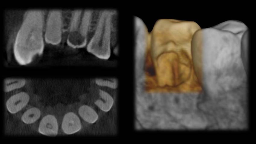
MINIMALLY INVASIVE IMMEDIATE IMPLANT TREATMENT FOR SINGLE-TOOTH REPLACEMENT Saeidi Pour et al

FIGURE 8. View of the teeth after decoronation.           FIGURE 9. Implant placement using a surgical guide.

                                                          FIGURE 11. Custom healing abutment installed.
FIGURE 10. Postimplantation occlusal view with the
remaining buccal fragment of the canine.

                                                          FIGURE 13. CAD/CAM surgical guide with implant position.

                                                          residual buccal root, allowing the additional fabrication
                                                          of the individual healing abutment (Figure 6). Figure 7
                                                          shows a preoperative lateral view of the upper left
FIGURE 12. Interim prosthesis inserted.
                                                          canine. The complex surgical implant placement
                                                          procedure was initiated by extraoral disinfection of the
abutment and interim prosthesis were produced; the        surgical site with a chlorhexidine solution for one
latter served as a provisional esthetic solution while    minute, application of local anesthesia, and
splinting the adjacent teeth to avoid any tooth           decoronation of the tooth with a diamond bur, leaving
movement.                                                 the tooth margins 1 mm above the gingival level
                                                          (Figure 8). The implant bed was prepared accord to
The use of a CAD/CAM surgical guide facilitated the       the manufacturer’s guidelines, with the remaining root
correct and precise positioning of the implant with the   in the alveolar socket (Figure 9). At this point, the

C 2017 Wiley Periodicals, Inc.
V                                DOI 10.1111/jerd.12291     Journal of Esthetic and Restorative Dentistry Vol 00  No 00  00^00  2017   5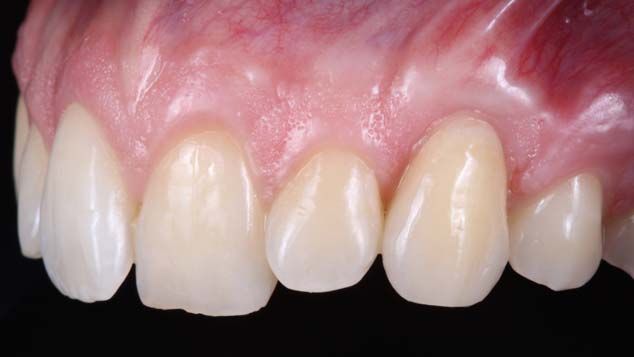
MINIMALLY INVASIVE IMMEDIATE IMPLANT TREATMENT FOR SINGLE-TOOTH REPLACEMENT Saeidi Pour et al

                                                                                  FIGURE 15. Ceramic restoration ready to be installed.
    FIGURE 14. Frontal view after three months of clinical
    implant service.

                                                                                  FIGURE 17. Frontal view after definite insertion of the
                                                                                  ceramic restoration.

                                                                                  system (Swiss Precision Implant; Thommen Medical,
                                                                                  Grenchen, Switzerland) and surgical guide. The implant
                                                                                  (14 mm/4 mm, Thommen SPI Element; Swiss Precision
    FIGURE 16. Postprosthetic radiographic evaluation.                            Implant) was placed according to the manufacturer’s
                                                                                  recommendation, situated closer to the palatal wall and
                                                                                  at the height of the buccal root segment (Figure 10).

                                                                                  For this implant system—again according to the
                                                                                  manufacturer’s guidelines—the final torque should
                                                                                  exceed 25 Ncm to for maximum primary stability.
                                                                                  Although the torque in the present case was lower than
                                                                                  25 Ncm, adequate primary stability was achieved and a
                                                                                  custom healing abutment (Figure 11) and an interim
                                                                                  prosthesis (Figure 12) could be inserted. The interim
                                                                                  prosthesis was connected to the adjacent teeth, not
                                                                                  placed directly on the implant, to shield it from
    FIGURE 18. Lateral view after definite insertion of the
    ceramic restoration.                                                          masticatory forces. During implant placement, a specific
                                                                                  point at the implant adapter ensures the correct buccal
    lingual, distal, and mesial fragments were carefully                          position. This point also exists for the laboratory analog.
    removed with minimal trauma, retaining the buccal part                        If there is no perfect agreement, small intraoral
    with approximately 4 to 5 mm on the socket,                                   modifications can still be made to the healing abutment.
    approximately 1 mm coronal to the buccal bone plate.
    An enamel matrix protein was applied and the                                  Thanks to this technique, no augmentation or
    implantation performed using the specified implant                             reconstructive surgical treatment was necessary.

6   Vol 00  No 00  00^00  2017 Journal of Esthetic and Restorative Dentistry                          DOI 10.1111/jerd.12291 V
                                                                                                                                C 2017 Wiley Periodicals, Inc.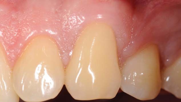
MINIMALLY INVASIVE IMMEDIATE IMPLANT TREATMENT FOR SINGLE-TOOTH REPLACEMENT Saeidi Pour et al

FIGURE 19. Postoperative smile.

FIGURE 20. Postoperative profile view.

Postoperative instructions were given to the patient, and       and 18). The implant screw channel was closed using
medication including an antibiotic and an analgesic was         gutta percha (VDW, Munich, Germany) and Tetric
prescribed, as well as the use of a chlorhexidine mouthrinse.   Evo Flow (Ivoclar Vivadent, Schaan, Liechtenstein).

The implantation was following by a bite registration           Figures 19 and 20 shows the patient’s smile and profile
using the existing surgical guide (Figure 13). The              with the final restoration.
impression coping was connected with the surgical
guide with light-curing resin (FRP-Resin; Bredent,
Senden, Germany) to allow accurate insertion of the             DISCUSSION
definitive implant analog into the former master cast
ahead of fabricating the final restoration.                      Recent studies have affirmed that the SST has the
                                                                potential to reduce bone resorption after tooth
Three months after implant placement, stable tissues            extraction and immediate implantation, mainly
around the custom healing abutment were observed                through the retention of the buccal/facial root
(Figure 14), allowing the installation of the previously        section.10,11,16,17 This is consistent with the present
fabricated definitive crown (Figure 15) with a torque of         case report that demonstrated soft- and hard-tissue
20 Ncm, resulting in an esthetic and functional                 stability around the implant, which is advantageous for
outcome with tissue stability preserved (Figures 16, 17,        lasting esthetic and functional outcomes.

C 2017 Wiley Periodicals, Inc.
V                                DOI 10.1111/jerd.12291          Journal of Esthetic and Restorative Dentistry Vol 00  No 00  00^00  2017   7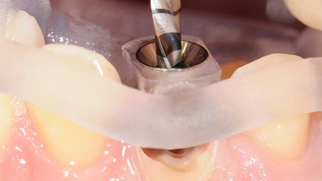
MINIMALLY INVASIVE IMMEDIATE IMPLANT TREATMENT FOR SINGLE-TOOTH REPLACEMENT Saeidi Pour et al

    The SST requires registration of a surgical index during                      low risk of inflammation. Based on histological and
    implantation ahead of fabricating the definite                                 clinical results, It also reduces resorption.
    restoration. Without this technique, it would be difficult
    to predict the shape and behavior of the remodeled soft                       It should be emphasized, however, that this is a sensitive
    tissue after completed healing. Especially the tooth                          technique that needs extensive planning. Its success
    extraction trauma, with its associated loss of periodontal                    greatly depends on the operator’s skills and ability to
    ligament and vascularization,16,17 results in                                 create a satisfying and long-lasting rehabilitation.
    unpredictable socket remodeling. Socket alterations have
    also been demonstrated with immediate implant                                 The present case report demonstrates the SST’s
    procedures and even with already osseointegrated                              potential for highly esthetic outcomes, with reduced
    implants, due to trauma to the bone wall.16                                   time and expense and less psychological stress for the
                                                                                  patient and the restorative team alike.
    Immediate as well as delayed implant placement are
    often associated with soft- or hard-tissue
    augmentation. This requires additional surgical
    interventions with a risk for resorption and infections,                      DISCLOSURE
    compromising the treatment result.17 The SST was
    developed to preserve the buccal/facial root sections                         The authors do not have any financial interest in the
    and to avoid trauma to the buccal wall during                                 companies whose materials are included in this article.
    extraction and implant preparation.

                                                                                  REFERENCES
    The concept of retaining a root to stabilize the alveolar
    ridge has been repeatedly described since 1950,2,18                           1.   Passoni BB, Marques de Castro DS, de Ara     ujo MA, et al.
    normally associated with pontic regions of fixed dental                             Influence of immediate/delayed implant placement and
    prostheses (FDP) and complete dentures where no                                    implant platform on the peri-implant bone formation. Clin
    inflammation was reported.2,18–20 Combined with the                                 Oral Implants Res 2016;5:1–8.
    SST, an enamel matrix derivative can also be indicated,                       2.   Araujo MG, Sukekava F, Wennstr€    om JL, Lindhe J. Tissue
    as in the present case.                                                            modeling following implant placement in fresh extraction
                                                                                       sockets. Clin Oral Implants Res 2006;17:615–24.
                                                                                  3.   Glocker M, Attin T, Schmidlin P. Ridge preservation with
    In conclusion, the most important advantages of SST                                modified “socket-shield” technique: a methodological case
    can be summarized as follows: no added cost for                                    series. Dent J 2014;2:11–21.
    materials; only a single surgical procedure; reduced                          4.   H€ammerle CH, Ara    ujo MG, Simion M, et al. Evidence-
    comorbidity; possibility of implant treatment in                                   based knowledge on the biology and treatment of extraction
    patients with previous endodontic pathology.                                       sockets. Clin Oral Implants Res 2012;23:80–2.
                                                                                  5.   Wilderman MN. Repair after a periosteal retention
                                                                                       procedure. J Periodont 1960;34:487–503.
    Disadvantages such as the need for tissue
                                                                                  6.   Wilderman MN, Wentz F, Orban BJ. Histogenesis of repair
    augmentation in several surgical steps (requiring                                  after mucogingival surgery. J Periodont 1963;31:283–99.
    additional time and putting added stress on the                               7.   Araujo MG, Sukekava F, Wennstr€    om JL, Lindhe J. Ridge
    patient) or increasing cost, or the difficult preservation                          alterations following implant placement in fresh extraction
    of the tissue architecture16,17 making the SST a                                   sockets: an experimental study in the dog. J Clin
    favorable option for dental practice. Also important is                            Periodontol 2005;32:645–52.
                                                                                  8.   Araujo MG, Wennstrom JL, Lindhe J. Modeling of the buccal
    the fact that the number of appointments is reduced,
                                                                                       and lingual bone walls of fresh extraction sites following
    which is desirable for patients and dentists alike.
                                                                                       implant installation. Clin Oral Implants Res 2006;17:606–14.
                                                                                  9.   Araujo MG, Silva CO, Misawa M, Sukekava F. Alveolar
    The SST offers a solution for tissue preservation directly                         socket healing: what can we learn?. Periodontol 2000 2015;
    after extraction and for implant osseointegration with a                           68:122–34.

8   Vol 00  No 00  00^00  2017 Journal of Esthetic and Restorative Dentistry                            DOI 10.1111/jerd.12291 V
                                                                                                                                  C 2017 Wiley Periodicals, Inc.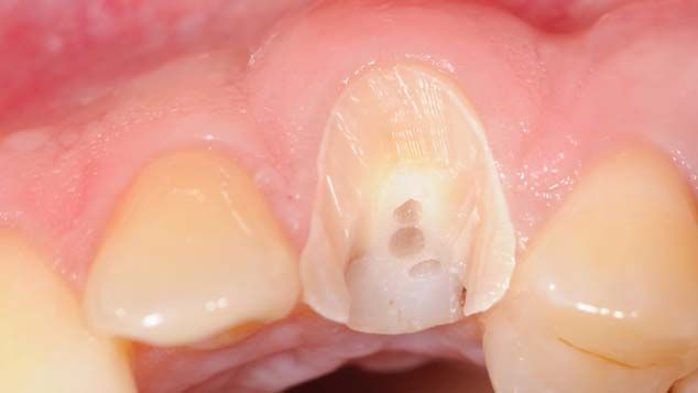
MINIMALLY INVASIVE IMMEDIATE IMPLANT TREATMENT FOR SINGLE-TOOTH REPLACEMENT Saeidi Pour et al

10. Hurzeler MB, Zuhr O, Schupbach P, et al. The socket-                16. Chen CL, Pan YH. Socket Shield technique for ridge
    shield technique: a proof-of-principle report. J Clin                   preservation: a case report. J Prost Implant 2013;2:16–21.
    Periodontol 2010;37:855–62.                                         17. Miller PA. Complete dentures supported by natural teeth.
11. B€aumer D, Zuhr O, Rebele S, et al. The socket-shield                   J Prosthet Dent 1958;8:924–8.
    technique: first histological, clinical, and volumetrical            18. Malmgren B, Cvek M, Lundberg M, Frykholm A.
    observations after separation of the buccal tooth                       Surgical treatment of ankylosed and infrapositioned
    segment – a pilot study. Clin Implant Dent Relat Res                    reimplanted incisors in adolescents. Scand J Dent Res
    2015;17:71–82.                                                          1984;92:391–9.
12. Guirado JL, Troiano M, L     opez-L opez, et al. Different        19. Andersson L, Emami-Kristiansen Z, Hogstrom J. Single-
    configuration of socket-shield technique in peri-implant                 tooth implant treatment in the anterior region of the
    bone preservation: an experimental study in dog mandible.               maxilla for treatment of tooth loss after trauma: a
    Ann Anat 2016;16:30124–8.                                               retrospective clinical and interview study. Dent Traumatol
13. Schneider D, Grunder U, Ender A, et al. Volume gain and                 2003;19:126–31.
    stability of peri-implant tissue following bone and soft tissue     20. Geng W, Liu C, Su Y, et al. Accuracy of different types of
    augmentation: 1-year results from a prospective cohort                  computer-aided design/computer-aided manufacturing
    study. Clin Oral Implants Res 2011;22:28–37.                            surgical guides for dental implant placement. Int J Clin Exp
14. Araujo MG, da Silva JC, de Mendonca AF, Lindhe J. Ridge                 Med 2015;8:8442–9.
    alterations following grafting of fresh sockets in man. A
    randomized clinical trial. Clin Oral Implants Res 2015;26:407–12.
15. Gluckman H, Du Toit J, Salama M. The socket-shield                  Reprint requests: Dr. Anja Liebermann,Goethestraße 70, 80336 Munich,
    technique to support the buccofacial tissues at immediate           Germany;Tel.: 149 89 4400 59571; Fax: 149 89 4400 59502; email:
    implant placement. Int Dentistry 2015;5:6–14.                       Anja.Liebermann@med.uni-muenchen.de

C 2017 Wiley Periodicals, Inc.
V                                DOI 10.1111/jerd.12291                   Journal of Esthetic and Restorative Dentistry Vol 00  No 00  00^00  2017   9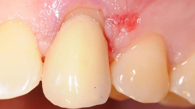
You can also read