Morphometric analysis of sella turcica in North Indian population: a radiological study
←
→
Page content transcription
If your browser does not render page correctly, please read the page content below
International Journal of Research in Medical Sciences
Chauhan P et al. Int J Res Med Sci. 2014 May;2(2):521-526
www.msjonline.org                                                                              pISSN 2320-6071 | eISSN 2320-6012

                                                                                   DOI: 10.5455/2320-6012.ijrms20140529
Research Article

      Morphometric analysis of sella turcica in North Indian population:
                            a radiological study
         Puja Chauhan1*, Sunita Kalra2, Shashi M. Mongia1, Sadakat Ali1, Anurag Anurag1

  1
   Department of Anatomy, Shri Guru Ram Rai Institute of Medical and Health Sciences, Dehradun, Uttarakhand, India
  2
   Department of Anatomy, University College of Medical Sciences, Delhi-110095, India

  Received: 21 January 2014
  Accepted: 2 February 2014

  *Correspondence:
  Dr. Puja Chauhan,
  E-mail: drpujachauhan@gmail.com

  © 2014 Chauhan P et al. This is an open-access article distributed under the terms of the Creative Commons
  Attribution Non-Commercial License, which permits unrestricted non-commercial use, distribution, and reproduction
  in any medium, provided the original work is properly cited.

   ABSTRACT

   Background: The purpose of this study was to describe the morphology and measure the size of the sella turcica in
   North Indian population.
   Methods: Lateral cephalometric radiographs of 180 individuals (90 males and 90 females) with an age range of 12 -
   65 years were taken. Morphology of sella turcica was studied and various measurements were taken to determine the
   shape of the sella. Statistical analysis was done to calculate differences in dimensions and to establish if any,
   relationship exists between age, sex and the morphometry of sella turcica.
   Results: The study found that sella turcica presented with a normal morphology in only 28 per cent of the subjects. A
   significant difference in linear dimensions between genders was found in sella height and width. When age was
   evaluated, some dimensions showed negative correlation with the age. Sella size of the older age group was as a rule
   larger than the younger age.
   Conclusion: Pathological enlargement of the pituitary fossa can be detected by this technique and may also be helpful
   in providing data in the assessment of racial, gender, age specific variation in the skull.

   Keywords: Morphometry, Sella turcica, Morphology

INTRODUCTION                                                          point. It is traceable for metric analysis which makes it an
                                                                      excellent source of information related to the
Sella turcica is located in the middle cranial fossa and lies         identification of pathologies of pituitary gland and
on the intracranial surface of the body of the sphenoid               various syndromes that affect the craniofacial region.
and consists of a central pituitary fossa bounded                     Knowledge of the normal radiological anatomy and
anteriorly by tuberculum sellae and posteriorly by                    variations in the morphometry of this area are helpful as a
dorsum sellae. Two anterior and two posterior clinoid                 tool to study growth in an individual, evaluation of
processes project over the pituitary fossa. The anterior              orthodontic treatment results and to recognize and further
clinoid processes are formed by the medial and anterior               investigate a variety of pathological situations.1-3
prolongations of the lesser wing of the sphenoid bone,
and the terminations of dorsum sellae are present in the              Normative data on the size of the sella turcica have been
form of posterior clinoid processes.                                  reported previously and typically range from 4 to 12mm
                                                                      for the vertical and 5 to 16 mm for the antero-posterior
Sella turcica is an important landmark readily seen on                dimension4-6 Changes in size of the sella turcica are
lateral cephalograms and acts as a noteworthy reference               frequently related to pathology; enlargement is the most

                                   International Journal of Research in Medical Sciences | April-June 2014 | Vol 2 | Issue 2   Page 521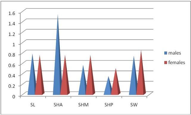
Chauhan P et al. Int J Res Med Sci. 2014 May;2(2):521-526

frequent finding but is frequently not accompanied by               The radiographs were scanned at 150 dpi and the points
bone erosion. Any abnormality or pathology of gland can             shown in Figure 1 were digitized with viewbox 3
manifest7 from an altered shape of sella turcica to a               software. The contour of the sella turcica was traced
disturbance in the regulation of secretion of various               between points tuberculum sella (TS) and posterior
glandular hormones leading to diverse problems like                 clinoid (PClin), and nine additional equally spaced points
acromegaly, gigantism, hyperthyroidism, amenorrhoea                 along this contour were located by the computer
etc. It has been suggested that cephalometric radiographs           software. The total of these 11 points defined a smooth
of subjects with these conditions may, in some instances,           curve that represented the outline of the sella turcica from
show an abnormal sella region, or vice versa i.e., subjects         TS to PClin, and these were used for shape analysis. In
with an abnormal sella turcica may actually have an                 addition, the outline was used to calculate the position of
undetected     underlying disease.8 The immense                     the most posterior point , the most anterior point and the
improvement of imaging methods has made it possible to              deepest point of the sella (sella floor), using the Frankfurt
perform research that defines the morphology of the                 plane (FH) as the horizontal reference direction.
human body structures. Morphology may vary from one
geographical region to other depending upon the type of             The following measurements were computed:
skull. The establishment of normal standards in Indian
population will aid in the process of eliminating any               1.   Sella width: largest antero-posterior dimension, as
abnormality in such an important part of cranium.                        measured parallel to the FH plane, from anterior to
Therefore, the purpose of this study was to analyze the                  posterior.
predominant morphological shape and measure the linear
dimensions of sella turcica to determine if any difference          2.   Sella length: distance from TS to PClin.
exists due to gender or age in north Indian population.
                                                                    3.   Sella height anterior: The vertical distance, as
METHODS                                                                  measured perpendicular to the FH plane, from TS to
                                                                         the sella floor.
This was a prospective study of patients seeking
orthodontic treatment in North India. Lateral                       4.   Sella height posterior: The vertical distance, as
cephalogram of 180 patients (90 males and 90 females)                    measured perpendicular to the FH plane, from PClin
aged 12 – 65 years were used in this study. All subjects                 to the sella floor.
were clinically healthy with no syndromes, clefts, or
other malformations and were treated by orthodontics                5.   Sella height median: The vertical distance, as
alone without surgical intervention. Radiographs showing                 measured perpendicular to the FH plane, from the
sella turcica area with maximum clarity were selected.                   sella floor to a point midway between PClin and TS.

The mid-saggital enlargement was 110 per cent and all               All measurements were taken after adjusting for the
linear measurements were corrected for magnification                magnification of the radiographs. The statistical tests
differences prior to the statistical analyses. The                  were run using SPSS software. Paired t-tests were
radiographs were divided into two groups according to               employed to evaluate the system error if any.
the subject’s age: pre-pubertal (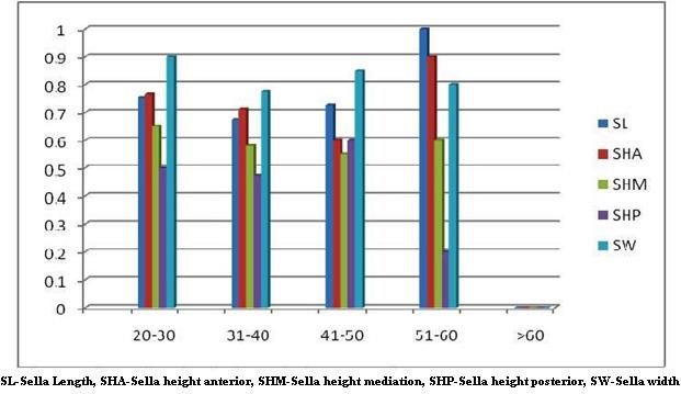
Chauhan P et al. Int J Res Med Sci. 2014 May;2(2):521-526

and reduced later. Whereas Sella width was maximum in             thereafter. Average sella width was 8.4mm being highest
the age group of 40-50 yrs (Figure 1, 2 and 3).                   in 20-30 yrs followed by a gradual decline.

                                                                  Morphology of sella turcica was found to be typical in
                                                                  just 28% of cases. Within the atypical sellae most had
                                                                  oblique anterior wall (23%), followed by irregular sellae
                                                                  (18%). None of the sella was seen to be pyramidal in type
                                                                  in this population (Figure 4).

Figure 1: Comparative analysis of various dimensions
      (mm) in different age groups in females.

                                                                    Figure 4: Incidence of different morphological types
                                                                                      of sella turcica.

                                                                  When Pearson’s coefficient of correlation was found to
                                                                  be negative for sella length and sella width in females
                                                                  whereas it was negative for sella height anterior and sella
                                                                  height posterior in males (Figure 5).

Figure 2: Comparative analysis of various dimensions
       (mm) in different age groups in males.

                                                                  Figure 5: Pearson’s coefficient of correlation in age vs.
                                                                      sella turcica dimensions in males and females.

                                                                  DISCUSSION

                                                                  Shape variation in the sella turcica have previously been
                                                                  reported by few researchers.11-13 Gordon and Bell (1922)
                                                                  examined the radiographs of children 1 - 12 years of age
Figure 3: Metric analysis of sella dimensions (mm) in
                                                                  and classified the sella turcica into circular, oval, and
                 males and females.
                                                                  flattened, or saucer shaped.11 They concluded that most of
                                                                  the subjects had either a circular or oval-shaped sella, and
In females average sella length was found to be 7.5mm
                                                                  noted that even with this broad classification, difficulty
and was largest in >60 yrs of age. Average sella height
                                                                  was found in placing some cases into one of the three
median was also 7.5mm and was seen to decline with age
                                                                  categories. Davidoff and Epstein (1950) used the term ‘J-
and in >50 yrs was again seen to increase in dimension.
                                                                  shaped sella’,14 while ‘omega sella’15 was introduced by
Average sella height posterior was 7.5mm, was
                                                                  Fournier and Denizet (1965). But these definitions were
maximum in the age 40-50 yrs and gradually declined
                                                                  later termed radiological myths by a study by Kier,16 who
                                                                  suggested disregarding both of these since these were

                               International Journal of Research in Medical Sciences | April-June 2014 | Vol 2 | Issue 2   Page 523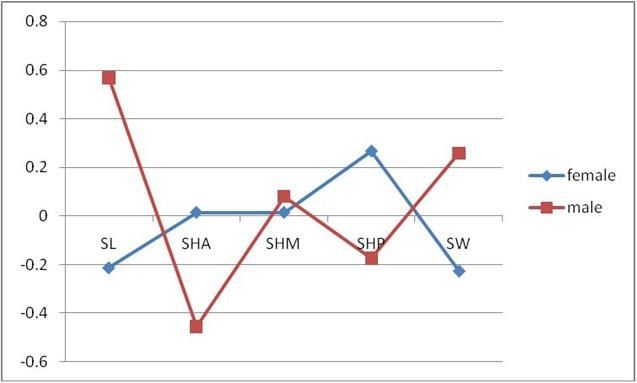
Chauhan P et al. Int J Res Med Sci. 2014 May;2(2):521-526

used to characterize abnormal pathology as well as                   The presence of a sella turcica bridge in normal
normal developmental patterns. Other descriptions of the             individuals has been shown to occur in 5.5 - 22 per cent
sella turcica have been proposed based on the appearance             of subjects,18 with an increased incidence in patients with
of flatness or concavity of the contours of the sella floor,         craniofacial 461 deviations.17 In the current study, a sella
the angles made by the contours of the tuberculum sella,             bridge (Figure 8) was found in the same range as other
the contours of the anterior and posterior clinoid                   studies (17%). An oblique anterior wall has also been
processes, and the fusion of both processes which is                 documented in normalas well as medically compromised
termed a ‘sella turcica bridge’.17                                   subjects such as children              with lumbosacral
                                                                     meningomyelocele and seckel syndrome.18-20 The current
In a study18 the shape of the sella turcica was categorized          study showed only 23% per cent of subjects with an
into six main types; normal sella turcica, oblique anterior          oblique anterior wall which was almost at par (26%) with
wall, double-contoured sella, sella turcica bridge,                  the study by Axelsson et al.18
irregularity (notching) in the posterior part of the sella,
and pyramidal shape of the dorsum sellae. Their results
show that normal sella turcica morphology was seen in
two-thirds of the subjects, while the remainder showed
variable morphological appearances.

In the current study, only 28% per cent of the subjects
appeared to have a normal shaped sella turcica, while
72% per cent presented with different shapes. The finding
of an irregular notching of the dorsum sella was
approximately 18% in the present study (Figure 6) which
was more frequent than other study,18 while a pyramidal
shape of the dorsum was reported more frequently in the
former investigation as compared to the present study
where there was no case detected with pyramidal dorsum
sella. A doubled contour floor was present in 7 per cent
of the subjects of the current study (Figure 7), which is
much higher than that reported by same study.
                                                                      Figure 8: “Irregular” sella turcica with same range as
                                                                                          other studies.

                                                                     When the linear dimensions of length, depth, and
                                                                     diameter of the sella turcica in the present study were
                                                                     compared with other investigations.18,21 significant
                                                                     difference between measurements was noted. Quakinine
                                                                     and Hardy (1987)21 performed a microsurgical anatomical
                                                                     study on 250 sphenoidal blocks obtained from cadavers
                                                                     of different ages. They found that the average transverse
          Figure 6: Bridging of Sella turcica.                       width of the sella turcica was 12mm, the length (antero-
                                                                     posterior diameter) 8mm, and the average height (vertical
                                                                     diameter) 6mm. Whereas average height was found to be
                                                                     more in the present study by approximately 1.7 mm
                                                                     (Table 1). But height of the gland was usually 2 mm
                                                                     shorter than the actual depth of the sella (the gland does
                                                                     not fill the whole volume of the sella turcica) and this
                                                                     should be taken into consideration during measurements.
                                                                     Similar results were also found in a Norwegian sample.18
                                                                     The linear dimensions in this Saudi sample were on
                                                                     average 2.02 - 2.73mm larger than those in the
                                                                     Norwegian subjects.

                                                                     In the current study there was highly significant
                                                                     difference between males and females in the sella turcica
                                                                     height posterior and sella width (p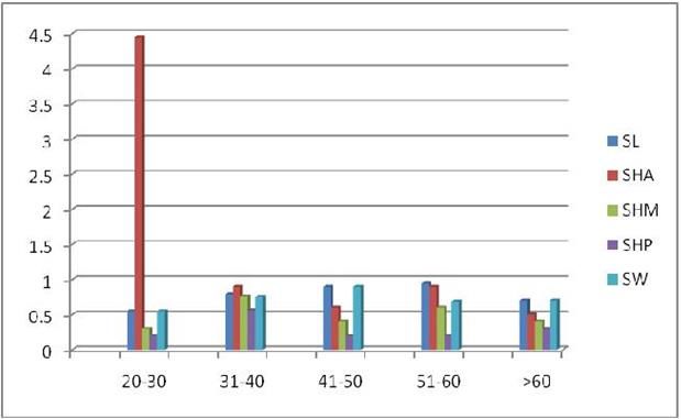
Chauhan P et al. Int J Res Med Sci. 2014 May;2(2):521-526

the pituitary fossa of males tended to be larger than that          identified with sufficient sensitivity once such values are
of females from about 1 - 13 years of age. Due to the               available. However, it is becoming more evident that
pubertal growth spurt in females which begins 2 years               what appear to be ‘abnormalities’ in shape may not
earlier than males, a significant change in pituitary fossa         always reflect underlying pathology. For example,
size occurs in females from 11 to 15 years of age.                  although asymmetry (double outline) and cortical erosion
Thereafter, growth acceleration in males, which is usually          of the sella floor are often considered signs of increased
2 - 3 years later than that in female, results in an                pathological significance,26 it is arguable because such
approximate equalization in sella area in both genders.             signs have been observed in a relatively large percentage
On the other hand, Haas (1954) compared the mean size               of asymptomatic subjects, without being related to the
in square millimeters of the sella area of boys and girls           presence of pituitary tumours. Conversely, pathology
aged 3 - 17 years and found some differences due to                 may exist without osseous manifestations. The largest
gender.22 He reported that the sella turcica of boys was            percentage of intrasellar tumours is microadenomas
greater than girls, but after 17 years of age, the sella of         (adenomas smaller than 10 mm in diameter), often too
females were slightly larger than that of males when the            small to cause sella enlargement or shape change. In fact,
effect of age on sella turcica size was studied, the sella          it is estimated that 10 - 20 per cent of the population may
sizes of the older age group in the present investigation           harbour microadenomas, most often asymptomatic.27
were more or less larger than the younger group. Similar            Moreover, even considerable enlargement of the pituitary
findings were reported by Preston (1979) who found a                gland may not produce osseous changes evident on
close correlation between the area of sella and age. His            routine films of the skull because the pituitary gland
findings on 182 lateral radiographs of individuals aged 5           occupies only part (approximately 80 per cent) of the
- 17 years revealed that the pituitary fossa increased in           volume of the sella turcica.28 Further studies are needed
size with age, which reflects the adolescent growth spurt           to assess the sensitivity and specificity of cephalograms
of females that occurs at an earlier age.23                         for detection of pituitary pathology. Of course,
                                                                    cephalograms do not constitute the radiological method
Choi et al. (2001) concluded that the linear dimensions of          of choice for diagnosis of a suspected pituitary tumour.
sella turcica had a positive linear tendency until 25 years         Computed tomography and magnetic resonance imaging
of age. After 26 years of age, no significant increase              provide much greater sensitivity. However, incidental
could be found in sella turcica size.24 Contrary to these           findings noted by the orthodontist may lead to further
findings, Elster et al. (1990), in a magnetic resonance             investigation of undiagnosed or subclinical conditions.1-3
imaging study of 169 patients aged 1 - 30 years, found              Although enlargement of the sella turcica may be a sign
that during childhood there was no difference between               of an intrasellar tumor or juxtasellar tumor asymptomatic
males and females, but that dramatic changes took place             enlargement of sella turcica may occur. Plain film
during puberty with swelling of the gland.12 When                   radiographs have a relatively high sensitivity for
studying the effects of gender and age, on the size of the          detecting sella change at between 67% and 77% of
sella turcica, the results show that age was significantly          positive findings and clinicians should be suspicious
related to the dimensions of sella, which were larger in            when any of the sella turcica dimensions exceed the
older subjects, irrespective of gender. No similar studies          upper limits of normal.
comparing these factors with sella size in north Indian
population could be found in the literature. The linear             It is well known that pathological enlargement of the
dimensions obtained from the current study can be used              pituitary fossa may often be distinguished at a glance.
to approximate the size of the pituitary gland, and may             Many such cases show erosion of the bone. In a study of
aid the clinician when confronted with an abnormally                lateral skull films of patients suffering from tuberculous
large sella turcica on lateral cephalograms. The                    meningitis, measurement has shown that in some adults
orthodontist should also be familiar with the different             the fossa increases in size to a degree which is significant
shapes of the sella area, in order to help distinguish              when the errors of the method are considered. In a few
pathology from normal developmental patterns.                       young children also, the fossa has been found to show a
                                                                    greater increase in size than might have been expected in
Choi et al. (2001) measured volume in addition to width             the normal growth processes. In none of these cases has
and height in a cross-sectional sample of orthodontic               there been any bony erosion, or any other sign of
patients.24 They found an increase in sella dimensions              enlargement which attracts the unaided eye. Therefore it
with age, from the 6 - 10 to the 21 - 25 age group.                 is concluded that, in doubtful cases, pathological
However, the change in height was minimal and probably              enlargement of the pituitary fossa may be detected by this
not statistically significant (no SD values were given). It         technique provided that serial films are taken while the
should be noted that an age-related increase of sella               enlargement is going on.
turcica size is expected because its contents, i.e. the
hypophysis, have been shown to increase in size with                Funding: No funding sources
age.25                                                              Conflict of interest: None declared
                                                                    Ethical approval: Not required
The search for establishment of normative values is based
on the assumption that pathological conditions will be

                                 International Journal of Research in Medical Sciences | April-June 2014 | Vol 2 | Issue 2   Page 525
Chauhan P et al. Int J Res Med Sci. 2014 May;2(2):521-526
REFERENCES 15. Fournier AM, Denizet D. Omega shaped sella
turcica. Marseille Med. 1965;102:503-9.
1. Friedland B, Meazzini MC. Incidental finding of an 16. Kier EL. ‘J’ and ‘omega’ shape of sella turcica:
enlarged sella turcica on a lateral cephalogram. Am anatomic classification of radiologic
J Orthodont Dentofac Orthoped. 1996;110:508-12. misconceptions. Act Radiol Diagnos. 1969;9:91-4.
2. Feldkamp J, Santen R, Harms E, Aulich A, Mödder 17. Becktor J, Einersen S, Kjær I. A sella turcica bridge
U, Scherbaum WA. Incidentally discovered in subjects with severe craniofacial deviations. Euro
pituitary lesions: high frequency of macroadenomas J Orthodont. 2000;22:69-74.
and hormone-secreting adenomas: results of a 18. Axelsson S, Storhaug K, Kjær I. Post-natal size and
prospective study. Clin Endocrinol. 1999;51:109-13. morphology of the sella turcica. Longitudinal
3. Alkofi de E. Pituitary adenoma: a cephalometric cephalometric standards for Norwegians between 6
finding. Am J Orthodont Dentofac Orthoped. and 21 years of age. Euro J Orthodont. 2004;26:597-
2001;120:559-62. 604.
4. Camp J. Normal and pathological anatomy of the 19. Kjær I, Hansen N, Becktor KB, Birebaek N, Balslev
sella turcica as revealed by roentgenograms. Am J T. Craniofacial morphology, dentition, and skeletal
Roentgenol. 1924;12:143-55. maturity in four siblings with seckel syndrome.
5. Silverman FN. Roentgen standards for size of the Cleft Palate Craniofac J 2001;38a:645-51.
pituitary fossa from infancy through adolescence. 20. Kjær I, Wagner A, Madsen P, Blichfeldt S,
Am J Roentgenol. 1957;78:451-60. Rasmussen K, Russell B. The sella turcica in
6. Chilton LA, Dorst JP, Garn SM. The volume of the children with lumbosacral myelomeningocele. Euro
sella turcica in children: new standards. Am J J Orthodont. 1998;20:443-8.
Roentgenol. 1983;140:797-801. 21. Quakinine GE, Hardy J. Microsurgical anatomy of
7. Elster AD. Imaging of the sella: anatomy and the pituitary gland and the sellar region: the pituitary
pathology. Seminars in Ultrasound, CT, and MRI. gland. Am Surg. 1987;53:285-90.
1993;14a:182-94. 22. Haas LL. The size of the sella turcica by age and
8. Weisberg LA, Zimmerman EA, Frantz A. Diagnosis sex. Am J Roentgenol Radium Therapy Nuclear
and evaluation of patients with enlarged sella. Am J Med. 1954;72:754-61.
Med. 1976;61:590-6. 23. Preston CB. Pituitary fossa size and facial typ. Am J
9. Israel H. Continuing growth in sella turcica with Orthodont. 1979;75:259-63.
age. Am J Roentgenol Radium Therapy Nuclear 24. Choi WJ, Hwang EH, Lee SR. The study of shape
Med. 1970;108:516-27. and size of normal sella turcica in cephalometric
10. Pisaneschi M, Kapoor G. Imaging of the sella and radiographs. Korean J Oral Maxillofac Radiol.
parasellar region. Neuroimag Clin North Am. 2001;31:43-9.
2005;15:203-19. 25. Argyropoulou M, Perignon F, Brunelle F, Brauner
11. Gordon MB, Bell AL. A roentgenographic study of R, Rappaport R. Height of normal pituitary gland as
the sella turcica in normal children. New York State a function of age evaluated by magnetic resonance
J Med. 1922;22:54-9. imaging in children. Pediatr Radiol. 1991;21:247-9.
12. Teal JS. Radiology of the adult sella turcica. Bull 26. Cook DM. Pituitary tumors: current concepts of
Los Angeles Neurolog Soc. 1977;42:111-7. diagnosis and therapy. Western J Med.
13. Tetradis S, Kantor ML. Prevalence of skeletal and 1980;133:189-96.
dental anomalies and normal variants seen in 27. Hall WA, Luciano MG, Doppman JL, Patronas NJ,
cephalometric and other radiographs of orthodontic Oldfield EH. Pituitary magnetic resonance imaging
patients. Am J Orthodont Dentofac Orthoped. in normal human volunteers: occult adenomas in the
1999;116:572-7. general population. Ann Intern Med.
14. Davidoff LM, Epstein BS. The abnormal 1994;120(10):817-20.
pneumoencephalogram. In: Davidoff LM, Epstein 28. Chang HP, Tseng YC, Chou TM. An enlarged sella
BS, eds. 1st ed. Philadelphia: Lea & Febiger; 1950: turcica on cephalometric radiograph.
388-394. Dentomaxillofac Radiol. 2005;34:308-12.
DOI: 10.5455/2320-6012.ijrms20140529
Cite this article as: Chauhan P, Kalra S, Mongia SM,
Ali S, Anurag A. Morphometric analysis of sella
turcica in North Indian population: a radiological
study. Int J Res Med Sci 2014;2:521-6.
International Journal of Research in Medical Sciences | April-June 2014 | Vol 2 | Issue 2 Page 526You can also read