Correlation of Cytological Grading in Lymphocytic Thyroiditis with Thyroid Hormones and Antibodies - A Retrospective Study in the Era of Bethesda ...
←
→
Page content transcription
If your browser does not render page correctly, please read the page content below
DOI: 10.7860/JCDR/2018/35123.11923
                                                                                                                                       Original Article

                            Correlation of Cytological Grading in
Pathology Section

                            Lymphocytic Thyroiditis with Thyroid
                                   Hormones and Antibodies - A
                      Retrospective Study in the Era of Bethesda
                                    System of Thyroid Reporting
SUPREETHA MEGALAMANE1, HEMALATHA ANANTHARAMAIAH2, SHASHIDHAR NAGARAJ KURPAD3

ABSTRACT                                                                          based on Bhatia A et al., criteria. Thyroid function tests were
Introduction: Thyroid disorders are the most common endocrine                     done in 98 cases and antithyroid antibody levels values was
disorders affecting about 42 million people in India. Among all                   available in 59 cases. Chi-square test and Pearson’s correlation
the disorders, Lymphocytic Thyroiditis (LT) is the second most                    coefficient were used and p≤0.05 was considered as significant
common thyroid lesion diagnosed on cytology next to goiter.                       for statistical analysis.
Aim: The present study was done to grade cytological smears                       Results: Female preponderance in the age group of 7-60 years
of LT and correlate grades with clinical presentation, Thyroid                    with diffuse thyroid enlargement was the common presentation.
Stimulating Hormone (TSH) and antithyroid antibodies.                             Maximum numbers of cases were of cytological grade 2 with
                                                                                  euthyroid and hypothyroid status. A p-value was significant
Materials and Methods: The present study was a retrospective
                                                                                  for correlation of all cytological grades with TSH values.
study of 185 cases from June 2014 to June 2016 conducted
                                                                                  Combination of Anti Thyroid Peroxidase (ATPO) and TSH
at Sri Devaraj Urs Medical College and Research centre, Kolar,
                                                                                  together had positive correlation with cytological grade 3.
Karnataka, India. Institutional ethical clearance was obtained
before the start of study. Patient’s clinical and demographic                     Conclusion: Cytological grading helps in assessing the severity
details, relevant cytology smears were retrieved and graded                       of the disease as it reflects the TSH levels and ATPO levels.

                                                                     Keywords: Cytological grade, Thyroid antibodies, Thyroid stimulating hormone

Introduction                                                                      Cytomorphological diagnosis may be superior in the initial stages
Thyroid disorders are the most common endocrine disorders                         of LT as antibody production may be confined to only intrathyroidal
affecting about 42 million people in India [1]. Among all the disorders           lymphocytes or localised areas without spillover into the blood
LT is the second most common thyroid lesion diagnosed on cytology                 [4,7,8]. Thus, there exists a wide range of values of TSH and
next to goiter [2].                                                               variability of presence of ATPO making early diagnosis of LT difficult
                                                                                  clinically and biochemically.
“Strauma Lymphomatosa” a chronic disorder of the thyroid gland is
                                                                                  However, a combination of cytomorphological, clinical and
characterized by diffuse lymphocytic infiltration, fibrosis, parenchymal
                                                                                  biochemical features helps in making a better diagnosis than using
atrophy and eosinophilic changes in some of the acinar cells (Hurthle
                                                                                  individual features alone [12].
cell change). The word LT is used synonymously with Hashimoto’s
thyroiditis or autoimmune thyroiditis which is again subclassified as             Not many studies have correlated LT grading on cytology with clinical,
atrophic and non goitrous thyroiditis on histopathology [3].                      hormonal status and thyroid auto antibodies. With this background
                                                                                  the aim of our study is to look into cytological features of LT and
LT typically presents with painless enlargement of thyroid gland,
                                                                                  grade it according to Bhatia A et al., [10], and also to correlate the
hypothyroidism, or both; 90% of LT patients also have High Anti-                  cytological grades of LT with clinical presentation, TSH, ATPO and
Thyroid Peroxidase (TPO) and anti-Thyroglobulin (Tg) antibodies                   Antithyroglobulin (ATG) levels.
[4].
On histopathology, the hallmark finding is lymphocytic infiltration               Materials and Methods
of thyroid follicles resulting in glandular destruction, formation                The present study was a retrospective study done from June 2014
of lymphoid follicles and Hurthle cell changes [5]. Fine Needle                   to June 2016 conducted at Sri Devaraj Urs Medical College and
Aspiration Cytology (FNAC) of thyroid provides a safe and accurate                Research centre. One hundred and eighty five cases of LT which
method for diagnosis with a sensitivity of 92% in predicting LT [6]. On           was diagnosed on cytology during this period were included in this
cytology presence of lymphocytic impingement of thyroid follicular                study. Institutional ethical clearance was obtained before start of
cells, presence of a mixed population of mature and transformed                   study. Demographic and clinical details along with stained slides
lymphocytes, Hurthle cells, follicular cells with fine chromatin, an              (Papanicolaou’s, Hematoxylin and Eosin and May Grunwald, Giemsa
iso-nucleosis, multinucleated giant cells, scanty or absence of                   stains) were obtained from the archives of our Pathology department.
colloid on aspirated smears are the hallmark of LT [6-11]. Hurthle                The diagnosis was confirmed and was graded according to criteria
cells appears as large cells forming small syncytial aggregates                   of Bhatia A et al., [10]. Any discrepancy was discussed and resolved
having well defined abundant finely granular eosinophilic cytoplasm               by the senior pathologist. All cases of LT confirmed on cytology
and large nucleus [7,12].                                                         were included and patients with co-existing malignancies were

Journal of Clinical and Diagnostic Research. 2018 Aug, Vol-12(8): EC01-EC04                                                                                1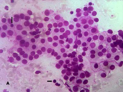
Supreetha Megalamane et al., Lymphocytic Thyroiditis and its Cytological Grading www.jcdr.ne
excluded. Patients with ischemic heart diseases, cerebrovascular
and neurological diseases, chronic renal impairment and pregnancy
were also excluded from our study as these conditions may cause
variation in thyroid hormone levels. Available data regarding age,
sex, presenting complaints, duration of symptoms, clinical signs of
hormonal variation, presence of nodular/diffuse enlargement were
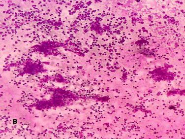
[Table/Fig-1]: Grade 1 Thyroiditis with mild lymphocytic infiltrate (arrows) (H&E;
obtained from our archives and entered in the master sheet. stain X 400). [Table/Fig-2]: Grade 2 Thyroiditis with moderate lymphocytic infiltrates
(H&E; X100). [Table/Fig-3]: Grade 3 Thyroiditis total destruction of follicle with dense
Cytological features of Lymphocytes: Epithelial cell ratio,
infiltration by lymphoid cells (MGG, X400) (Figure D: Inset shows Hurthle cell change
follicular atypia, Hurthle cells, plasma cells and eosinophils were – MGG; X1000). (Images from left to right)
documented.
All cases were graded as per Bhatia A et al., grading system Cytological grading No. of Cases
which is as follows [10]: Grade I (Mild): Few lymphoid cells Grade 1 45
infiltrating the follicles/increased number of lymphocytes in the Grade 2 74
background [Table/Fig-1].
Grade 3 66
Grade II (Moderate): Moderate lymphocytic infiltration or mild
Total 185
lymphocytic infiltration with Hurthle cell change/giant cells/an
[Table/Fig-4]: Grading of lymphocytic thyroiditis.
isonucleosis [Table/Fig-2].
Grade III (Severe): Florid lymphocytic inflammation with germinal Thyroid stimulating Antibodies (IU/mL) (n=59cases)
centre formation, very few follicular cells left [Table/Fig-3]. Hormone (0.27-5.5 IU/
mL) (n=98) ATPO(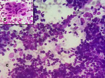
www.jcdr.net                                                                  Supreetha Megalamane et al., Lymphocytic Thyroiditis and its Cytological Grading

with ATPO values when compared alone, So combination of raised                In our study euthyroid were predominant in grade 1 with 73.1%,
ATPO and increased TSH or alone increased TSH was significantly               which was in contrast to studies by Uma P et al., and Kudva R et al.,
better in predicting grade 3 than only raised ATPO value. Relation            where they showed balanced distribution of hypothyroid cases in all
of cytological grading of LT with values of TSH and ATPO are                  the grades [14,20]. This can be because of geographical variations
represented in [Table/Fig-5].                                                 in prevalence of hypothyroidism.
                                                                              Raised ATPO levels was also described by Anila KR et al., wherein
DISCUSSION                                                                    majority of cases (41.6%) were of grade 1, grade 2 (31.7%) and
The pathogenesis of LT is a complex multistep process, comprising             grade 3 in 27% [12]. We also found similar distribution [Table/Fig-5].
of various genetic, environmental and immunological factors. Initially        These findings supports that our patient presents at an early stage
in active phase there is antibody mediated destruction of the thyroid         with raised ATPO levels, even before there is serological evidence of
follicles and intrathyroidal lymphocytic infiltration. Later in the chronic   hormonal imbalance [12,21].
phase, there will be only minimal residual, atrophic follicles with
                                                                              The relationship between grades, TSH levels and auto antibodies
fibrosis of the thyroid parenchyma, Depending on the stage, the
                                                                              can be explained as follows. Alteration of autoantibodies causes
patient present with different clinical features, hormonal status and
                                                                              destruction of thyrocytes which in turn leads to abnormalities in
thyroid autoantibodies levels. The cytological grades also depends
                                                                              the hormonal levels. Early stages, autoantibodies will be elevated
on the stage of the disease process [10,12].
                                                                              without much destruction of thyrocytes, hence normal hormonal
In the present study the age of occurrence of LT ranged from 7-80             levels [10,12]. Thus, on cytology these changes are reflected as
years with female predominance. Majority of the patients were in              grade 1 wherein only lymphocytes, without much Hurthle cells and
the age group of 25-45 years, this age distribution was comparable            follicular atypia are seen. No follicular cell infiltration by lymphocytes
to the observation made by Bhatia A et al., [10]. Contrast to our             is seen.
findings, the study from United Kingdom showed that patients were
                                                                              Once the destruction is progressive, as in grade 3 with abundant
predominantly older women. This change in distribution of cases
                                                                              lymphoctyes, germinal follicle form, numerous Hurthle cells and
may be due to geographical variations in thyroid disorders [12].
                                                                              destruction of follicles are seen. This destruction also results in
In our study, the prevalence of juvenile LT (0-18 years) was 13.5%.           hormonal abnormalities which is in agreement with our observation
The incidence of juvenile LT was comparable with studies done by              of Sood N et al., Anila KR et al., Uma P et al., and Baker JR et al.,
other authors [13,14].                                                        [2,12,14,21].
The contrasting theories have been put forwarded to describe the              Out of six cases of hyperthyroid three cases showed increased
hypothesis of prevalence of LT. This disparity in age distribution among      APTO. In that two cases were of grade 1 and one case was of grade
India and foreign studies is because of onset of thyroiditis in younger       2. This phenomenon can be explained as follows; in early stage
age group, due to high prevalence of iodine deficiency in India [12,15].      of the disease acute autoantibody mediated destruction of thyroid
One more school of thought has proposed that high iodine intake               follicular cells results in exudation of hormones into the circulation
may be an important risk factor for onset of thyroiditis. These patients      resulting in hyperthyroidism [12,21]. Similar to the present study,
with ATPO and ATG levels positive at baseline are more likely to be           Chandanwale SS et al., and Bagchi N et al., showed 23% and
prone for thyroid dysfunction than seronegative patients [16].                8.1% respectively hyperthyroid cases where there was no clinical
Our findings of clinical presentation of 72.4% with diffuse                   presentation but on cytology showed features of LT. They showed
enlargement and 27.6% of patients with nodular presentation was               that most of the patients recover from hyperthyroidism [3,22].
similar by Bhatia A et al., and Kumar N et al., [10,17]. Contrast to
our findings Friedman M et al., have found nodular presentation in            LIMITATION
80% of cases [18]. There is a controversial opinion that nodules              The number of cases of hyperthyroidism was very less to arrive at any
represent early stages of the disease even before the clinical and            logical conclusion and all cases of FNAC did not have biochemical
biochemical changes have set in [10,12,17].                                   parameters for correlation.
Lymphoid: Epithelial ratio is important feature for diagnosis of LT
ranging from 2:1 to 10:1 on cytology smear in florid cases mimicking          CONCLUSION
reactive lymphoid hyperplasia [8,18,19]. In our study, 120 cases              Cytological grading of LT reflects the hormonal and antithyroid antibodies
(64.8%) showed high ratio.                                                    levels. Diagnosis at early stage (grade 1) will help the treating clinicians
Hurthle cell change was seen in 74.5% of patients which was                   to start early treatment and decrease the disease burden by preventing
comparable with other studies. [10,17,19]. Friedman M et al., showed          the patients going into hypothyroid state. Fine needle aspiration studies
that 98% cases having Hurthle cell change and characteristically              are affordable when compared with thyroid antibody testing. Hence,
seen in LT on cytological smears [18].                                        cytological grading can be considered as a basic investigation even
In our study follicular atypia was seen in 16.2% which is lesser than         when biochemical parameters are not available.
that seen in other studies [8,19].
Plasma cells were present in 37 cases (20%) of LT. Probable reason            REFERENCES
may be that T-cells are the predominant population of intrathyroidal           [1] Unnikrishnan GA, Menon UV. Thyroid disorders in India: An epidemiological
                                                                                   perspective. Indian J Endocr Metab. 2011;15:S78-81.
lymphocytes in LT with B-cells confined to germinal centres, making            [2] Sood N, Nigam JS. Correlation of fine needle aspiration cytology findings
plasma cells relatively infrequent on cytological smears. Studies                  with thyroid function test in cases of lymphocytic thyroiditis. J Thyroid Res.
conducted by Jayaram G et al., have also reported 23%, and Rathi                   2014;2014:430510.
M et al., 18% of LT [7,19].                                                    [3] Chandanwale SS, Gore CR, Bamanikar SA, Gupta N, Gupta K. Cytomorphologic
                                                                                   spectrum of Hashimoto's thyroiditis and its clinical correlation: A retrospective
Though none of these features were statistically significant, presence             study of 52 patients. Cytojournal. 2014;1:9.
of these cytological features may help us in rising suspicion of LT on         [4] Sanyal D. Spectrum of Hashimoto’s thyroiditis: Clinical, biochemical &
                                                                                   cytomorphologic profile. Indian J Med Res. 2014;140:710-12.
FNAC when few cells are yielded on aspiration of thyroid.
                                                                               [5] Parvathaneni A, Fischman D, Cheriyath P. A New Look at Hypothyroidism. In:
Grading of LT was done according to Bhatia A et al., criteria [10]. In             Drahomira Springer. Hashimoto’s Thyroiditis. [Internet]. Croatia: In Tech; DOI:
our study majority of patients presented with grade 2 (40%) which                  10.5772/30288. [cited 2012 Feb 17]. Available from: http://www.intechopen.
                                                                                   com/books/a-new-look-at-hypothyroidism/hashimoto-s-disease.
was in tandem with Bhatia A et al., who reported 44% of grade 2                [6] Ahmad F, Kumar A, Khatri J, Mittal A, Awasthi S, Dutta S. Cytological diagnosis
cases [10]. Sood N et al., (40%) and Kumar N et al., (38.1%) also                  of hashimoto’s thyroiditis revealing the increased frequency than expected: a
have reported similar findings [2,17].                                             retrospective study of 750 thyroid aspirates. Int J Med Res Prof. 2016;2:143-46.

Journal of Clinical and Diagnostic Research. 2018 Aug, Vol-12(8): EC01-EC04                                                                                            3
Supreetha Megalamane et al., Lymphocytic Thyroiditis and its Cytological Grading www.jcdr.ne
[7] Jayaram G, Marwaha RK, Gupta RK, Sharma SK. Cytomorphologic aspects of [15] Kalra S, Kalra B, Sawhney K. Usage of non-iodized saltin North West India.
thyroiditis. A study of 51 cases with functional, immunologic and ultrasonographic Thyroid Res Pract. 2013;10:12-14.
data. Acta Cytol. 1987;3:687-93. [16] Li Y, Teng D, Shan Z, Teng X, Guan H, Xiaohui YU, et al. Antithyroperoxidase and
[8] Poropatich C, Marcus D, Oertel YC. Hashimoto's thyroiditis: Fine-needle Antithyroglobulin Antibodies in a Five-Year Follow-Up Survey of Populations with
aspirations of 50 asymptomatic cases. Diagn Cytopathol. 1994;11:141-45. Different Iodine Intakes. J Clin Endocrinol Metab. 2008;93:1751–57.
[9] Jayaram G, Orell SR; Thyroid. In: Orell SR, Sterrett GF. (Eds): Fine Needle [17] Kumar N, Ray C, Jain S. Aspiration cytology of Hashimoto's thyroiditis in an
Aspiration Cytology. 5th edition. Churchill Livingstone, Elsevier, 2012;118-55. endemic area. Cytopathol. 2002;13:31-39.
[10] Bhatia A, Rajwanshi A, Dash RJ, Mittal BR, Saxena AK. Lymphocytic thyroiditis – [18] Friedman M, Shimaoka K, Rao U, Tsukada Y, Gavigan M, Tamura K. Diagnosis of
is cytological grading significant? A correlation of grades with clinical, biochemical, chronic lymphocytic thyroiditis (nodular presentation) by needle aspiration. Acta
ultrasonographic and radionuclide parameters. Cyto Journal. 2007;4:10. Cytol. 1981;25:513-22.
[11] Sanchez MA, Stahl RE. The Thyroid, Parathyroid and Neck masses other than [19] Rathi M, Ahmad F, Budania SK, Awasthi S, Kumar A, Dutta S. Cytomorphological
Lymph nodes. In: Koss LG, Melamed MR. (Eds): Koss’ Diagnostic cytologyandits aspects of hashimoto’s thyroiditis: our experience at a tertiary center. Clinical
Histopathologic Bases. 5th Edition. Lippincott Williams & Wilkins, 2006, 1149-1177. Medicine Insights: Pathology. 2014;7:1–5.
[12] Anila KR, Nayak N, Jayasree K. Cytomorphologic spectrum of lymphocytic [20] Kudva R, Kishore M. Hashimoto’s thyroiditis: a correlation of cytolomorphology
thyroiditis and correlation between cytological grading and biochemical with clinical, biochemical & radiological findings. IJSR. 2105;4:1771-74.
parameters. J Cytol. 2016;33:145-49. [21] Baker JR, Saunders NB, Wartofsky L, Tseng YC, Burman KD. Seronegative
[13] Marwaha RK, Tandon N, Garg MK, Desai A, Kanwar R, Sastry A, et al. Thyroid Hashimoto thyroiditis with thyroid autoantibody production localized to the
status two decades after salt iodization: Country-wide data in school children thyroid. Ann Intern Med. 1988;108:26-30.
from India. Clin Endocrinol. 2012;76:905–10. [22] Bagchi N, Brown TR, Parish RF. Thyroid dysfunction in adults over age 55
[14] Uma P, Kartheek BVS, Himaja S, Lekha LC, Babu AK, Lakshmi AB. Lymphocytic years. A study in an Urban US community. Arch Intern Med. 1990;150(4):785-
thyroiditis: a correlation of cytological grades with clinical, biochemical and 87.
ultrasound findings. Int J Res Med Sci. 2013;1:523-31.
PARTICULARS OF CONTRIBUTORS:
1. Assistant Professor, Department of Pathology, Sri Devaraj Urs Medical College, Kolar, Karnataka, India.
2. Associate Professor, Department of Pathology, Sri Devaraj Urs Medical College, Kolar, Karnataka, India.
3. Professor and Head, Department of Biochemistry, Sri Devaraj Urs Medical College, Kolar, Karnataka, India.
NAME, ADDRESS, E-MAIL ID OF THE CORRESPONDING AUTHOR:
Dr. Hemalatha Anantharamaiah,
Associate Professor, Department of Pathology, Sri Devaraj Urs Medical College, Tamaka, Kolar-563101, Karnataka, India. Date of Submission: Dec 11, 2017
E-mail: drhemashashi@gmail.com Date of Peer Review: Feb 26, 2018
Date of Acceptance: May 26, 2018
Financial OR OTHER COMPETING INTERESTS: None. Date of Publishing: Aug 01, 2018
4 Journal of Clinical and Diagnostic Research. 2018 Aug, Vol-12(8): EC01-EC04You can also read