Diagnostic utility of simple hematologic markers in acute gastroenteritis patients admitted to the emergency department
←
→
Page content transcription
If your browser does not render page correctly, please read the page content below
Available online at www.medicinescience.org

                                                                                                                                      Medicine Science
                                                                 ORIGINAL ARTICLE                                                     International
                                                                                                                                      Medical Journal
                                                         Medicine Science 2020;9(2):376-80

Diagnostic utility of simple hematologic markers in acute gastroenteritis patients admitted
                               to the emergency department

                            Okan Bardakci1,            Murat Das1,       Gokhan Akdur1, Okhan Akdur1                     Yavuz Beyazit2
                                    1
                                        Canakkale Onsekiz Mart University, Department of Emergency Medicine, Canakkale, Turkey
                                        2
                                          Canakkale Onsekiz Mart University, Department of Internal Medicine, Canakkale, Turkey

                                                       Received 15 November 2019; Accepted 27 February 2020
                                                Available online 16.05.2020 with doi: 10.5455/medscience.2019.08.9213

Abstract

The contributions of hematologic parameters to the inflammatory response via different leukocyte and platelet pathways are well known. However, the diagnostic yield of
these parameters in acute gastroenteritis (AGE) is not yet well understood. This study was planned to investigate the diagnostic value of simple hematological markers,
including mean platelet volume (MPV), neutrophil-to-lymphocyte ratio (NLR), red cell distribution width (RDW), platelet distribution width (PDW), and platelet-to-lym-
phocyte ratio (PLR), in patients with AGE admitted to the emergency department. A total of 57 patients with AGE of either viral or bacterial origin and 69 age and sex-
matched control subjects were studied. NLR, PLR, MPV, PDW, and RDW values in all patients were calculated and recorded from complete blood cell counts. A total of
126 patients (57 men [45.2%] and 69 women [54.8%]) were included in the study. The mean NLR and PLR values of AGE patients were significantly higher than those
of health controls (NLR = 4.44 ± 4.1 for AGE patients and 2.22 ± 1.2 for controls [P < 0.001]; PLR = 160.4 ± 102.4 for AGE patients and 113.8 ± 42.6 for controls [P =
0.02]. ROC curve analysis suggested that the optimum NLR cut-off point for AGE was 2.08, with a sensitivity, specificity, PPV, and NPV of 70%, 65%, 62%, and 72%,
respectively (AUC = 0.704). The optimum PLR cut-off point for AGE was 105.55, with sensitivity, specificity, PPV, and NPV of 71%, 50%, 54%, and 68%, respectively
(AUC = 0.648). We demonstrated that NLR and PLR levels are elevated in AGE patients. Thus, NLR and PLR levels can be considered a valuable tool to differentiate
acute gastroenteritis from other non-inflammatory emergent conditions.

Keywords: Acute gastroenteritis, neutrophil-to-lymphocyte ratio, platelet-to-lymphocyte ratio

Introduction                                                                         the early phases of AGE apart from abdominal tenderness or pain.
                                                                                     For this reason, in emergency medicine settings, the diagnosis and
Acute gastroenteritis (AGE) is a common and costly clinical                          follow-up of these patients must be performed with caution using
problem in both children and adults with various etiologies.                         a multidisciplinary approach.
AGE can be defined as the inflammation of the gastrointestinal
system and is usually characterized by diarrhea, fever, nausea, and                  Recently, different hematological parameters have been proposed
vomiting [1]. AGE affects 3 to 5 billion children each year, with                    to evaluate the inflammatory status of distinct disease states.
1.5 million visits to primary care providers each year and 220000                    [5-7]. In this context, mean platelet volume (MPV), neutrophil-
hospital admissions for children under 5 years of age. Moerover,                     to-lymphocyte ratio (NLR), red cell distribution width (RDW),
hospital admissions in adult AGE patients appear to be increasing                    platelet distribution width (PDW), and platelet-to-lymphocyte
in recent years [2]. Although there is no single specific test to                    ratio (PLR) have been found to be effective as a reflection of
diagnose AGE, history and physical examination have paramount                        inflammatory burden and disease activity in several disorders,
importance. Unfortunately, these findings and complaints                             including hyperemesis gravidarum, acute appendicitis, ulcerative
usually resolve within several days of the initial complaint, and                    colitis, chronic viral hepatitis, hepatocellular carcinoma, and
diarrhea and abdominal tenderness may not be present [3]. This                       cardiovascular diseases [5,8-13]. Unfortunately, there is scarce
could confuse the differential diagnosis, which includes acute                       evidence that demonstrates the value of these hematologic markers
appendicitis, diverticulitis, ovarian cyst rupture, and adnexitis [4].               in patients with AGE. Moreover, these studies have shown
Moreover, it should be noted that no symptoms may be present in                      conflicting results; thus, no definitive conclusions can be made
                                                                                     [13, 14]. With this background, in this study we aimed to analyze
                                                                                     the diagnostic value of MPV, PDW, PLR, NLR, and RDW in AGE
*Coresponding Author: Murat Das, Canakkale Onsekiz Mart University,
                                                                                     patients.
Department of Emergency Medicine, Canakkale, Turkey
E-mail: muratdas58@gmail.com

                                                                                                                                                                376
doi: 10.5455/medscience.2019.08.9213					                                                                   Med Science 2020;9(2):376-80

Materials and Methods                                                 variables. The correlation between classification of the patient
                                                                      groups separated by cutoff values was calculated according to the
Patient selection                                                     variables, and real classification was expressed by examination of
                                                                      sensitivity and specificity
This is a case-control study conducted in patients who had
been diagnosed with AGE, either bacterial or viral, in the Adult      Results
Emergency Department at Canakkale Onsekiz Mart University
                                                                      A total of 126 patients (57 men [45.2%] and 69 women [54.8%] )
Hospital, Çanakkale, Turkey, between January 1, 2017 and
                                                                      were included in the study. Table 1 summarizes the demographic
December 31, 2017. After the approval from the ethics committee
                                                                      and laboratory characteristics of the AGE patients and control
of Canakkale Onsekiz Mart University (2011-KAEK-27/2019-
                                                                      group. The mean age of the AGE patients and control group
1900040923) a total of 57 patients with AGE and 69 age-
                                                                      were 49.9 ± 18.5 years and 47.2 ± 16.4 years, respectively. The
matched healthy controls without any complaints were recruited
                                                                      two groups were not significantly different with regard to age or
for this study. The power of the study was 80% associated with
                                                                      distribution of gender.
95% confidence interval. The AGE group included 57 patients
who presented to the emergency department with complaints of
                                                                       Table 1. Comparison of clinical and laboratory parameters between patients
abdominal pain, vomiting, and diarrhea, on whom a complete blood       and controls
cell (CBC) count was performed, and who were discharged from                                         Gastroenteritis     Healthy Controls
emergency department with a final diagnosis of AGE (International                                                                                    p
                                                                                                         (n:57)               (n:69)
Classification of Diseases, 10th revision, code A08.4-A08.5) after                                      Mean±SD              Mean±SD
exclusion of acute abdomen diagnosis. Patients who were younger
                                                                       Age(year)                        49.9±18.5            47.2±16.4          0.398
than 18 years of age, had incomplete hospital data, had used
                                                                       Gender
previous medications, or had chronic inflammatory bowel diseases,
malabsorption syndromes, immunodeficiency and malnutrition, or            Female(%)                      33 (57.8)            36 (52.1)
chronical hematologic and malignant disease were not included in          Male(%)                        24 (42.2)            33 (47.9)
the present study. The control subjects were people who presented      WBC (mm3×103)                      9.5±3.1              7.5±1.7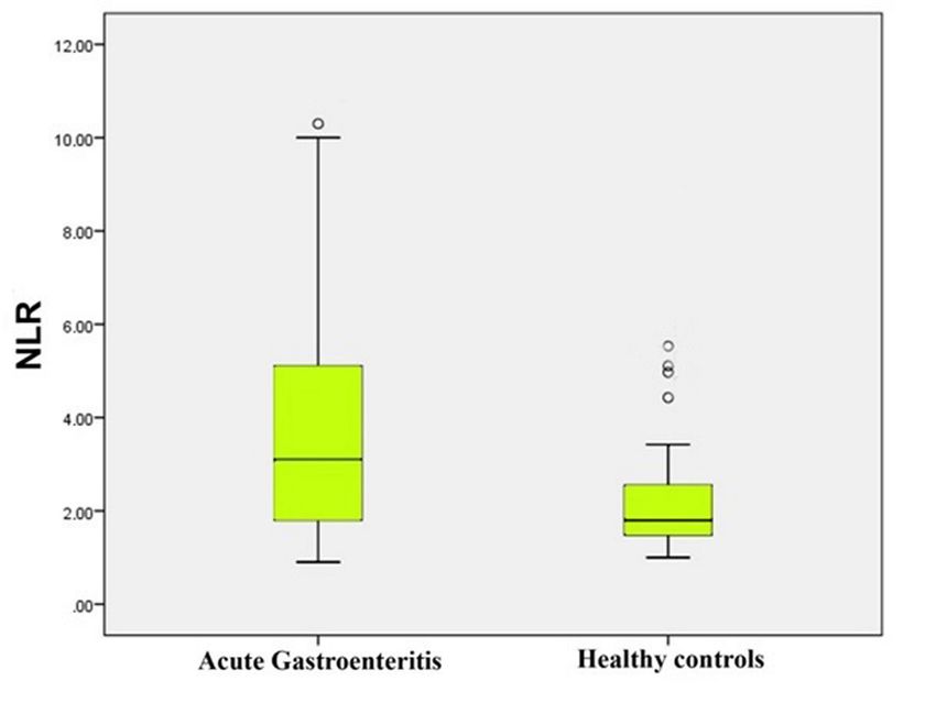
doi: 10.5455/medscience.2019.08.9213 Med Science 2020;9(2):
The ROC curve analysis was performed to evaluate the predictive There were no statistically significant differences in MPV, PDW,
ability of hemogram parameters to diagnose AGE. Sensitivity, and RDW between the two groups (Table 1).
specificity, and area under the curve values for hemogram Discussion
parameters were estimated based on cutoff values determined by
ROC analyses. In this study, the diagnostic value of MPV, NLR, PLR, RDW,
and PDW was investigated in AGE patients. The findings of the
present study revealed that only NLR and PLR were higher in AGE
patients compared with controls. NLR values were found to have
high sensitivity, specificity, and predictive value in differentiating
AGE patients from controls.
AGE is the inflammation of gastrointestinal system, usually
trigged by a bacterial or viral infection and causing abdominal
cramps, diarrhea, and vomiting. Although the role of inflammation
in AGE is unclear, pathophysiology usually differs depending the
underlying condition. In response to the presence of an irritant
or infectious agent, it is not surprising to encounter systemic
hematologic responses in peripheral blood data.
The unique role of platelets in inflammatory conditions has
Figure 2. PLR levels of acute gastroenteritis patients and healthy controls
been outlined in a number of studies in which the link between
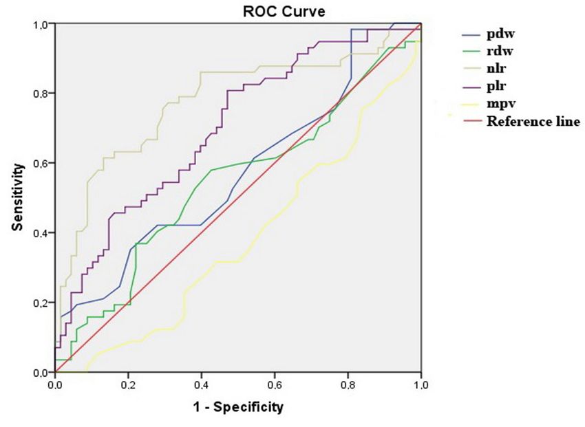
The ROC curve analysis suggested that the optimum NLR cut-off platelet activation and the pathophysiology of the diseases with
point for AGE was 2.08, with a sensitivity, specificity, PPV, and inflammation was detected [15]. In this context, preliminary
NPV of 70%, 65%, 62%, and 72%, respectively (AUC = 0.704; Fig. evidence suggests that MPV levels are significantly affected in
3). The optimum PLR cut-off point for AGE was 105.55, with a patients with gastroenteritis [13,14]. Although a majority of these
sensitivity, specificity, PPV, and NPV of 71%, 50%, 54%, and 68%, studies were performed in children, all of them demonstrated a
respectively (AUC = 0.648; Fig. 3).The results of the same analysis significant decrease in MPV levels. Mete et al [13] recently
for the other hematologic parameters are summarized in Table 2. showed that MPV levels were lower in children with rotavirus
gastroenteritis compared with healthy children. Similarly, a study
Table 2. ROC analyses of NLR and PLR with other hematologic indices to by Matowicka-Karna et al. [16] demonstrated that that MPV levels
differentiate acute gastroenteritis from healthy controls. in patients infected with Entamoeba histolytica were lower than in
Sensitivity Specificity controls. Interestingly, Çelik et al. [17] showed that MPV levels
Cutoff AUC PPV(%) NPV(%) were increased in amebiasis patients with gastroenteritis. Authors
(%) (%)
NLR 2.08 0.704 70 65 62 72 speculated that the discrepancies between studies was likely
associated with the severity of the systemic inflammation. In this
PLR 105.55 0.648 71 50 54 68
study, we found no significant difference in MPV levels between
MPV 8.35 0.376 49 66 54 60
AGE patients and controls. This might be due to several reasons,
PDW 16.4 0,571 66 48 53 64 including the severity of systemic inflammation and the relatively
RDW 13.6 0,550 68 46 50 60 small number of study participants.
NLR. neutrophil-to-lymphocyte ratio; PLR: platelet to lymphocyte ratio; MPV: The other platelet indices, such as PDW, may present valuable
mean platelet volume; PDW: platelet distribution width; RDW: red cell distri-
bution width; AUC: area under curve; PPV: positive predictive value; NPV:
clinical data in inflammatory diseases. A number of studies
negative predictive value correlated blood PDW values to many diseases including
appendicitis, pre-eclampsia, recurrent miscarriages, and dementia
[18-22]. PDW is an index that reflects the heterogeneity of platelets.
In inflammatory conditions, activation of platelets causes platelet
shape alterations, with anisocytosis leading to an increase in PDW
values [23-24]. Similar to MPV levels, we observed no alterations
in PDW values between study groups.
NLR is another parameter investigated in the present study. NLR
is a simple, widely used, and inexpensive index of systemic
inflammatory burden that correlates with prognosis in distinct
disease conditions [11]. Although there are no studies that have
specifically investigated NLR in AGE patients, there are a number
of studies that investigated NLR in inflammatory and neoplastic
conditions including ulcerative colitis, acute appendicitis,
colorectal cancer, hepatocellular, nasopharyngeal, and metastatic
renal cell carcinoma [11,25-28]. Considering that the primary agent
in gastroenteritis is infectious microorganisms, it can be predicted
that neutrophil count, which is highly sensitive for the detection
Figure 3. Receiver operating characteristic (ROC) curveys of NLR and PLR with of infectious conditions, and its ratio to lymphocyte count would
other hematologic indices provide more valuable information.
378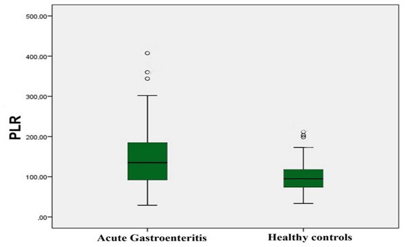
doi: 10.5455/medscience.2019.08.9213				                                                              		                    Med Science 2020;9(2):376-80

The elevated PLR and NLR levels that have been demonstrated in                        6.    Wu M, Zhou L, Zhu D, et al. Hematological indices as simple, inexpensive
the current study could also be a result of a physiological immune                          and practical severity markers of obstructive sleep apnea syndrome: a meta-
response of circulating leucocytes and platelets to gastroenteritis-                        analysis. J Thorac Dis. 2018;10:6509-21.
associated inflammatory conditions, which results in amplification                    7.    Yildirim M, Turkyilmaz E, Avsar AF. Preoperative neutrophil-to-lymphocyte
of neutrophils and platelets and a decrease in lymphocyte counts.                           ratio has a better predictive capacity in diagnosing tubo-ovarian abscess.
Similar to NLR, PLR is another hematologic parameter that                                   Gynecol Obstet Invest. 2015;80:234–9
is suggested to have a key value in the diagnosis of systemic
inflammation [29]. Although PLR alterations in gastroenteritis                        8.    Purnak T, Olmez S, Torun S, et al. Mean platelet volume is increased in chronic
                                                                                            hepatitis C patients with advanced fibrosis. Clin Res Hepatol Gastroenterol.
patients have not been demonstrated previously, literature data
                                                                                            2013;37:41-6.
suggests elevated PLR levels in distinct disease conditions. In a
recent study by Çınar et al. [30], PLR values were found to be                        9.    Suvak B, Torun S, Tas A, et al. Mean platelet volume is a useful indicator of
significantly higher in acute appendicitis during pregnancy.                                systemic inflammation in cirrhotic patients with ascitic fluid infection. Ann
Similarly, Akpinar et al. [31] demonstrated the predictive value                            Hepatol. 2013;12:294–300.
of PLR in ulcerative colitis patients. In this context, the finding of
                                                                                      10.   Akbas EM, Demirtas L, Ozcicek A, et al. Association of epicardial adipose
elevated NLR and PLR in gastroenteritis patients adds significant
                                                                                            tissue, neutrophil-to-lymphocyte ratio and platelet-to-lymphocyte ratio with
value to the diagnostic evaluation of AGE patients in emergency                             diabetic nephropathy. Int J Clin Exp Med. 2014;7:1794–801.
clinical settings.
                                                                                      11.   Torun S, Tunc BD, Suvak B, et al. Assessment of neutrophil-lymphocyte ratio
We recognize some limitations inherent to our study. The                                    in ulcerative colitis: a promising marker in predicting disease severity. Clin
first limitation is the negligence of other well-demonstrated                               Res Hepatol Gastroenterol. 2012;36:491–7.
inflammatory markers such as TNF-α, and IL-6 and IL-1β. It would
be noteworthy to evaluate serum levels of these pro-inflammatory                      12.   Beyazit Y, Sayilir A, Torun S, et al. Mean platelet volume as an indicator
cytokines and correlate these cytokine levels with our test results.                        of disease severity in patients with acute pancreatitis. Clin Res Hepatol
                                                                                            Gastroenterol. 2012;36:162-8.
Secondly, despite the findings of the present study, our results
should be interpreted cautiously because of the relatively moderate                   13.   Mete E, Akelma AZ, Cizmeci MN, et al. Decreased mean platelet volume in
sample size which limited the power of the study. And third, it                             children with acute rotavirus gastroenteritis. Platelets. 2014;25:51-4.
would have been useful to evaluate the same AGE patients after
clinical and laboratory remission was achieved was achieved.                          14.   Tanju C, Ekrem G, Berksoy Emel A, et al. Mean platelet volume as a negative
                                                                                            marker of inflammation in children with rotavirus gastroenteritis. Iran J
Conclusion                                                                                  Pediatr. 2014;24:617-22.

                                                                                      15.   Boshnak N, Boshnaq M, Elgohary H. Evaluation of platelet indices and red cell
Clinicians may find it difficult to distinguish AGE from a number
                                                                                            distribution width as new biomarkers for the diagnosis of acute appendicitis. J
of acute inflammatory disease conditions despite appropriate
                                                                                            Invest Surg. 2018;31:121-9.
medical evaluations in emergency clinics. Therefore, easy-to-use
hematologic markers, especially NLR and PLR, can be considered                        16.   Matowicka-Karna J, Panasiuk A. Does anti-parasitic treatment normalize
as a valuable tool to differentiate acute gastroenteritis from other                        platelets morphology in patients infested with Entamoeba histolytica? Rocz
non-inflammatory emergent conditions.                                                       Akad Med Bialymst. 1996;41:258-67.

Competing interests                                                                   17.   Çelik T, Güler E, Berksoy EA, et al. Mean platelet volume in children with
We declare that we have no conflict of interest.                                            acute gastroenteritis caused by entamoeba histolytica. Turk. Parazit Derg.
                                                                                            2015;39:205-8.
Financial Disclosure
The financial support for this study was provided by the investigators themselves.    18.   Sitotaw C, Asrie F, Melku M. Evaluation of platelet and white cell parameters
                                                                                            among pregnant women with Preeclampsia in Gondar, Northwest Ethiopia: A
Ethical approval
                                                                                            comparative cross-sectional study. Pregnancy Hypertens. 2018;13:242-7.
Ethical approval was received from the ethics committee of Canakkale Onsekiz Mart
University (2011-KAEK-27/2019-1900040923)                                             19.   Yang SW, Cho SH, Kwon HS, et al. Significance of the platelet distribution
                                                                                            width as a severity marker for the development of preeclampsia. Eur J Obstet
References                                                                                  Gynecol Reprod Biol. 2014;175:107-11.
1.   Chow CM, Leung AK, Hon KL. Acute gastroenteritis: from guidelines to real        20.   Boshnak N, Boshnaq M, Elgohary H. Evaluation of platelet indices and red cell
     life. Clin Exp Gastroenterol. 2010;3:97-112.                                           distribution width as new biomarkers for the diagnosis of acute appendicitis. J
                                                                                            Invest Surg. 2018;31:121-9.
2.   Dalby-Payne J, Elliott E. Gastroenteritis in children. Clin Evid. 2005;13:343-
     53.                                                                              21.   Liang QC, Jin D, Li Y, et al. Mean platelet volume and platelet distribution
                                                                                            width in vascular dementia and Alzheimer’s disease. Platelets. 2014;25:433-8.
3.   Degerli V. Diagnostic value of haematological parameters in differentiation
     between acute appendicitis and acute gastroenteritis. Med J Izmir Hospital.      22.   Mete Ural U, Bayoglu Tekin Y, Balik G, et al. Could platelet distribution
     2018;22:171-7.                                                                         width be a predictive marker for unexplained recurrent miscarriage? Arch
                                                                                            Gynecol Obstet. 2014;290:233-6.
4.   Reust CE, Williams A. Acute abdominal pain in children. Am Fam Physician.
     2016;93:830-6.                                                                   23.   Budak YU, Polat M, Huysal K. The use of platelet indices, plateletcrit,
                                                                                            mean platelet volume and platelet distribution width in emergency non-
5.   Beyazit F, Ozturk FH, Pek E, et al. Evaluation of the hematologic system as a
                                                                                            traumatic abdominal surgery: a systematic review. Biochem Med (Zagreb).
     marker of subclinical inflammation in hyperemesis gravidarum: a case control
                                                                                            2016;26:178-93.
     study. Ginekol Pol. 2017;88:315-9.

                                                                                                                                                                    379
doi: 10.5455/medscience.2019.08.9213 Med Science 2020;9(2):376-80
24. Mukker P, Haridas A, Kallinkeel N, et al. Comparative study of platelet 28. Markar SR, Karthikesalingam A, Falzon A, et al. The diagnostic value of
indices in cirrhosis, cirrhosis with sepsis and normal population. Int J Res neutrophil: lymphocyte ratio in adults with suspected acute appendicitis. Acta
Med Sci. 2016;4:1423-8. Chir Belg. 2010;110:543-7.
25. Halazun KJ, Aldoori A, Malik HZ, et al. Elevated preoperative neutrophil to 29. Kawamura Y, Takeshita S, Kanai T, et al. The combined usefulness of the
lymphocyte ratio predicts survival following hepatic resection for colorectal neutrophil-to-lymphocyte and platelet-to-lymphocyte ratios in predicting
liver metastases. Eur J Surg Oncol. 2008;34:55-60. intravenous immunoglobulin resistance with Kawasaki disease. J Pediatr.
2016;178:281-4.
26. Kishi Y, Kopetz S, Chun YS, et al. Blood neutrophil-to-lymphocyte ratio
predicts survival in patients with colorectal liver metastases treated with 30. Cinar H, Aygun A, Derebey M, et al. Significance of hemogram on diagnosis
systemic chemotherapy. Ann Surg Oncol. 2009;16:614-22. of acute appendicitis during pregnancy. Ulus Travma Acil Cerrahi Derg.
2018;24:423-8.
27. Tamhane UU, Aneja S, Montgomery D, et al. Association between admission
neutrophil to lymphocyte ratio and outcomes in patients with acute coronary 31. Akpinar MY, Ozin YO, Kaplan M, et al. Platelet-to-lymphocyte ratio and
syndrome. Am J Cardiol. 2008;102:653-7. neutrophil-to-lymphocyte ratio predict mucosal disease severity in ulcerative
colitis. J Med Biochem. 2018;37:155-62.
380You can also read