DO YOU KNOW HOW YOUNG YOUR HEART IS? - De Gruyter
←
→
Page content transcription
If your browser does not render page correctly, please read the page content below
ARS Medica Tomitana - 2018; 2(24): pag. 66 - 71
                                                                                 doi: 10.2478/arsm-2018-0012

                     DO YOU KNOW HOW YOUNG YOUR HEART IS?

Tase Cristina Ramona1,2, Cojocaru Lucia1,3, Rusali Andrei1,3, Suta Cristina1,4

1
  Faculty of Medicine, University "Ovidius" of Constanta
2
  Emergency Clinical County Hospital of Constanta, Emergency Department
3
  Emergency Clinical County Hospital of Constanta, Cardiology Clinic
4
  Emergency Clinical County Hospital of Constanta, Internal Medicine Clinic

                                                                                                             Lucia Cojocaru
                                                                   Emergency Clinical Hospital of Constanta, Cardiology Clinic,
                                                                                Tomis Boulevard No 145, Constanta, Romania
                                                                                               email: bostanlucia@yahoo.com
                                                                                                       phone: +40 722370635
ABSTRACT

    We present the case of a 25 years old patient who was submitted to our unit with a first time acute coronary
syndrome. Despite his young age he had multiple cardiovascular risk factors. Although the chest pain was
atypical and the electrocardiogram on presentation had unspecific changes, repeated investigations established
the diagnosis of anterolateral myocardial infarction. Per primam angioplasty with stent implantation in the
proximal segment of left anterior descending artery was performed, with good clinical outcome. Awareness
is the key in establishing the diagnosis of myocardial infarction in young patients.

    Keywords: myocardial infarction, young adults, low HDL-cholesterol, family history

Introduction                                                       As a clinician, there are real challenges in
                                                            the proper approach and management skills in
        Coronary heart disease (CHD) is the leading         prevention and treatment of CHD among young
cause of death worldwide, being responsible                 individuals, the majority of them being unaware
for 20% of all deaths in Europe alone (1-3).                of their risk factors, presenting atypical symptoms
Although CHD primarily occurs in patients over              that may delay presentation in the emergency
40, younger people can be affected (4,5).                   department and have higher rates of medication
        In the past two decades, due to the                 non-adherence (9-12). The different risk factor
primary and secondary prevention strategies,                profiles, clinical presentations and prognoses
there has been a reduction in the incidence of              should be taken into consideration when treating
cardiovascular (CV) events and mortality in                 young patients with CHD (4,5).
the general population (6,7). However, in the
young adults, the incidence of acute myocardial
infarction (MI) remained stable and CV disease
is still a major cause of death (6-8).

                                                                                                     Unauthenticated        66
                                                                                       Download Date | 2/2/20 8:43 PM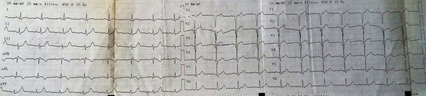
Clinical Case half of interventricular septum. The repeated
ECG during the pain showed Q waves in V1-V3,
We present the case of a 25-years old 1mm ST elevation in V3 and 0.5mm elevation
male patient, smoker (3.5 pack-year), without in DI, aVL, V2, with ST depression in DII, DIII,
past medical history, but with a positive family aVL and negative T wave in aVL (Figure 2);
history for cardiovascular disease (father with this rapidly evolved to significant elevated ST
sudden death after MI at 32 years old), presented segment in DI, aVL, V2-V5 (Figure 3).
to the emergency department for severe
precordial stabbing chest pain. The pain occurred
at rest, lasted for approximately 3 hours and was
relieved after one tablet of nitroglycerin given by
the ambulance crew. He described having had,
in the previous 3 months, repetitive episodes Figure 2. Electrocardiogram during chest pain – sinus
of stabbing left sided chest pain during effort rhythm, 75bpm, Q waves in V1-V3, 1mm ST elevation
in V3 and 0.5mm elevation in DI, aVL, V2, with ST
with spontaneous remission at rest (in than 5 depression in DII, DIII, aVL and negative T wave in aVL
minutes) for which he had an medical exam that
established the diagnosis of atypical chest pain
and recommended an stress test, which he didn’t
performed.
On the admission, the patient was free of
pain, in good clinical condition. The clinical
exam revealed a normoponderal patient with a
blood pressure of 120/60 mmHg in both arms, Figure 3. Electrocardiogram during chest pain (prior to
angiography) – sinus rhythm, 62bpm, QS in V1-V3 and
a heart rate of 56 bpm, without heart murmurs, poor R in V4, Q in aVL, elevated ST segment in DI, aVL,
gallop or pericardial friction rub and without V2-V5 that encompasses the T wave in V2-V4, negative T
signs of pulmonary or systemic congestion. wave in DI, aVL
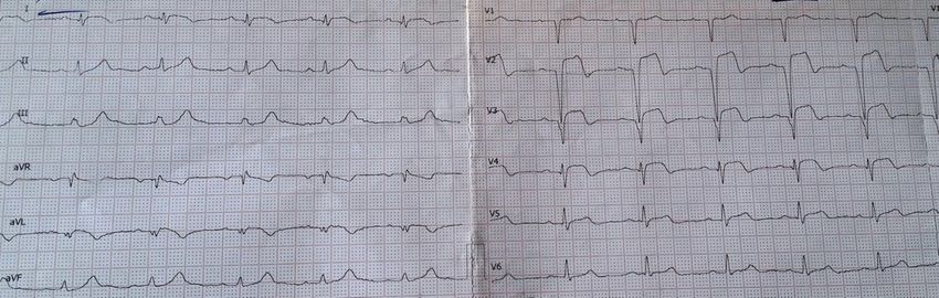
Electrocardiogram (ECG) on admission
revealed sinus bradycardia, 52 bpm, QRS axis Laboratory tests revealed positive
= + 30⁰, slight ST elevation in V2 (0.5mm), myocardial necrosis markers (with typical
biphasic T waves in V1-V2 and negative T wave myocardial necrosis-associated rise during
in aVL (Figure 1). hospital stay, peak CK-MB=209U/L and peak
TnT 2488ng/mL), mixed dyslipidemia with
normal LDL-cholesterol (82mg/dl), low HDL-
cholesterol (27mg/dl) and high triglycerides
levels (196 mg/dl), and mild leukocytosis with
Figure 1: Electrocardiogram on admission - sinus neutrophilia. The liver function tests, renal
bradycardia, 52 bpm, QRS axis = + 30⁰, slight ST
function tests and ionogram were in normal
elevation in V2 (0.5mm), biphasic T waves in V1-V2 and
negative T wave in aVL. limits.
The diagnosis of acute anterolateral MI was
Emergency echocardiography revealed established and the patient received a loading dose
normal cardiac chambers size, normal left of unfractioned heparin and double antiplatelet
ventricular (LV) systolic and diastolic function and was sent to the cath lab. The coronarography
(left ventricle ejection fraction - LVEF of revealed a 50-70% stenosis in the proximal left
65%), no significant valvulopathy, and no signs anterior descending artery (LAD) with the aspect
of pulmonary hypertension, no pericardial of a ruptured plaque (Figure 4) and less than 50%
fluid and no signs of aortic dissection. During stenosis in the medial segment of LAD and ostium
the echocardiographic exam the patient had of first diagonal artery. Emergency angioplasty
another episode of precordial stabbing pain with stenting (drug-eluting stent) of the culprit
accompanied by akinesis of LV apex and apical lesion was performed and post-interventional
67 Unauthenticated
Download Date | 2/2/20 8:43 PM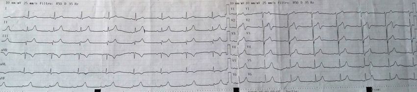
TIMI 3 flow was established (Figure 5).                       is in good clinical condition, asymptomatic, with
                                                              a proper adherence to treatment but he resumed
                                                              smoking and has a sedentary lifestyle. The
                                                              laboratory tests reveal optimal LDL-cholesterol
                                                              (68mg/dL) and triglyceride (90mg/dL) levels,
                                                              but the HDL-cholesterol remains low (30mg/
                                                              dL). The ECG shows no R waves in V1-V3 and
                                                              at echocardiography there is a mild LV apical
                                                              hypokinesis (LVEF of 55%) as his only evidence
                                                              of the previous MI; the ECG stress test is
                                                              negative for ischemia. Given his family history,
                                                              smoking habit, sedentary lifestyle and low HDL-
                                                              cholesterol the patient remains at high-risk of
                                                              subsequent ischaemic events.

                                                              Discussion

                                                                     The prevalence of CHD in younger subjects
                                                              is difficult to establish because is frequently a
Figure 4. Angiography - 50-70% stenosis in the proximal
   left anterior descending artery with the aspect of a       silent process (4,5). There are limited data on
                 ruptured plaque (arrow)                      the frequency of MI in younger subjects, studies
                                                              showing that 4 to 10% of patients with MI were
                                                              under 40 or 45 years of age (4,5,13-15).
                                                                     Although CHD is an uncommon entity in
                                                              young patients, it is an important public-health
                                                              issue given the negative impact on physical,
                                                              mental, social, and financial health, and greater
                                                              healthcare utilization among affected individuals
                                                              (4-6).
                                                                     Young patients with MI usually have
                                                              multiple risk factors for CHD (5). Studies
                                                              found that 90 to 97% of them had one or more
                                                              traditional risk factors for atherosclerosis
                                                              (4,5,16-18). Cigarette smoking is the most
                                                              common and most modifiable risk factor in
                                                              young patients, being present in 65 to 92 percent
                                                              of young patients with MI, compared to 24 to
                                                              56 percent of patients older than 45 years of
                                                              age (4,5,17). Younger patients with CHD more
 Figure 5. Angioplasty with stenting (arrow) of the culprit
lesion in the proximal left anterior descending artery with
                                                              often have a family history of premature CHD
                    distal TIMI 3 flow                        (41 to 64 percent) and the offspring of patients
                                                              with premature CHD are more likely to have
       During hospitalization in the cardiology               coronary risk factors than those without such
unit, under pharmacological treatment with double             family history (4,5,17,19,20). This association
antiplatelet therapy, beta-blocker, angiotensin-              between family history and premature CHD can
converting-enzyme inhibitor and statin, the                   be due to both genetic and environmental factors
clinical evolution was good and the patient was               (4,5). Lipid abnormalities are another risk factor
discharged free of pain, without signs of heart               for CHD in young patients. When compared to
failure. Three years after the acute MI the patient           older patients, young patients have lower HDL-
                                                              cholesterol concentrations, higher triglycerides
                                                                                                 Unauthenticated    68
                                                                                   Download Date | 2/2/20 8:43 PM
and the same prevalence of hypercholesterolemia the patient, in accordance to current clinical
(4,5,21). Arterial hypertension and diabetes practice guidelines. Young patients with acute
mellitus appear to be less common in young ST-elevation MI should be treated with primary
patients with MI (4,5,19,22). However, young angioplasty or, if not available, thrombolytic
patients frequently have subtle problems with therapy (4,26). When compared to older patients,
glucose metabolism, more than half of them young patients do better regardless of the type of
having decreased oral glucose tolerance and reperfusion they received (4). The angiographic
hyperinsulinemic response to oral glucose findings in young patients also differ from
challenge that is also a risk factor for CHD (4,5). those of older ones. Younger patients have a
Obesity is another important coronary risk factor higher incidence of normal coronary arteries,
and it appears to be an independent risk factor for mild luminal irregularities, and single vessel
coronary atherosclerosis, at least in young men coronary artery disease than do older patients
(4,5). Other risk factors that have been identified (4,5,18,21,22,27). Spontaneous coronary artery
in young patients with MI are: oral contraceptive dissection and Kawasaki disease are two rare
use, factor V Leiden, frequent cocaine use, causes of MI that occur more commonly in the
smoking marijuana and psychosocial factors, young and must be taken into consideration when
such anger (4,5). In our patient, risk factors for assessing these patients (4).
CHD have been smoking, the positive family When compared to older patients, younger
history for premature CHD and low HDL- patients have lower in-hospital mortality, better
cholesterol levels. long-term outcome and same rate of reinfarction
Current CV risk calculators are less (4). In the treatment of MI survivors, risk factor
applicable to younger patients where they reduction plays a central role (4).
underestimate the risk (6,23). The YOUNG-MI
study gave criteria for new risk factor calculators Conclusions
and awareness of CHD events in patients < 50
years, knowing that most patients do not cross Although symptomatic CHD is
the threshold for primary prevention defined by uncommon in young people, with increasing
current guidelines and consequently they are not rates of traditional CV risk factors we can expect
considered candidates for preventive therapies CHD to become even more prevalent in this age
(6,23). group (6). We must keep in mind that current
When considering the clinical presentation, available risk calculators underestimate the CV
young adults are more likely to have atypical risk in young population and most of them will
symptoms, as was the case of our patient (4,5). not benefit from primary prevention, increasing
When compared to older patients with CHD, the incidence of CV events. No matter the
a higher proportion of young patients do not patient’s age, the age of his heart will be given by
experience stable angina, and, in the majority the cluster of his CV risk factors.
of cases, an acute coronary syndrome is the
first manifestation of CHD (4,5,13,21). Among Acknowledgements
those who have preceding chest pain, the firs
episodes often occur only in the week prior to MI We thank all members of the medical team
(4,13). The most important differential diagnosis and the patient.
of MI in young patients is acute myocarditis.
This disorder can mimic the MI and must be References
considered in young patients with a clinical
presentation of acute coronary syndrome but 1. Jernberg T, Hasvold P, Henriksson M, Hjelm
with normal coronary angiogram (4,5,24,25). H, Thuresson M, Janzon M. Cardiovascular
The management of young patients with risk in post-myocardial infarction patients:
acute MI varies with the type: ST-elevation nationwide real world data demonstrate
or non-ST elevation. The overall approach the importance of a long-term perspective.
is generally the same regardless the age of European heart journal. 2015 Jan
69 Unauthenticated
Download Date | 2/2/20 8:43 PM13;36(19):1163-70. analysis. Annals of Pharmacotherapy.
2. Mathers CD, Loncar D. Projections of 2010;44(9):1410-21.
global mortality and burden of disease from 12. Yeh RW, Sidney S, Chandra M, Sorel M,
2002 to 2030. PLoS medicine. 2006 Nov Selby JV, Go AS. Population trends in the
28;3(11):e442. incidence and outcomes of acute myocardial
3. Nichols M, Townsend N, Scarborough infarction. N Engl J Med. 2010;362(23):2155-
P, Rayner M. Cardiovascular disease in 65.
Europe: epidemiological update. European 13. Fournier JA, Sanchez A, Quero J, Fernandez-
heart journal. 2013 Sep 7;34(39):3028-34. Cortacero JA, Gonzalez-Barrero A.
4. Azar RR. Coronary heart disease and Myocardial infarction in men aged 40 years
myocardial infarction in young men and or less: a prospective clinical-angiographic
women. In: UpToDate, Verheught, F (Ed), study. Clin Cardiol. 1996;19(8):631-6.
UpToDate, (Accessed on February 11, 2018). 14. Doughty M, Mehta R, Bruckman D, Das S,
5. Razaghani A, Hafeez-Ul-Hassanvirk. Triple Karavite D, Tsai T, et al. Acute myocardial
Vessel Coronary Artery Disease in Young infarction in the young--The University
Female. GJMR. 2014;14(5):1-5. of Michigan experience. Am Heart J.
6. Singh A, Collins B, Qamar A, Gupta A, 2002;143(1):56-62.
Fatima A, Divakaran S, Klein J, Hainer J, 15. Greenland P, Reicher-Reiss H, Goldbourt U,
Jarolim P, Shah RV, Nasir K. Study of young Behar S. In-hospital and 1-year mortality in
patients with myocardial infarction: Design 1,524 women after myocardial infarction.
and rationale of the YOUNG‐MI Registry. Comparison with 4,315 men. Circulation.
Clinical cardiology. 2017 Nov 1;40(11):955- 1991;83(2):484-91.
61. 16. Chouhan L, Hajar HA, Pomposiello JC.
7. Roth GA, Huffman MD, Moran AE, Feigin Comparison of thrombolytic therapy for
V, Mensah GA, Naghavi M, et al. Global acute myocardial infarction in patients
and regional patterns in cardiovascular aged < 35 and > 55 years. Am J Cardiol.
mortality from 1990 to 2013. Circulation. 1993;71(2):157-9.
2015;132(17):1667-78. 17. Hoit BD, Gilpin EA, Henning H, Maisel
8. Gupta A, Wang Y, Spertus JA, Geda M, AA, Dittrich H, Carlisle J, et al. Myocardial
Lorenze N, Nkonde-Price C, et al. Trends in infarction in young patients: an analysis by
acute myocardial infarction in young patients age subsets. Circulation. 1986;74(4):712-21.
and differences by sex and race, 2001 to 18. al-Koubaisy OK, Mehdi RS, Arem FD,
2010. J Am Coll Cardiol. 2014;64(4):337-45. Ahmed IT. Cine angiographic findings in
9. Goff DC, Jr., Lloyd-Jones DM, Bennett G, young Iraqi men with first acute myocardial
Coady S, D’Agostino RB, Gibbons R, et al. infarction. Cathet Cardiovasc Diagn.
2013 ACC/AHA guideline on the assessment 1990;19(2):87-90.
of cardiovascular risk: a report of the 19. Cole JH, Miller JI, 3rd, Sperling LS,
American College of Cardiology/American Weintraub WS. Long-term follow-up of
Heart Association Task Force on Practice coronary artery disease presenting in young
Guidelines. Circulation. 2014;129(25 Suppl adults. J Am Coll Cardiol. 2003;41(4):521-8.
2):S49-73. 20. Bao W, Srinivasan SR, Wattigney WA,
10. Canto JG, Rogers WJ, Goldberg RJ, Berenson GS. The relation of parental
Peterson ED, Wenger NK, Vaccarino V, et al. cardiovascular disease to risk factors in
Association of age and sex with myocardial children and young adults. The Bogalusa
infarction symptom presentation and in- Heart Study. Circulation. 1995;91(2):365-
hospital mortality. JAMA. 2012;307(8):813- 71.
22. 21. Chen L, Chester M, Kaski JC. Clinical
11. Mann DM, Woodward M, Muntner P, Falzon factors and angiographic features associated
L, Kronish I. Predictors of nonadherence with premature coronary artery disease.
to statins: a systematic review and meta- Chest. 1995;108(2):364-9.
Unauthenticated 70
Download Date | 2/2/20 8:43 PM22. Zimmerman FH, Cameron A, Fisher LD, 26. Ibanez B, James S, Agewall S, Antunes MJ,
Ng G. Myocardial infarction in young Bucciarelli-Ducci C, Bueno H, et al. 2017
adults: angiographic characterization, risk ESC Guidelines for the management of acute
factors and prognosis (Coronary Artery myocardial infarction in patients presenting
Surgery Study Registry). J Am Coll Cardiol. with ST-segment elevationThe Task Force
1995;26(3):654-61. for the management of acute myocardial
23. Akosah KO, Schaper A, Cogbill C, infarction in patients presenting with ST-
Schoenfeld P. Preventing myocardial segment elevation of the European Society of
infarction in the young adult in the first place: Cardiology (ESC). European Heart Journal.
how do the National Cholesterol Education 2018;39(2):119-77.
Panel III guidelines perform? J Am Coll 27. Wolfe CM, Vacek JL. Myocardial infarction
Cardiol. 2003;41(9):1475-9. in the young: angiographic features and risk
24. Sarda L, Colin P, Boccara F, Daou D, Lebtahi factor analysis of patients with myocardial
R, Faraggi M, et al. Myocarditis in patients infarction at or before the age of 35 years.
with clinical presentation of myocardial Chest. 1988 Nov 1;94(5):926-30.
infarction and normal coronary angiograms.
J Am Coll Cardiol. 2001;37(3):786-92.
25. Karjalainen J, Heikkila J. Incidence of three
presentations of acute myocarditis in young
men in military service. A 20-year experience.
Eur Heart J. 1999;20(15):1120-5.
71 Unauthenticated
Download Date | 2/2/20 8:43 PMYou can also read