Clinical diagnostic value of telomere length measurement in inherited bone marrow failure syndromes
←
→
Page content transcription
If your browser does not render page correctly, please read the page content below
Clinical diagnostic value of telomere length measurement
in inherited bone marrow failure syndromes

by Shunsuke Miwata, Atsushi Narita, Yusuke Okuno, Kyogo Suzuki, Motoharu Hamada,
Taro Yoshida, Masayuki Imaya, Ayako Yamamori, Manabu Wakamatsu, Kotaro Narita,
Hironobu Kitazawa, Daisuke Ichikawa, Rieko Taniguchi, Nozomu Kawashima, Eri Nishikawa,
Nobuhiro Nishio, Seiji Kojima, Hideki Muramatsu, and Yoshiyuki Takahashi

Haematologica 2021 [Epub ahead of print]

Citation: Shunsuke Miwata, Atsushi Narita, Yusuke Okuno, Kyogo Suzuki, Motoharu Hamada,
Taro Yoshida, Masayuki Imaya, Ayako Yamamori, Manabu Wakamatsu, Kotaro Narita,
Hironobu Kitazawa, Daisuke Ichikawa, Rieko Taniguchi, Nozomu Kawashima,
Eri Nishikawa, Nobuhiro Nishio, Seiji Kojima, Hideki Muramatsu, and Yoshiyuki Takahashi.
Clinical diagnostic value of telomere length measurement in inherited bone marrow failure syndromes.
Haematologica. 2021; 106:xxx
doi:10.3324/haematol.2021.278334

Publisher's Disclaimer.
E-publishing ahead of print is increasingly important for the rapid dissemination of science.
Haematologica is, therefore, E-publishing PDF files of an early version of manuscripts that
have completed a regular peer review and have been accepted for publication. E-publishing
of this PDF file has been approved by the authors. After having E-published Ahead of Print,
manuscripts will then undergo technical and English editing, typesetting, proof correction and
be presented for the authors' final approval; the final version of the manuscript will then
appear in print on a regular issue of the journal. All legal disclaimers that apply to the
journal also pertain to this production process.
Clinical diagnostic value of telomere length measurement in inherited bone marrow failure

syndromes

Shunsuke Miwata1, Atsushi Narita1, Yusuke Okuno1,2, Kyogo Suzuki1, Motoharu Hamada1, Taro

Yoshida1, Masayuki Imaya1, Ayako Yamamori1, Manabu Wakamatsu1, Kotaro Narita1, Hironobu

Kitazawa1, Daisuke Ichikawa1, Rieko Taniguchi1, Nozomu Kawashima1, Eri Nishikawa1, Nobuhiro

Nishio1,2, Seiji Kojima1, Hideki Muramatsu*1, Yoshiyuki Takahashi*1

1
    Department of Pediatrics, Nagoya University Graduate School of Medicine, Nagoya, Japan.
2Center for Advanced Medicine and Clinical Research, Nagoya University Hospital, Nagoya,

Japan.*Corresponding authors

Corresponding authors:

Hideki Muramatsu, M.D., Ph.D.

Department of Pediatrics, Nagoya University Graduate School of Medicine, 65 Tsurumai-cho,

Showa-ku, Nagoya, Aichi 466-8560, Japan

Tel: +81-52-744-2294;

Fax: +81-52-744-2974;

Email: hideki-muramatsu@med.nagoya-u.ac.jp

Yoshiyuki Takahashi, M.D., Ph.D.

Department of Pediatrics, Nagoya University Graduate School of Medicine, 65 Tsurumai-cho,

Showa-ku, Nagoya, Aichi 466-8560, Japan

Tel: +81-52-744-2294;

Fax: +81-52-744-2974;

Email: ytakaha@med.nagoya-u.ac.jp

                                                 1
Acknowledgments
The authors acknowledge all clinicians, the patients, and their families. The authors thank Ms.
Yoshie Miura and Ms. Hiroko Ono for their valuable assistance.
Authorship
SM, AN, HM performed laboratory analyses, gathered clinical information, designed and conducted
the research, analysed data, and wrote the paper. MI, AY, MW, KN, HK, DI, RT and YO performed
laboratory analyses; MH, YT, and gathered clinical information; KS, NK, EN, NN and SK conducted
the research; YT directed the research and wrote the paper.
Conflict-of-interest disclosure
All authors declare no competing financial interests.
Short Running title: USEFULNESS OF TELOMERE LENGTH MEASUREMENT FOR IBMFS
Manuscript word count: 1493
References: 15
Table: 1
Figures: 2
Supplementary Tables: 3
2Bone marrow failure (BMF) is characterized by a hypocellular marrow and encompasses a diverse
group of inherited and acquired disorders. Inherited bone marrow failure syndromes (IBMFS) occur
in approximately 5%–30% of patients with BMF in a pediatric cohort and consist of more than 25
defined disease entities, including dyskeratosis congenita (DC), Fanconi anemia (FA),
Diamond–Blackfan anemia (DBA), and Shwachman–Diamond syndrome (SDS).1 IBMFS are a
heterogeneous group of disorders in which BMF is usually associated with physical abnormalities.
The diagnosis of IBMFS previously relied on the recognition of characteristic clinical features.
Recent diagnostic advances using next-generation sequencing have revealed that some patients
initially diagnosed with idiopathic aplastic anemia (AA) had cryptic presentations of IBMFS.2 This
issue is important as a more accurate diagnosis may improve treatment outcomes.
Telomeres are the end segments of chromosomes, composed of long DNA repeats and a
protein complex, and are essential for genome integrity. Germline mutations in telomere biology
genes can result in significantly short peripheral blood lymphocyte telomere length (TL) in patients
with DC.3 Although there is a consensus on the usefulness of TL for screening DC but not for other
IBMFS, several investigators have demonstrated that TL is excessively short in patients with AA4
and non-DC IBMFS,5 including FA, SDS, and DBA. To assess the diagnostic value of TL, we
measured TL in 133 patients with BMF and compared it in patients with DC, non-DC IBMFS, and
AA.
We retrospectively studied 133 patients (68 male and 65 female) with BMF in Japan
between 2013 and 2018. We collected peripheral blood samples at diagnosis from all patients,
3measured TL from peripheral blood lymphocytes, and performed targeted sequencing analysis
covering 184 genes associated with IBMFS (Supplemental Table 1), as described in our previous
studies.2,4 TL was measured by flow-fluorescence in situ hybridization (flow-FISH) using Telomere
PNA Kit (Dako Cytomation, Glostrup, Denmark) according to the manufacturer’s instructions. We
calculated the age-adjusted relative TL in terms of the standard deviation (SD) from 71 normal,
age-matched, healthy controls (median age, 29 years; range, 1–47 years) as previously
described.4 In 112 of 133 (84%) patients, paroxysmal nocturnal hemoglobinuria (PNH)-type
granulocytes and red blood cells were also evaluated by flow cytometry.4 As thresholds for
determining the presence of minor PNH clones, we defined >0.020% and >0.037% for CD11b+
CD55– CD59+ granulocytes and glycophorin A+ CD55– CD59– erythrocytes, respectively.
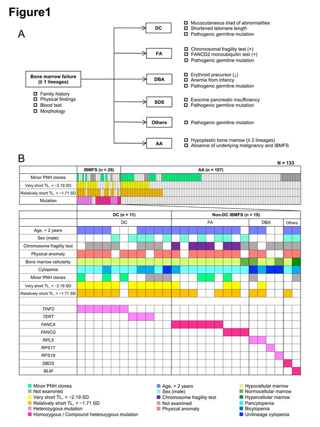
We diagnosed patients using a diagnostic flowchart (Figure 1A) developed using published
diagnostic criteria for specific IBMFS and acquired AA.6,7 The severity of cytopenia was determined
according to the Camitta severity criteria for AA.8 We divided 133 patients into three groups: DC,
non-DC IBMFS, and AA. All statistical analyses were performed using EZR (Saitama Medical
Center, Jichi Medical University, Saitama, Japan).9 Written informed consent was obtained from
patients or their legal guardians. This study was approved by the ethics committee of the Nagoya
University Graduate School of Medicine.
Table 1 shows the clinical characteristics of patients included in this study. The median age
at diagnosis of the total cohort was 7 years (range, 0–22 years). Of the 133 patients, 105, 24, and 4
were diagnosed with pancytopenia, bicytopenia, and unilineage cytopenia (3 anemia and 1
4thrombocytopenia), respectively. In patients with pancytopenia or bicytopenia, severity was
assessed as very severe, severe, and moderate in 27, 35, and 67 patients, respectively. The
median TL in all 133 patients was –0.96 SD (range, −5.73 to +4.00 SD).
Using targeted sequencing, we detected from 24 patients (18%) 31 pathogenic variants (3
nonsense, 14 missense, 5 frameshift, 4 splice site, and 4 deletion) of known causative IBMFS
including TINF2 (n = 6), TERT (n = 3), FANCA (n = 6), FANCG (n = 3), RPL5 (n = 2), RPS19 (n = 1),
RPS17 (n = 1), SBDS (n = 1), and BLM (n = 1). Homozygous mutations were found in 3 patients (2
in FANCC and 1 in FANCA), compound heterozygous in 4 patients (2 in FANCA and 1 each in
FANCG and SBDS), hemizygous in 3 patients in FANCA, and heterozygous in 14 patients (6 in
TINF2, 3 in TERT, 2 in RPL5, and 1 each in RPS17, RPS19, and BLM). Each patient’s genetic
variants are shown in Supplementary Table 2.
Out of the 133 patients and following the diagnostic flowchart (Figure 1A), 11 were
diagnosed with DC (8%), 15 with non-DC IBMFS (11%), and 107 with AA (81%). The schematic
representation of the results of gene analysis and the clinical features of IBMFS are shown in
Figure 1B. Of the 11 patients with DC, 9 were genetically diagnosed (6 in TINF2 and 3 in TERT),
and those without diagnostic genetic mutations were diagnosed following clinical diagnostic criteria.
The 15 non-DC IBMFS cases consisted of 9 FA, 4 DBA, 1 SDS, and 1 Bloom syndrome. All of
these diagnoses were confirmed by the presence of germline mutations in IBMFS-related genes.
Physical anomalies were observed in 11 of 15 (73%) patients. The individual clinical features and
genetic results of the patients with IBMFS are shown in Supplementary Table 2.
5We compared the clinical characteristics of patients with DC, non-DC IBMFS, and AA
(Table 1). Median age and gender distribution did not show significant differences among the three
groups. Severe or very severe cytopenia was significantly more frequent (P = 0.024) in AA cases
(57/107, 53%) compared with DC (3/10, 30%) and non-DC IBMFS cases (2/12, 17%).
Median TL in the patients with DC, non-DC IBMFS, and AA were −3.50 SD (range, −5.73 to
+0.83 SD), −1.89 SD (range, −4.74 to +2.05 SD), and −0.84 SD (range, −4.27 to +4.00 SD),
respectively (Figure 2A). Patients with DC showed significantly shorter TL compared with those
with non-DC IBMFS (P = 0.031) and AA (P < 0.001). Furthermore, patients with non-DC IBMFS
tended to show shorter TL than those with AA (P = 0.096).
To validate the efficacy of TL measurement in diagnosing DC and IBMFS, receiver
operating characteristic curves identified two cut-off values with the optimum sensitivity and false
positive rate (1-specificity) combination,been reported (Supplementary Table 3).3,5,10–15 Alter et al.5 previously reported that very short telomeres (
This study confirms that a relatively short TL was found in a significant proportion of

patients with DC and non-DC IBMFS, indicating the clinical diagnostic value of TL measurement in

identifying patients who need further testing, particularly comprehensive genetic analysis.

References

1.    Chhabra P, Bhatia P, Singh M, et al. Pediatric bone marrow failure: Clinical, hematological

      and targeted next generation sequencing data. Blood Cells Mol Dis. 2021;87:102510.

2.    Muramatsu H, Okuno Y, Yoshida K, et al. Clinical utility of next-generation sequencing for

      inherited bone marrow failure syndromes. Genet Med. 2017;19(7):796-802.

3.    Du HY, Pumbo E, Ivanovich J, et al. TERC and TERT gene mutations in patients with bone

      marrow failure and the significance of telomere length measurements. Blood.

      2009;113(2):309-316.

4.    Sakaguchi H, Nishio N, Hama A, et al. Peripheral blood lymphocyte telomere length as a

      predictor of response to immunosuppressive therapy in childhood aplastic anemia.

      Haematologica. 2014;99(8):1312-1316.

5.    Alter BP, Giri N, Savage SA, Rosenberg PS. Telomere length in inherited bone marrow

      failure syndromes. Haematologica. 2015;100(1):49-54.

6.    Marsh JCW, Ball SE, Cavenagh J, et al. Guidelines for the diagnosis and management of

      aplastic anaemia. Br J Haematol. 2009;147(1):43-70.

7.    Shimamura A, Alter BP. Pathophysiology and management of inherited bone marrow failure

      syndromes. Blood Rev. 2010;24(3):101-122.

8.    Camitta BM, Storb R, Thomas ED. Aplastic anemia (second of two parts): pathogenesis,

      diagnosis, treatment, and prognosis. N Engl J Med. 1982;306(12):712-718.

9.    Kanda Y. Investigation of the freely available easy-to-use software “EZR” for medical

      statistics. Bone Marrow Transplant. 2013;48(3):452-458.

                                                  8
10. Ball SE, Gibson FM, Rizzo S, Tooze JA, Marsh JCW, Gordon-Smith EC. Progressive
telomere shortening in aplastic anemia. Blood. 1998;91(10):3582-3592.
11. Hanson H, Mathew CG, Docherty Z, Mackie Ogilvie C. Telomere shortening in Fanconi
anaemia demonstrated by a direct FISH approach. Cytogen Cell Genet.
2001;93(3-4):203-206.
12. Thornley I, Dror Y, Sung L, Wynn RF, Freedman MH. Abnormal telomere shortening in
leucocytes of children with Shwachman-Diamond syndrome. Br J Haematol.
2002;117(1):189-192.
13. Li X, Leteurtre F, Rocha V, et al. Abnormal telomere metabolism in Fanconi’s anaemia
correlates with genomic instability and the probability of developing severe aplastic anaemia.
Br J Haematol. 2003;120(5):836-845.
14. Pavesi E, Avondo F, Aspesi A, et al. Analysis of telomeres in peripheral blood cells from
patients with bone marrow failure. Pediatr Blood Cancer. 2009;53(3):411-416.
15. Ong SY, Li ST, Wong GC, Ho AYL, Nagarajan C, Ngeow J. Delayed diagnosis of
Shwachman diamond syndrome with short telomeres and a review of cases in Asia. Leuk
Res Rep. 2018;9:54-57.
9Table 1. Clinical characteristics and laboratory findings of patients with bone marrow failure
(BMF).
All patients DC Non-DC IBMFS AA P
(N = 133) (n = 11) (n = 15) (n = 107) value
Age, years, median (range) 7 (0–22) 7 (1–19) 6 (0–15) 7 (0–22) 0.675
Gender, n (%)
Male 68 (50) 5 (45) 7 (47) 56 (52) 0.851
Female 65 (49) 6 (55) 8 (53) 51 (48)
Cytopenia, n (%)
Unilineage cytopenia 4 (3) 1 (9) 3 (20) 0 (0)Figure Legends Figure 1. Diagnostic flowchart and profiles of patients with bone marrow failure (BMF). (A) Diagnostic flowchart for inherited bone marrow failure syndromes (IBMFS) and aplastic anemia (AA). Diagnosis was based on clinical criteria, syndrome-specific laboratory tests, and genetic analysis using targeted sequencing. (B) Clinical and genetic profiles of 133 patients with bone marrow failure (BMF). Each column indicates one patient. DC, dyskeratosis congenita; FA, Fanconi anemia; DBA, Diamond–Blackfan anemia; SDS, Shwachman–Diamond syndrome; PNH, paroximal nocturnal hemogrobinuria; TL, telomere length. Figure 2. Comparison of peripheral blood lymphocyte telomere length (TL) in patients with bone marrow failure (BMF). (A) Comparison of standard deviations (SD) in TL in patients with dyskeratosis congenita (DC), non-DC inherited bone marrow failure syndromes (IBMFS), and aplastic anemia (AA). Kruskall–Wallis and Holm’s tests were used to investigate the relationships among the three groups. P-values < 0.05 were considered statistically significant. (B, C) The cut-off values for TL were set according to the optimal combination of sensitivity and false positive rate (1-specificity) derived from receiver operating characteristic (ROC) curves, which determined
Supplementary Table 1. Target 184 gene list.

             Disease categories                                              Gene
               Aplastic anemia                  PRF1      TERF1     TERF2
 Congenital amegakaryocytic thrombocytopenia     MPL
     Congenital dyserythropoietic anemia       CDAN1       KLF1    SEC23B
       Chromosome fragile syndromes             ATM        BLM     DCLRE1C           LIG4     NBN     RAD50
                                               GATA1      RPL31     RPS10           RPS19    RPS29
          Diamond-Blackfan anemia               RPL11     RPL35A    RPS14           RPS24     RPS7
                                                RPL26      RPL5     RPS17           RPS26

                                               C16orf57    DKC1     NOP10           TERC     TINF2     POT1
           Dyskeratosis congenita
                                                CTC1       NHP2     RTEL1            TERT    WRAP53   TERF2IP
                                               BRCA2      FANCB     FANCE           FANCI    PALB2
               Fanconi anemia                   BRIP1     FANCC     FANCF           FANCL    RAD51C
                                               FANCA      FANCD2    FANCG           FANCM     SLX4
                                                 ADA       ENO1      GPI             HBB      PGK1     SPTB
                                                ADD1      EPB41     GPX1             HK1     PIEZO1    TPI1
              Hemolytic anemia                   AK1      EPB42      GSR            NT5C3     PKLR
                                               ALDOA       G6PD      GSS            PFKM     SLC4A1
                                                ANK1      GCLC      HBA1             PGD     SPTA1
                                               ASXL1       FLT3     KRAS            NRAS     SETBP1
      Juvenile myelomonocytic leukemia
                                                 CBL       JAK3      NF1            PTPN11
                                               AEBP2      CSF3R     EZH2             KIT     SF3B1    U2AF1
                                                ATRX       CTCF     FBXW7           LAMB4    SH2B3    U2AF2
                                                 B2M       CUX1     GNAS            LUC7L2   SMC1A    UMODL1
                                                BCOR       DAXX    GPRC5A           MAP3K4   SMC3      WT1
                                               BCORL1     DCAF7      IDH1           NCOR2    SRP72    ZRSR2
         Hematological Malignancies             BRAF      DIDO1      IDH2           NPM1     SRSF2    ZSWIM4
                                               BRCC3       DIS3      IRF1            PHF6    STAG2
                                               CDH23      DNMT3A     JAK1           PRPF8    STAT3
                                               CEBPA       EED      JARID2          RAD21    SUZ12
                                               CREBBP     ETNK1    KANSL1            RB1      TET2
                                               CSMD1       ETV6     KDM6A            RIT1     TP53
            MonoMAC syndrome                   GATA2
          Myeloproliferative disorder           JAK2
     Neuronal ceroid lipofuscinosis type 2      TPP1
                Pancytopenia                     AK2       IKZF1
                                               ACTN1      GP1BA     ITGA2B          MYH9     TUBB1
         Congenital thrombocytopenia
                                                 FLI1      GP9      ITGB3           RUNX1     VWF
     Paroximal nocturnal hemogrobinuria         PIGA
                                               ABCB7      GLRX5    SLC19A2          YARS2
            Sideroblastic anemia
                                               ALAS2       PUS1    SLC25A38
        Severe congenital neutropenia          ELANE      G6PC3      GFI1            HAX1    VPS45
       Shwachman-Diamond syndrome               SBDS
          Wiskott-Aldrich syndrome              WAS
               WHIM syndrome                   CXCR4
    X-linked lymphoproliferative syndrome      SH2D1A      XIAP
Supplementary Table 2. Clinical characteristics of patients with inherited bone marrow
failure syndromes (IBMFS).
Age Triad of DC Physical Amino acid Allelic frequency
UPN Diagnosis Gene Nucleotide change Zygosity Clinvar ID / Reference TL (SD)
Sex Nail dystrophy Skin pigmentation Leuko-plakia anomaly change (GnomAD)
1 DC 2F + − − + TINF2 c.845G>A p.R282H Hetero Not reported VCV000005625.1 0.83
2 DC 1F − − − + TINF2 c.845G>A p.R282H Hetero Not reported VCV000005625.1 −5.73
3 DC 5F + − + + TINF2 c.847C>T p.P283S Hetero Not reported VCV000038920.2 −2.40
4 DC 7F − + − + TINF2 c.851C>G p.T284R Hetero Not reported Alder et al., 2015 −4.45
5 DC 11 F − − − + TINF2 c.845G>A p.R282H Hetero Not reported VCV000005625.1 −3.55
6 DC 2M − − − − TINF2 c.844C>T p.R282C Hetero Not reported VCV000005627.3 −3.61
7 DC 17 M − + − + TERT c.1892G>A p.R631Q Hetero Not reported VCV000029899.1 −2.64
8 DC 6M − − − + TERT c.2701C>T p.R901W Hetero Not reported VCV000029901.2 −2.19
9 DC 19 M − − − − TERT Deletion − Hetero Not reported − −3.50
10 DC 12 F − + − + − − − − − − −2.50
11 DC 11 M + + − + − − − − − − −3.82
12 FA 10 M − − − − FANCA c.2546delC p.S849fs*40 Homo 3.98E-06 VCV000408166.3 −1.84
FANCA c.2546delC p.S849fs*40 3.98E-06 VCV000408166.3
13 FA 8F − − − + Hemi −1.89
FANCA Deletion − Not reported −
FANCA c.2470T>C p.C824R Hetero Not reported Novel
14 FA 13 M − − − + 0.83
FANCA c.1418T>C p.L473P Hetero Not reported Novel
FANCA c.2470T>C p.C824R Not reported Novel
15 FA 6F − − − + Hemi −0.26
FANCA Deletion − Not reported −
FANCA c.2546delC p.S849fs*40 3.98E-06 VCV000408166.3
16 FA 3F − − − + Hemi −3.29
FANCA Deletion − Not reported −
FANCA c.2778+1G>A − Hetero Not reported VCV000635518.5
17 FA 15 M − − − + −1.71
FANCA c.2210C>T p.A737V Hetero Not reported VCV000456093.1
18 FA 2M − − − − FANCG c.307+1G>C − Homo Not reported VCV000006714.5 2.05
19 FA 10 M − − − + FANCG c.307+1G>C − Homo Not reported VCV000006714.5 −4.74
FANCG c.1066C>T p.Q356X Hetero Not reported VCV000006715.6
20 FA 5F − − − + −3.58
FANCG c.194delC p.65fs*7 Hetero Not reported VCV000660043.2
21 DBA 1F − − − − RPS19 c.301C>T p.R101C Hetero 3.99E-06 Ilenia et al., 2010 0.32
22 DBA 0F − − − − RPS17 Deletion − Hetero Not reported − 1.21
23 DBA 6F − − − + RPL5 c.3+1G>A − Hetero Not reported Novel −2.10
24 DBA 13 F − − − + RPL5 c.657C>G p.Y219X Hetero Not reported Novel −2.83
SBDS c.184A>T p.K62X Hetero 0.000258 VCV000449095.5
25 SDS 6M − − − + −1.99
SBDS c.258+2T>C − Hetero Not reported VCV000003196.15
26* BS 13 M − − − + BLM c.557_559 delCAA p.S186X Homo Not reported VCV000005455.2 −1.55
*UPN26: A 13-year-old boy presented with anemia and thrombocytopenia. He was receiving growth hormone replacement therapy
for short stature associated with extremely low birth weight. General examination revealed a characteristic facial appearance
including an erythematous rash on the nose and cheeks, cryptorchidism, urethral malformation, facial stenosis, and a small nose.
Bone marrow aspiration showed hypercellularity with moderate dysplasia. Genetic analysis revealed a homozygous mutation in the
BLM gene, leading to the diagnosis of Bloom’s syndrome (BS). IBMFS, inherited bone marrow failure syndromes; UPN, unique
patient number; DC, dyskeratosis congenita; TL, telomere length; SD, standard deviation; FA, Fanconi anemia; DBA, Diamond–
Blackfan anemia; SDS, Shwachman–Diamond syndrome; BS, Bloom syndrome.Supplementary Table 3. Summary of reports analyzed on TL of inherited bone marrow
failure syndromes (IBMFS).
Age, Median Patients Patients of TL
Reference Method Threshold
(range) (N) shortening (%)
Fanconi anemia
Ball, 1998 ND (4–18) 6 4 (67) Southern, TRFYou can also read