Comparative study on prevalence of diastasis recti in primipara and multipara undergone full term normal delivery - JMPAS
←
→
Page content transcription
If your browser does not render page correctly, please read the page content below

ISSN NO. 2320–7418 DOI: 10.22270/jmpas.V10I4.1298
Research article
Comparative study on prevalence of diastasis recti in primipara and multipara undergone full term
normal delivery
Simran Jaiswal, Shalaka Dhankar, Shruti Deshpande*, Sakshi P Arora
Ravi Nair Physiotherapy College, Datta Meghe Institute of Medical Sciences, Wardha, Maharashtra, India.
ABSTRACT
After pregnancy, Diastasis Recti is fairly frequent in women. It happens when the rectus abdominis muscles separate along the linea alba, causing the
linea alba to expand. Diastasis recti is caused by a number of reasons. Multiple pregnancies and hormonal factors are the first and most apparent.
Because of the elevated levels of relaxin, progesterone, and oestrogen during pregnancy, the linea alba weakens due to connective tissue softening.
Diastasis recti should be treated as soon as possible in women. Dial calipers are used to assess diastasis recti in women who have just given birth. A
dial caliper is a basic and quick measurement tool. The aim of the present study was to find out “Prevalence of Diastasis Recti in Primipara and
Multipara Undergone Full Term Normal Delivery”. This study was carried out in Physiotherapy OPD, Ravi Nair Physiotherapy College and AVBRH,
(Sawangi) Wardha. Diastasis recti was evaluated in full term normal delivery females. Diastasis recti was compared in primipara and multipara using
dial calliper. The total number of full term normal delivery females in this study was 80 from the age group 22-37, of which 36 were primipara and 44
were multipara, and it showed that inter-rectal distance is greater in multipara while it was less in primipara.. From the present study we can conclude
that there is presence of diastasis recti more in multipara than compared to primipara undergone full term normal delivery.
Keywords: Diastasis Recti, Pregnancy, Full Term Normal Delivery, Primipara, Multipara
Received - 17/06/2021, Reviewed - 03/07/2021, Revised/ Accepted- 29/07/2021
Correspondence: Shruti Deshpande* shruti.rnpc@dmimsu.edu.in
Assistant Professor, Department of Community Health Physiotherapy, Ravi Nair Physiotherapy College, Datta Meghe Institute of Medical Sciences,
Sawangi, Wardha, Maharashtra, India.
INTRODUCTION
caused by the expanding uterus, the anterior abdominal wall weakens,
One of the most common pregnancy complication is
resulting in diastasis recti. As the pregnancy progresses, the rectus
diastasis recti. The separation of two bellies, the rectus abdominis,
abdominis muscle becomes stretched and elongated around the
along the linea alba and the enlargement of linea Alba causes diastasis
growing uterus. The abdominal musculature's main function is trunk
recti. Weakening of the linea Alba results because of softening of the
control [1] .
connective tissue as there is increased level of relaxin, progesterone
Finger width method, ultrasonography, magnetic resonance
and estrogen during pregnancy. Low back pain is also caused by
imaging (MRI), computed tomography (CT) scan, callipers, and tape
diastasis recti abdominis, which can be corrected surgically. Pregnant
measurement are some of the methods used to measure diastasis recti
women are advised to exercise since it has numerous benefits,
abdominis. Diastasis recti abdominis can be treated conservatively by
including preserving strength, muscular tone, and endurance. Exercises
focusing on postnatal exercises, which restrict diastasis recti abdominis
also promote the patient's well-being by reducing labor discomfort and
advancement, improve the patient's well-being, facilitate weight loss,
low back pain. Because the abdominal musculature is stretched
and improve the patient's cardiovascular endurance. If a woman is
repeatedly and for a long time in multiparous women, they are more
unable to perform optimal activities such as weight transfer through the
likely to develop diastasis recti. Pregnant women's abdominals are
pelvic girdle, surgical intervention (abdominoplasty) is required.
weaker than non-pregnant women's. Exercises can assist in
Parity can increase chances of diastasis recti. Diastasis recti abdominis
improvement of muscle’s strength and tone. It aids in the reduction and
occurs in both primipara and multipara women, however the
shrinking of diastasis recti abdominis size. Because of the tension
Journal of medical pharmaceutical and allied sciences, Volume 10 - Issue 4, 1298, July - August 2021, Page – 3249 - 3253 3249
ISSN NO. 2320–7418                                                                                                          DOI: 10.22270/jmpas.V10I4.1298
prevalence of diastasis recti abdominis below the umbilicus is higher                           In this situation, a woman requires abdominal surgery,
in multiparous women, whereas diastasis recti abdominis at the level                referred as abdominoplasty, which is performed after birth since the
of the umbilicus is the same in both primipara and multipara. Diastasis             abdominal muscles and core fascia structures have been damaged for a
recti is more common in multiparous women than in primiparous                       long time, preventing correct functioning. Strengthening the abdominal
women Urinary incontinence, faecal incontinence, uterus prolapse,                   muscles before pregnancy reduces the likelihood of diastasis rectus
myofascial pelvic pain, rectal prolapse, and bladder prolapse are all               abdominis, and if it does occur, the diastasis is usually mild. It is also
linked to diastasis recti abdominis. Diastasis recti abdominis has also             claimed that doing abdominal strengthening exercises before
been linked to lumbo-pelvic pain. Because of their cost effectiveness               pregnancy and continuing them during pregnancy will reduce the
and accessibility, tape measurement and the finger-width method are                 likelihood of a caesarean section and influence a more effective
designed to examine diastasis recti abdominis. In the assessment of                 delivery [5]. Because of stretch in the linea alba or thinning of the linea
diastasis recti, a digital calliper is designed to measure the widening of          alba, most women experience an increase in inter-rectal distance in the
the inter rectal distance by more than 2.5 cm at one or more evaluation             abdominal muscle during or after pregnancy. If the diastasis recti
points [2]. Diastasis recti is more prevalent in multiparous women than             widen by more than 2.7 cm over the umbilical level, it is considered
in primiparous women       [3].   Obesity, multiparity, are more likely to          pathologically positive. Diastasis of the rectus abdominis usually
cause diastasis recti abdominis. Including multiple births diastasis recti          occurs in the second trimester of pregnancy, but it is most common in
abdominis can also be found in women who have narrow pelvis as in                   the third trimester. There have been a few studies that suggest that the
pregnancy location of the baby is more anteriorly. Diastasis recti                  inter-rectal distance increases at the 14th week of pregnancy and
abdominis can start at the umbilicus, extend above and below it, or                 continues to increase till delivery. Recovery of the diastasis of the
even include the linea alba   [4].
                                                                                    rectus abdominis occurs occur between 1 day and 8 weeks after
            There is not much understanding about diastasis rectus                  delivery.
abdominis in today’s era. The majority of women are unaware of                                  The rectus abdominis diastasis rises from 66 percent to 100
diastasis recti and have no idea if they have it. These women are also              percent throughout the third trimester of pregnancy, then drops to 53
unaware of several exercises that can be practiced. The abdominal                   percent following delivery. Diastasis recti can be treated surgically
muscles are targeted in these workouts, which can be done during                    which can reduce its effect such as back pain. If regular workouts, such
pregnancy. For women who led a sedentary lifestyle prior to being                   as core muscle strengthening, are done before and during pregnancy,
pregnant,    a   pregnancy        without   complications   may   be   an           the likelihood of developing diastasis recti is lowered, and if diastasis
encouragement for them. Women who have sedentary lifestyles should                  recti does develop, the size of the diastasis recti is reduced. Women
be encouraged to incorporate adequate aerobic and strengthening                     with rectus abdominis diastasis are advised abdominal exercises after
workouts into their daily routine. Women who were physically active                 delivery. Back care and postural training, aerobic exercises, and
prior to pregnancy should maintain their lifestyle, which includes                  external support such as corsets and tubigrips are all non-surgical
working out on a daily basis, doing aerobics and strengthening                      options for treating diastasis recti. If the exercises are performed, the
exercises. Exercises help to improve physical pain response, improve                tone, strength and control of the abdominal muscle can be maintained,
body posture, and lessen pain in the lumbar spine area provided the                 as exercise lowers the tension on the linea alba. Women who exercise
exercises are properly chosen. This helps after delivery and also taking            consistently during pregnancy have a lower risk of developing diastasis
care of the new born. After pregnancy, any other woman may develop                  recti than women who have a sedentary lifestyle and do not exercise
diastasis recti abdominis, but with the right therapy, the inter rectal             on a regular basis. Exercises conducted during pregnancy help to speed
distance can be reduced.                                                            up the recovery of diastasis recti abdominis. The most reliable methods
            The nature of diastasis rectus abdominis, its predisposing              for measuring the diastasis recti abdominal width are ultrasound and
factors, it’s consequences, and physiotherapy treatment must all be                 digital callipers. Palpation and finger-width method are also used to
made known to women. After pregnancy, diastasis rectus abdominis                    determine the diastasis recti abdominal width, although they are not
affects two groups of women. The first group includes women who can                 considered reliable or valid for determining the exact inter-
re-establish adequate load transmission via the abdominal wall,                     rectal distance [6]. When multiparous mothers lift and carry their young
whether they have diastasis recti or not. In the second group of women              children regularly, they put additional strain on their already weakened
diastasis rectus abdominis is larger than the normal ones but they are              abdominal muscles, which can lead to the development of diastasis
unable to transfer load through abdominal wall. Squatting, standing on              recti. Many women do the Valsalva manoeuvre while lifting. This
one leg, changing positions such as sitting to standing, climbing stairs,           causes pressure on the abdominal muscles, which can cause straining
and walking are not possible for women in this category.                            and widening of the DRA. Fatigue also makes women assume poor

Journal of medical pharmaceutical and allied sciences, Volume 10 - Issue 4, 1298, July - August 2021, Page – 3249 - 3253                                 3250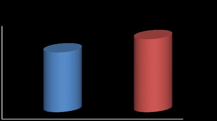
ISSN NO. 2320–7418                                                                                                                                 DOI: 10.22270/jmpas.V10I4.1298
posture and also make use of inadequate body mechanics. In the                                                       Figure 1: Distribution of women according to their age

primiparous women when the separation exists to the mild degree,
increased diastasis can be because of mechanical stresses on the                                                     50%                                                    37.50%
                                                                                                                                                     33.75%
                                                                                                                     40%          28.75%

                                                                                                      % of women
abdominal wall with pregnancy and also additional hormonal effects.
                                                                                                                     30%
Obesity, multiparity. foetal macrosomia, flaccid abdominal muscles                                                   20%

and multiple pregnancies are the main risk factors of the rectus                                                     10%
                                                                                                                      0%
abdominis diastasis [7].                                                                                                      22-26 yrs          27-31 yrs          32-37 yrs

          Several studies have been conducted to determine the
                                                                                                                                               Age Group(years)
prevalence of diastasis recti in multipara, but only a few investigations
have found the existence of diastasis recti in primipara and in females                                              Distribution of women according to age: In the present study
who delivered at full term and without complications. As a result, the              80 participants were included and divided into two groups as per
current study is being conducted to determine the prevalence of                     convenient sampling into 36 and 44 participants in each group. In this
diastasis recti in females who have had a full-term normal delivery and             study, the age of participants allotted in both the group primipara and
to compare it between primipara and multipara women. The objectives                 multipara were between 22 to 34 years. The percentage of age of
of the study included to evaluate the Diastasis recti in Full Term                  participating individual was 28.75% of age 22-26 years, 33.75% of age
Normal Delivery Females and to compare the Diastasis Recti in                       group 27-31 years and 37.5% of age group 32-37 years.
Primipara and Multipara.                                                                                             Figure 2: Distribution of women according to gravidity

MATERIALS AND METHODS
      The observational study was carried out at the Department of                                                                                                   55%
                                                                                                                      70%
community health sciences, Ravi Nair Physiotherapy College and                                                                            45%
                                                                                                                      60%

                                                                                               % of women
AVBRH Hospital Sawangi (M), Wardha. The inclusion criteria were                                                       50%
                                                                                                                      40%
framed as the Primipara with Full Term Normal Delivery, Multipara                                                     30%
with Full Term Normal Delivery and up to 6 months postpartum                                                          20%
                                                                                                                      10%
between the age 20 to 35 years. The exclusion criteria included female                                                 0%
                                                                                                                                Primigravida                 Multigravida
undergone C–section, female with previous history of abdominal
surgeries and female with BMI > 40 kg/m2. The variables concentrated                                                                            Gravidity

on the width of diastasis recti. The biased criteria included Age and
                                                                                                                     Distribution of women according to gravidity: In the present
other Anthropometric factors between the two groups will be matched
                                                                                    study 80 participants were included and allotted into two groups i.e.
and subjection not fulfilling the selection criteria will be excluded to
                                                                                    primipara and multipara. There were 36 primipara and 44 multiparas.
prevent bias. The sample size was 80 participants and calculated using
                                                                                    45% were primipara and 55% were multipara.
convenient sampling method.
                                                                                    Outcome measure:
RESULT
          The present study titled “Prevalence of Diastasis recti in               1. Comparison of inter-rectal distance among the gravidity of women:
primipara and multipara undergone full term normal delivery” which                  Test for normality of inter-rectal distance using dial caliper among
comprised of 80 primipara and multipara from age group 22-37 out of                 gravidity of women shows mean value of 1.84 in 36 primipara and
which 36 were primipara and 44 were multipara.                                      mean value of 2.37 in 44 multipara with 18.30 t value and p=0.0001,
Statistical analysis                                                                S. Standard deviation of 36 primipara is 0.08 and standard deviation of
          Statistical analysis was done by using descriptive and                    44 multipara is 2.37 which indicates that inter-rectal distance is more
inferential statistics using student’s unpaired t test and one way                  in multipara when compared among the gravidity of the women.
ANOVA and software used in the analysis was SPSS 27.0 version. For                     Figure 3: Comparison of Inter-rectal distance among gravidity of women
the purpose, all the data collected was entered into an excel sheet,                                                  3                                                     2.37
                                                                                        Mean Inter-rectal distance

tabulated and subjected to statistical analysis.      Several statistical                                            2.5
                                                                                                                                     1.84
measures were used such as mean, standard deviation of mean,                                                          2
                                                                                                 and SD

                                                                                                                     1.5
standard error mean, student’s unpaired t test data from subject’s
                                                                                                                      1
demography details i.e. age. Comparison between Prevalence of
                                                                                                                     0.5
diastasis recti in primipara and multipara with respect to outcome
                                                                                                                      0
measures of dial caliper used to measure inter-rectal distance was done                                                          Primigravida                        Multigravida

by student’s unpaired t-test.

Journal of medical pharmaceutical and allied sciences, Volume 10 - Issue 4, 1298, July - August 2021, Page – 3249 - 3253                                                             3251
ISSN NO. 2320–7418 DOI: 10.22270/jmpas.V10I4.1298
2. Age wise comparison of inter-rectal distance among women: accurate, and useful tool for measuring diastasis recti [8].
Test for normality of inter-rectal distance using dial caliper among age Van de Water AT, Benjamin DR conducted a systematic
of the women shows standard deviation of 0.08 in 23 women of the age review and meta-analysis to determine the best methods for detecting
group 22-26. In the age group of 27-31 of 27 women standard diastasis recti abdominis width, comparing dial calliper and ultrasound.
deviations was 0.30. In the women of age group 32-37 and standard They discovered that a dial calliper is a simple and practical method,
deviation was 0.21. Total mean value of 80 participants is 2.13 and although ultrasonography has feasibility issues [9].
total standard deviation is 0.31. F value is 40.49 and p=0.0001, S. This Turan V et al. did a study to determine the prevalence of
indicates that prevalence of diastasis recti is more in increased age and diastasis recti abdominis in multiparous adults and concluded that
prevalence of diastasis recti is reduced in younger age. greater parity and recurrent abdominal operations are the causes of
Figure 4: Age wise comparison of Inter-rectal distance among women higher DRA. The current study therefore is carried out to find out
3 prevalence of DRA in primipara to find out whether musculoskeletal
2.39
changes during pregnancy can contribute to diastasis recti and to
Mean Inter-rectal distance and SD
2.5 2.11
1.83
compare it with multiparous in females with full term normal delivery
2
[10]. In our study result showed that multiparous women have more of
1.5 Diastasis rectus abdominis than primiparous women.
1 CONCLUSION
From the present study we can concluded that there is presence of
0.5 diastasis recti more in multipara than compared to primipara undergone
0 full term normal delivery. This study will help people to understand
22-26 yrs 27-31 yrs 32-37 yrs
that there is increased inter-rectal distance when the participants age is
increased and when the age of the participant less than there is reduced
DISCUSSION
The goal of this study was to compare the prevalence of inter-rectal distance is. Hence core-strengthening and regular exercise
diastasis recti in primipara and multipara women. Several studies have is very important as it helps reduce the chance of diastasis recti after
found that diastasis recti are common in females who have had a C- pregnancy in both primipara and multipara irrespective of any age.
section, whether primipara or multipara, due to the separation of the Limitation
1. It was difficult to convince patient for being a part of this study
rectus abdominis muscle after surgery. However, diastasis can occur
2. Due to SARS COVID-19 pandemic the number of patient were
due to the separation of two bellies of the recti muscle to accommodate
reduced in the hospital
the growing size of the uterus, but the gap is eventually reduced after
3. Most of the participants were afraid to be a part of the research due
delivery in the postpartum period when the structures of the abdomen
to SARS-COVID 19 pandemic.
and pelvis return to their normal position, but diastasis may still persist
4. Due to SARS COVID-19 pandemic the duration of study was
and show different features in females who have had multiple
reduced from 6 months to 5 months hence we reduced our sample size
deliveries. Therefore, current study aimed to compare the prevalence
from 175 to 80 considering the pandemic scenario
and severity of diastasis recti in primipara and multipara with Full
Author’s Contribution
Term Normal Delivery.
All authors contributed equally to the manuscript.
The results show that there is presence of Diastasis recti more
Conflict of Interest
in multipara compared to primipara undergone full term normal
The authors declare no conflict of interest.
delivery. The comparison of prevalence of diastasis recti in primipara
Funding Source
and multipara was reported with reduced inter-rectal distance in The research has not received any external funding.
primipara and increased inter-rectal distance in multipara using dial Acknowledgement
We thank the patients who participated and contributed samples to the
caliper. In younger women prevalence of diastasis recti is less than
study.
compared to women of older age after pregnancy.
REFERENCES
To our knowledge the study showing prevalence of diastasis
1. Chiarello CM, Falzone LA, McCaslin KE, Patel MN, Ulery KR,
recti in primipara and multipara undergone full term normal delivery 2005. “The Effects of an Exercise Program on Diastasis Recti
using dial caliper is very few. Boxer SE and Jones S conducted an inter- Abdominis in Pregnant Women”. J Women’s Health Phys Ther.
29(1), 11–6.
rater reliability research and found that rectus abdominis diastasis may
2. Kn SS, 2020. “An Overview of the Studies on Diastasis Recti
be quantified with a high degree of reliability using a dial calliper Abdominis in Postpartum Women”. J Gynecol Womens Health
during the postpartum period. It has been shown to be a simple, 14(5).
3. Adkitte R, Bhatt K, Yeole U, Gawali P, Gharote G, 2016.
Journal of medical pharmaceutical and allied sciences, Volume 10 - Issue 4, 1298, July - August 2021, Page – 3249 - 3253 3252ISSN NO. 2320–7418 DOI: 10.22270/jmpas.V10I4.1298
“Prevalence of diastasis of rectus abdominis muscle in immediate
post-partum women of urban and rural areas”. 3(5), 460-62.
4. Demartini E, Deon KC, Fonseca EG de J, Portela BS, Demartini E,
Deon KC, 2016. “Diastasis of the rectus abdominis muscle
prevalence in postpartum”. Fisioter Em Mov. 29(2), 279–86.
5. P Bobowik, A Dabek, 2018. “Physiotherapy in women with
diastasis of the rectus abdominis muscles”. Advances in
Rehabilitation. 3, 11-18.
6. Benjamin DR, van de Water ATM, Peiris CL, 2014. “Effects of
exercise on diastasis of the rectus abdominis muscle in the
antenatal and postnatal periods: a systematic review”.
Physiotherapy. 100(1), 1–8.
7. Fei H, Liu Y, Li M, He J, Liu L, Li J, 2021. “The relationship of
severity in diastasis recti abdominis and pelvic floor dysfunction:
a retrospective cohort study”. BMC Women Health. 21(1), 68.
8. S. Boxer, S Jones, 1997. “Intra-rater reliability of rectus abdominis
diastasis measurement using dial calipers”. Aust J Physiother.
43(2), 109–14.
9. Benjamin DR, 2016. “Measurement methods to assess diastasis of
the rectus abdominis muscle (DRAM): A systematic review of
their measurement properties and meta-analytic reliability
generalisation”. Man Ther. 21, 41–53.
10. Turan V, Colluoglu C, Turkyilmaz E, Korucuoglu U, 2011.
“Prevalence of diastasis recti abdominis in the population of young
multiparous adults in Turkey”. Ginekol Pol. 82(11), 817–21.
How to cite this article
Simran J, Shalaka D, Shruti D, Sakshi P. A, 2021. “Comparative
study on Prevalence of Diastasis Recti in Primipara and
Multipara Undergone Full Term Normal Delivery”. Jour. of
Med. P’ceutical & Allied Sci. V 10 - I 3, 1298 P-3249 - 3253.
doi: 10.22270/jmpas.V10I4.1298.
Journal of medical pharmaceutical and allied sciences, Volume 10 - Issue 4, 1298, July - August 2021, Page – 3249 - 3253 3253You can also read