Building a Web-Augmented Reality application for demonstration of kidney pathology for veterinary education
←
→
Page content transcription
If your browser does not render page correctly, please read the page content below
Polish Journal of Veterinary Sciences Vol. 24, No. 3 (2021), 345–350

DOI 10.24425/pjvs.2021.137671

                                                                                                    Original article

         Building a Web-Augmented Reality
       application for demonstration of kidney
         pathology for veterinary education

                                        H.T. Atmaca1, O.S. Terzi2

                          Department of Pathology, Faculty of Veterinary Medicine,
                           1

                      Balikesir University, Cagis Yerleskesi, 10145, Balikesir, Turkey
                    2
                      Department of Internal Medicine, Faculty of Veterinary Medicine,
    Ankara University, Ziraat Mahallesi Sehit Omer Halisdemir Bulvari, 06110, Altindag, Ankara, Turkey

                                                      Abstract
                Three-dimensional (3D) models created with computers and educational applications
            designed using such models are used in the medical field every day. However, there is a lack
            of macroscopic demonstration applications built with digital 3D models in the field of veterinary
            pathology. The aim is to build a fully interactive 3D educational web-based augmented reality
            application, to demonstrate macroscopic lesions in kidneys for educational purposes. We used
            open source and free software for all 3D modelling, Augmented Reality and website building.
            Sixteen 3D kidney pathology models were created. Kidney models modelled in 3D and published
            as WebAR are as follows: normal kidney, unilateral neurogenic shutdown with atrophy, hydrone-
            phrosis, hypercalcemia of malignancy tubular nephrosis, interstitial corticomedullary nephritis,
            renal infarct, multifocal petechial hemorrhages, polycystic kidneys, renal masses, multifocal
            nephritis, pigmentary nephrosis, papillary necrosis, glucose-related rapid autolysis (pulpy
            kidney), pyelonephritis, renomegaly and kidney stones. With the workflow shown here, it has
            been presented as a feasible model application for human pathology and presented to educators,
            researchers and developers who have 3D models and AR in their field of interest. To the best
            of the authors’ knowledge, this is the first study on Web-Augmented Reality application for
            veterinary pathology education.

            Key words: veterinary pathology, education, three dimensional, augmented reality, kidney

                   Introduction                                (PLC Informa 2020). Although these applications are
                                                               mostly in the field of human anatomy, some researchers
    Three-dimensional (3D) models created with com-            and companies have also developed similar applica-
puters and educational applications designed based             tions for veterinary anatomy. Although each application
on them are used in the medical field every day. These         has a different interface and interactive software
applications, with their wide utility range, take place        engine, a wide variety of demonstration methods and
online and are also accessible in university libraries         interactive material presentation have been developed,
Correspondence to: H.T. Atmaca, e-mail: ht_atmaca@yahoo.com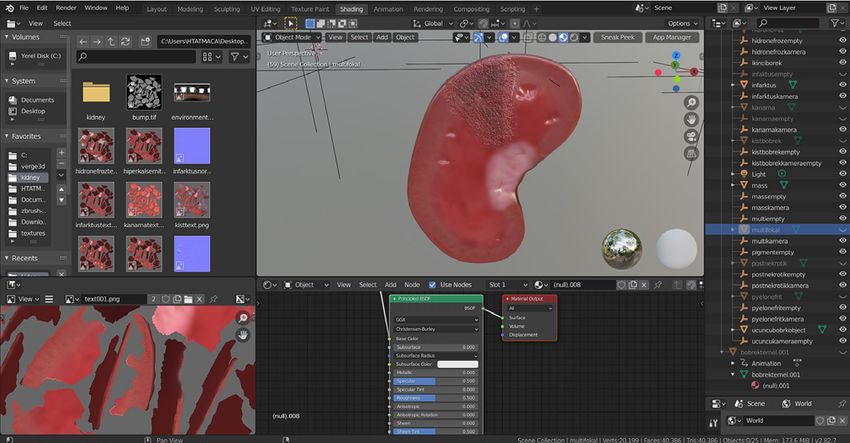
346                                                                                                H.T. Atmaca, O.S. Terzi

Fig. 1. Demonstration of workflow for 3D modeling and interactive application.

mainly to assist the teachers and students. Digital                  ground and a sense of reality is created by adding texts,
microscopic and macroscopic image archives and                       pictures, sounds, animations or 3D objects to the video
applications are the principal educational materials                 image of the real world (Billinghurst et al. 2001).
used in pathology education. Virtual microscopy appli-                   In this study, we built a fully interactive 3D educa-
cations (European College of Veterinary Pathologists –               tional WebAR application demonstrating macroscopic
Histology Slide Database 2020, The Joint Pathology                   lesions in kidneys. This application was built using
Center 2020) and static gross pathology photos                       open source software that anyone can use. It is hoped
(Necropsy Show and Tell 2021, Orvalho and Peleteiro                  that this will aid the understanding of gross kidney
2021) on different websites are very important in                    pathology by veterinary students. The workflow
pathology education. These digital microscopy data-                  methodology created here could inspire further research
bases play an important role in the educational process              in this field, to help educators develop certified digital
of veterinary medicine worldwide. It is indeed a fact                educational materials for their students.
that macroscopy for morphological diagnosis is one
of the first and important stages in pathology.
    Although there are 3D applications developed for                              Materials and Methods
veterinary anatomy education, there is a lack of macro-
scopic demonstration applications built with digital 3D                                     Modelling
models in the field of veterinary pathology. This makes                  A single basic demonstrative 3D kidney anatomy
imperative the need for such 3D applications in the                  was modelled for the application. The workflow for this
veterinary curriculum for education and training.                    application is shown in Fig. 1. Modelling was done
The application described in this paper will undoubted-              in open source and free Blender 2.82 software. First,
ly be a valuable tool in veterinary pathology education.             the Blender software was run and a 3d UV sphere was
Mobile augmented reality (mAR) technology, which                     added to the scene (Fig. 2). We then started to outline
has become widespread in recent years, permits stu-                  the kidney model in which we would show the lesions.
dents to enjoy flexible learning wherever and whenever               We used a cross section kidney structure in the kidney
they want (Bujak et al. 2013). Therefore, we designed                model (Fig. 3). Thus, we showed the cortex, medulla,
this application for use on both the web and mAR, and                renal pelvis, and the capsule.
named it a WebAR application. As is known, in AR
technology, the real environment is used as a back-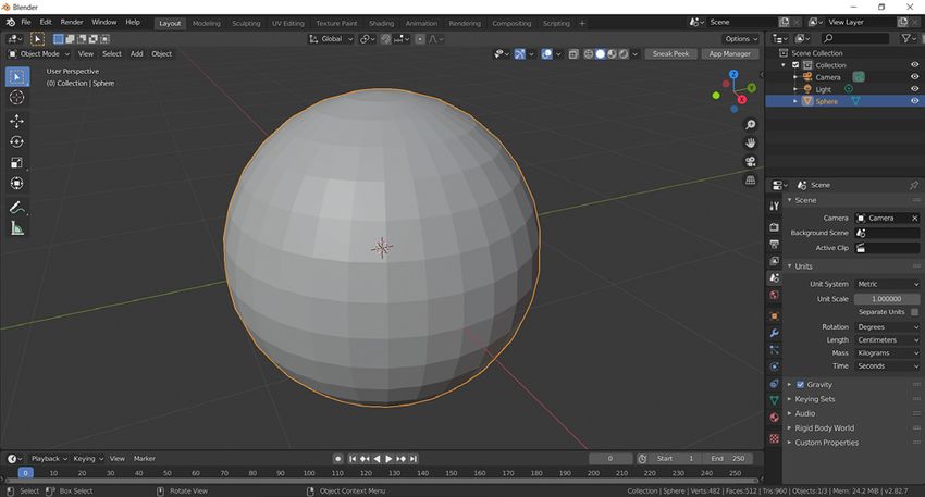
Building a Web-Augmented Reality application ...                                                                     347

Fig. 2. Modelling interface in Blender. A UV mesh sphere was used to create a kidney model using sculpting mode.

Fig. 3. Cross section kidney structure created using sculpting mode in Blender from a UV mesh sphere.

      Sculpting, texture painting and shading                                        Augmented Reality (AR)
    After the modelling process, sculpting was per-                      The 3D kidney models were saved as a single file
formed on the 3D kidney model in order to understand                 in the GLB format, including the texture images. GLB
the different macroscopic findings (Fig. 4). Following               is the binary file format representation of 3D models
the literature, photorealism was applied with texture                saved in the GL Transmission Format (glTF). GLB files
painting and shading onto the 3D kidney model (Fig. 5).              were used for AR applications on both web and android
Lesion description and demonstration of the models                   applications. GLB files for AR on IOS devices were
were made as indicated in the literature (King et al.                converted to USDZ format in the Reality composer
2014, Cianciolo and Mohr 2015).                                      program on the MacOS device and saved. Whichever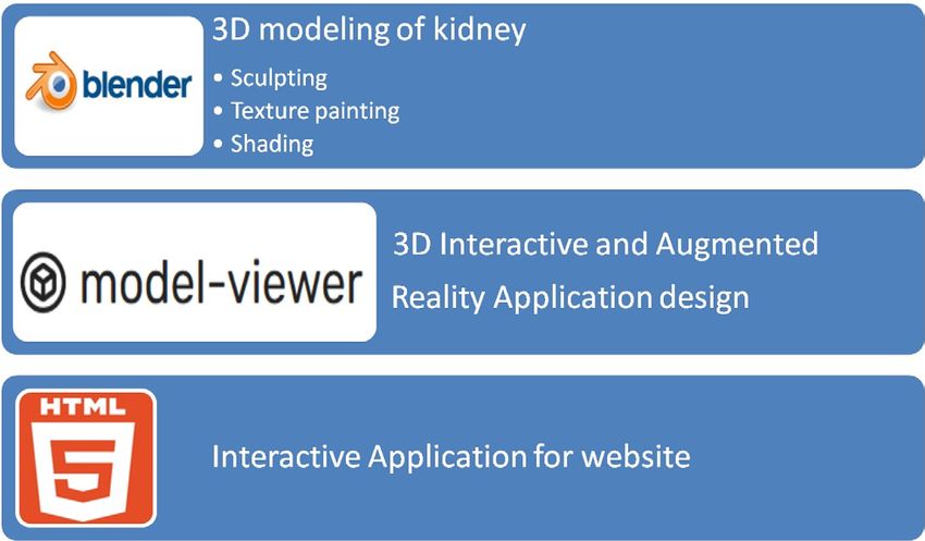
348 H.T. Atmaca, O.S. Terzi
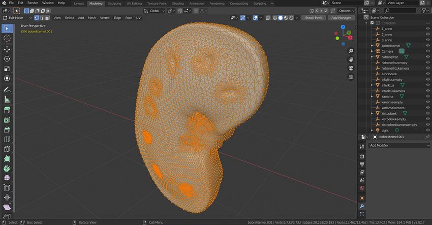
Fig. 4. Sculpting interface in Blender. Working on cystic kidney. With sculpting mode, adding cysts with blob and inflate tools in Blender.
Fig. 5. Texture painting and shading interface in Blender.
Working on infarct. With referring some macroscopic image databases for veterinary pathology, lesions illustrated with painting
and texturing. We do not use original macroscopic images for texturing, we use painting and shading to simulate the natural
appearance of the lesions and as described in textbooks for student.
device is used for AR view, the appropriate file format Creating interactivity
for that device is automatically displayed. If you run the
application with an Android mobile device, you will The 3D kidney models were then converted into
view the file in GLB format, if you run it with an IOS a web-based application using HTML codes. The codes
device, you will view the USDZ format file with the that are required for the WebAR application are open
same 3D kidney models. source codes and were obtained from github (GitHub -
google/model-viewer 2020). The web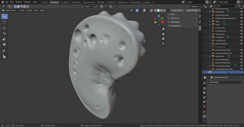
Building a Web-Augmented Reality application ...                                                                  349

component can be used to view and act together with          odology used in this study can be considered successful
3D models on the web, and it smoothly transitions            since the main macroscopic lesions shown. Further
to placing, and acting together with, such 3D models         development of the current work by including other
in Augmented Reality on the web (Developer/Arcore,           organ pathologies or histopathology could be the basis
Google 2021). Annotations that make it easier for stu-       for future research. 3D and AR applications showing
dents to understand were added. These annotations            macroscopic lesions of all organs and tissues were
were added on each web page in the application and           envisaged, but due to the lack of appropriate staff and
on the 3D models. These annotations were made using          the shortage of time we could allocate for this research,
the GLB editor. We also made the basic webpage con-          we decided to confine ourselves to kidney pathology.
tents, such as listing the 3D kidney models, providing       We hope other researchers may be motivated by our
information about the application and the contact page,      effort to create similar 3D pathology applications
with free HTML website builder softwares.                    of other organs and tissues. The anatomy of kidneys
                                                             in domestic animals in veterinary medicine education
                                                             is variable. Therefore, the lesions were shown on a com-
                        Results                              mon illustrated model.
                                                                 AR technology, which has gained popularity
    Sixteen 3D kidney pathology models, with a total         in many areas, has been used frequently in education in
file size of 19.9MB, were created in the software            recent years. AR applications are reportedly quite effec-
and saved, according to the lesions. The following 3D        tive in facilitating meaningful learning, as they contrib-
models were created: normal kidney, unilateral neuro-        ute to the understanding of the content by visualising
genic shutdown with atrophy, hydronephrosis, hyper-          virtual 3D structures and clarifying complex issues
calcemia of malignancy tubular nephrosis, interstitial       (Wu et al. 2013). Interacting with the 3D view of
corticomedullary nephritis, renal infarcts, multifocal       objects from different perspectives improves students’
petechial haemorrhages, polycystic kidneys, renal            spatial and practical skills (Kerawalla et al. 2006, Cheng
masses, multifocal nephritis, pigmentary nephrosis,          and Tsai 2013). Additionally, AR technology provides
papillary necrosis, glucose-related rapid autolysis          instant feedback through real-time interactions that
(pulpy kidney), pyelonephritis, renomegaly and kidney        enable students to control their own learning processes
stones.                                                      (Bujak et al. 2013).
    These models were uploaded to the internet server.           Our application aims at imparting learning by
Thereafter, the website was activated immediately. It is     demonstrating basic macroscopic lesions with the logic
available at https://www.veterinarypathology3d.com.          of illustration, based on 3D models prepared on the
All models, including annotation and information, can        computer. Of course, this educational workflow could be
be viewed on the desktop from any computer through           done with other 3D techniques too. Digital 3D models
any browser. When accessing our website from any             (Photometry) obtained by photogrammetry, digital 3D
mobile device, models can be viewed exactly as from          models obtained with a 3D scanner, or 3D printed
desktop browsers. Optionally, the AR feature can be          tissues and organs obtained from these 3D models, can
activated and the model displayed on a surface with          be cited as examples of 3D techniques. There are
a real world background.                                     numerous advantages to using digital 3D models
                                                             in pathology, some of which can also be obtained in 3D
                                                             printing (McMenamin et al. 2014, Mahmoud and Ben-
                     Discussion
                                                             nett 2015, Lim et al. 2016, Salazar-Gamarra et al. 2016).
    In our study, macroscopic lesions in the kidneys         Each of these has an advantage over the other. There are
were designed as an interactive 3D application for edu-      disadvantages to 3D printing too, such as a lack of per-
cation and training. We have presented the workflow          sonnel and expensiveness of the devices. Providing an-
methodology with the application created as both             notations on the digital 3D models will support the stu-
a web-based and AR application. This application             dents working alone, by adding notes they should pay
is built with widely used open source software and pub-      attention to, and by indicating the way the lesions are
lic WebGL codes and is launched for problem-free use.        seen. It is of course true that the lesions are exhibited
    The purpose of this 3D interactive application is to     in real organs in the pathology museum, and that the
create an additional resource for veterinary pathology       students obtain sufficient macroscopic morphological
education. The next step will be user verification of this   information from them. However, unfortunately, not
resource. Moreover, a study on the impact of this prac-      every faculty of institutes offering veterinary pathology
tice on students will have particular value in proving its   education supports these museums. To establish
place in the veterinary medicine curriculum. The meth-       a pathology museum, it is necessary to carry out numer-
350 H.T. Atmaca, O.S. Terzi
ous necropsies, collect, fix and store tissues with European College of Veterinary Pathologists. Histology Slide
lesions. This process requires many years for newly Database [accessed August 31, 2020]. https://www.ecvpath.
established institutions that do not handle a wide range org/histology-database
Google. GitHub - google/model-viewer: Easily display inter-
of cases. It is therefore an important development for active 3D models on the web and in AR! [accessed
students to be able to see the macroscopic lesions in 3D August 31, 2020]. https://github.com/google/model-viewer
and obtain additional information with this application, Hua-quan R, Jun J, Zhao-fang Z (2014) Study on Digitalized
as if they were physically in the pathology museum at a 3D Specimen Making of Pathologic Gross Specimen
time and location of their convenience. Basing on Object Panorama. In: Li S., Jin Q., Jiang X.,
In addition, it has been observed that the informa- Park J. (eds) Frontier and Future Development of Infor-
mation Technology in Medicine and Education. Lecture
tion displayed on 3D models is always more permanent
Notes in Electrical Engineering, vol 269. Springer,
than that on two-dimensional (2D) images. This situa- Dordrecht.
tion has paved the way for the development of educa- Kerawalla L, Luckin R, Seljeflot S, Woolard A (2006)
tion for students. The interaction arising from these 3D “Making it real”: exploring the potential of augmented
models provides better clinico-pathological correlation reality for teaching primary school science. Virtual Real
for the physician than a 2D image and a better learning 10: 163-174.
environment for the student (McMenamin et al. 2014, King JM, Roth-Johnson L, Dodd DC, Newsom ME (2014)
The Necropsy Book: A Guide for Veterinary Students,
Hua-quan et al. 2014, Allen et al. 2016, Peterson and Residents, Clinicians, Pathologists, and Biological
Mlynarczyk 2016). Researchers. 7th ed., The Internet-First University Press.
The aim of this study was to clearly demonstrate Lim KH, Loo ZY, Goldie SJ, Adams JW, McMenamin PG
how widely available software can be used to build (2016) Use of 3D printed models in medical education:
a fully interactive 3D package to increase the under- A randomized control trial comparing 3D prints versus
standing of macroscopic lesions in the kidney, thus cre- cadaveric materials for learning external cardiac anatomy.
Anat Sci Educ 9: 213-221.
ating a utilitarian application for education and training.
Mahmoud A, Bennett M (2015) Introducing 3-Dimensional
Of course, further validation and participation by Printing of a Human Anatomic Pathology Specimen:
the end users – students and veterinary clinicians – will Potential Benefits for Undergraduate and Postgraduate
be required to fully assess the impact of this educational Education and Anatomic Pathology Practice. Arch Pathol
application. Through this process, we have created Lab Med 139: 1048-1051.
a foundation on which future veterinary educators Cianciolo RE, Mohr FC (2015) Chapter 4 – Urinary System
and students can build tailor-made packages for use in Pages, 376-464 In: Maxie M.G. (Ed.), Jubb, Kennedy and
Palmer’s Pathology of Domestic Animals. Vol.2. 6th ed.
veterinary pathology education. Actually, this is not Elsevier Saunders, Philadelphia.
limited to veterinarians and veterinary students. With McMenamin PG, Quayle MR, McHenry CR, Adams JW
the workflow shown here, it has been developed as (2014) The production of anatomical teaching resources
a universally feasible model application for human using three-dimensional (3D) printing technology. Anat
pathology and is presented to educators, researchers Sci Educ 7: 479-486.
and developers who have 3D models and AR as their Necropsy Show and Tell. Necropsy Show and Tell: Veterinary
Pathology Images. [accessed January 4, 2021]. https://
fields of interest.
secure.vet.cornell.edu/nst/nst.asp
Orvalho JS, Peleteiro MC. Online Veterinary Pathology Atlas.
[accessed March 31, 2021]. http://www.fmv.ulisboa.pt/
References atlas/atlas_ing.htm
Peterson DC, Mlynarczyk GS (2016) Analysis of traditional
Allen LK, Eagleson R, de Ribaupierre S (2016) Evaluation versus three-dimensional augmented curriculum on ana-
of an online three-dimensional interactive resource for tomical learning outcome measures. Anat Sci Educ
undergraduate neuroanatomy education. Anat Sci Educ 9: 529-536.
9: 431-439. PLC Informa. anatomy.tv (3D anatomy source) [accessed
Billinghurst M, Kato H, Poupyrev I (2001) The magic- August 31, 2020]. https://www.anatomy.tv/resources
book-moving seamlessly between reality and virtuality. Salazar-Gamarra R, Seelaus R, da Silva JV, da Silva AM,
IEEE Comput Graph Appl 21(3): 6-8. Dib LL (2016) Monoscopic photogrammetry to obtain 3D
Bujak KR, Radu I, Catrambone R, MacIntyre B, Zheng R, models by a mobile device: a method for making facial
Golubski G (2013) A psychological perspective on aug- prostheses. J Otolaryngol Head Neck Surg 45: 33.
mented reality in the mathematics classroom. Comput The Joint Pathology Center. The Joint Pathology Center (JPC)
Educ 68: 536-544. – Veterinary Pathology Services. [accessed August
Cheng K-H, Tsai C-C (2013) Affordances of Augmented 31, 2020]. https://www.askjpc.org/vetpath/
Reality in Science Learning: Suggestions for Future Wu HK, Lee SW, Chang HY, Liang JC (2013) Current status,
Research. J Sci Educ Technol 22: 449-462. opportunities and challenges of augmented reality in edu-
Developer/Arcore, Google. Model-viewer. [accessed August cation. Comput Educ 62: 41-49.
31, 2020]. https://developers.google.com/ar/develop/
webxr/model-viewerYou can also read