Study on reflection of human skin with liquid paraffin as the penetration enhancer by spectroscopy
←
→
Page content transcription
If your browser does not render page correctly, please read the page content below
Study on reflection of human skin with
                 liquid paraffin as the penetration
                 enhancer by spectroscopy

                 Kun Chen
                 Yanmei Liang
                 Yuan Zhang

Downloaded From: https://www.spiedigitallibrary.org/journals/Journal-of-Biomedical-Optics on 04 Nov 2021
Terms of Use: https://www.spiedigitallibrary.org/terms-of-use
Journal of Biomedical Optics 18(10), 105001 (October 2013)

                Study on reflection of human skin with liquid paraffin as
                the penetration enhancer by spectroscopy
                Kun Chen, Yanmei Liang, and Yuan Zhang
                Nankai University, Institute of Modern Optics, Key Laboratory of Optical Information Science and Technology, Ministry of Education, Tianjin 300071,
                China

                              Abstract. Optical clearing agents can improve tissue optical transmittance by reducing the diffuse reflection. The
                              reflection on in vivo human skin before and after applying anhydrous glycerol and 30 to 50% liquid paraffin glyc-
                              erol mixed solution are investigated in this paper. From their visible and near-infrared reflection spectroscopy, all of
                              their diffuse reflections are reduced after applying the agents. It is found that the three mixed solutions show stronger
                              effect than that of anhydrous glycerol. These results further prove liquid paraffin can enhance the percutaneous
                              penetration of glycerol and take synergistically optical clearing effect with glycerol over visible and near-infrared
                              wave bands. © The Authors. Published by SPIE under a Creative Commons Attribution 3.0 Unported License. Distribution or reproduction of this work in
                              whole or in part requires full attribution of the original publication, including its DOI. [DOI: 10.1117/1.JBO.18.10.105001]

                              Keywords: reflection; spectroscopy; skin; liquid paraffin; glycerol.
                              Paper 130340RR received May 13, 2013; revised manuscript received Aug. 28, 2013; accepted for publication Sep. 9, 2013; published
                              online Oct. 3, 2013.

                1    Introduction                                                                     lipophilic agents has been added in OCAs to improve the delivery
                Motivated by the growing maturity of laser treatment and optical                      of agents in skin so as to achieve a better optical clearing effect,
                imaging diagnosis, optical techniques, such as optical coherence                      such as polypropylene glycol-based polymers,17 dimethyl sulfox-
                                                                                                      ide (DMSO),19,22 oleic acid,19 azone,20 thiazone,24,25 and liquid
                tomography (OCT), confocal microscopy, nonlinear micros-
                                                                                                      paraffin.26–28
                copy, and laser spectroscopic methods, are widely used in
                                                                                                          Optical wave bands used by different optical technologies are
                many fields. However, the complicated morphological nature
                                                                                                      usually different. Many studies have been reported that biotissue
                of human tissue and variations of the refractive indices with
                                                                                                      in vivo and in vitro has different absorption coefficient (μa ), scat-
                internal different components make biotissues become a high
                                                                                                      tering coefficient (μs ), and refractive index (n) for different light
                scattering medium for visible and near-infrared wavelengths,
                                                                                                      wavelengths.4 Therefore, it is necessary to evaluate the optical
                i.e., the therapeutic and diagnostic optical window.1–5
                                                                                                      clearing effect of an OCA thoroughly over the therapeutic and
                Multiple scattering and absorption attenuate the effective
                                                                                                      diagnostic optical window. Because of the excellent comprehen-
                light intensity of reaching internal tissue and diminish the
                                                                                                      sive characteristics of liquid paraffin, its synergistic effect as the
                detecting depth. Therefore, they limit the clinical application                       penetration enhancer of glycerol is further studied by spectros-
                of optical imaging techniques.                                                        copy in this paper, whose purpose is to provide its anticipative
                    Currently, osmotic chemical agents used for optical clearing                      result when it will be used in different optical technologies.
                of biotissue have become a considerable interest. Optical clear-                          A commercial spectrometer was used to measure the surface
                ing technique has been successfully developed to reduce the                           reflection of human skin before and after applying liquid par-
                scattering properties and improve the light penetration depth                         affin glycerol mixtures, and then the reduction of diffuse reflec-
                by application of optical clearing agents (OCAs) with hyperos-                        tance was calculated. It was shown that anhydrous glycerol and
                molarity and biocompatibility.6 It has the significant potential to                   different concentrations of liquid paraffin glycerol mixtures had
                improve the application of spectroscopic and optical imaging                          different optical clearing effects on in vivo fingers for the spectra
                techniques in clinic.                                                                 ranging from 600 to 1400 nm. It was also found that the mix-
                    OCAs, such as polyethylene glycol,7 glucose,8,9 glycerol,10–22                    tures had different effects for light with different wavelengths
                propylene glycol,16 and dextran,23 can reduce the scattering and                      from the fluctuation of spectra.
                enhance light penetration in biotissue. Generally, they are almost
                hydrophilic agents. However, it is a relatively slow process for
                these hydrophilic agents to penetrate the stratum corneum,                            2     Materials and Methods
                when they are applied on the surface of skin. The agents with
                high concentration can achieve good osmosis effect, but they                          2.1     Samples and Chemical Agents
                have their limitations in clinical applications considering the                       In our experiment, 12 volunteers’ fingers were measured,
                safety. A noninvasive way of incorporating a permeation enhancer                      including 10 males and 2 females, whose ages are from 22
                can improve the osmosis effect in the stratum corneum. Some                           to 43. All of them are healthy without any aberrance, such as
                                                                                                      scar, fibroma, pigmented nevus, or other diseases on their
                                                                                                      fingers.
                Address all correspondence to: Yanmei Liang, Nankai University, Institute of
                Modern Optics, Tianjin 300071, China. Tel: 86-22-23503121; Fax: 86-22-                   According to Ref. 26, when liquid paraffin is mixed with
                23503121; E-mail: ymliang@nankai.edu.cn                                               anhydrous glycerol by volume ratio 3∶7 to 5∶5, the mixed

                Journal of Biomedical Optics                                                 105001-1                                                 October 2013   •   Vol. 18(10)

Downloaded From: https://www.spiedigitallibrary.org/journals/Journal-of-Biomedical-Optics on 04 Nov 2021
Terms of Use: https://www.spiedigitallibrary.org/terms-of-use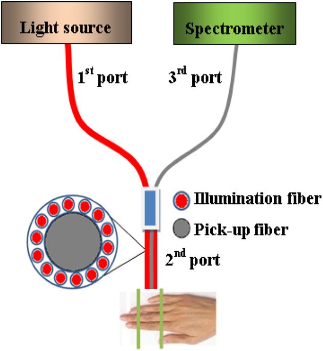
Chen, Liang, and Zhang: Study on reflection of human skin with liquid paraffin. . .
solution displays the best optical clearing effect for in vivo ratio of reflectance (ΔR) similar to Ref. 29 is used in
samples. In addition, it can keep the structure of the tissue our paper to calculate quantitatively the reduction of diffuse
from deforming, which means it can balance between dehydra- reflectance before and after treatment with agents. It is defined
tion and moisture retention over a long time. Therefore, the same as follows:
concentration range was used in our experiment. That is, four
kinds of solution were used in this study, including anhydrous Rmin − Rcontrol
ΔR ¼ × 100%; (1)
glycerol and three different concentrations of liquid paraffin Rcontrol
whose volume fractions in mixed solutions are 30, 40, and
50%, respectively. Glycerol (WEICHEN Brand) and liquid par- where Rcontrol is the measured diffuse reflectance at 1054.4 nm
affin (WENDA Brand), made by Tianjin Weichen Chemical before treatment with agents and Rmin is the minimal measured
Reagents Company Limited and Tianjin Yingda Rare diffuse reflectance in the whole measurement process at
Chemical Reagents Factory, Tianjin, China, respectively, were 1054.4 nm. The reason for choosing 1054.4 nm for the assess-
used in our study. ment of reflectance is because the spectrum analyzer detects the
maximal power of the reflection at this wavelength.
2.2 Experimental System
2.4 Measurement Method
A fiber-based spectrum analyzer system was set up in our
experiment, whose sketch map is shown in Fig. 1. A specially The experiment was carried out at 22°C room temperature.
designed Y-type fiber bundle was used in the system. Its first During the measuring process, the finger was fixed on the plat-
port is a fiber bundle composed of 14 same fibers whose form. In order to calculate the reduction of diffuse reflectance,
core diameter is 100 μm, which is connected with a halogen the reflection spectra without the agent were first collected three
tungsten lamp through a fiber optic connector of SMA 905. times and the average of values was regarded as the baseline for
Its second port is also a fiber bundle composed of an inner each sample. Then, the agent was topically applied onto the sur-
fiber with a core diameter of 600 μm and the 14 fibers evenly face of the finger, and spectra were collected at time intervals of
distributed all around, as shown in Fig. 1. The broadband light 5 min. In order to evaluate the reflectance exactly, the distance
from the halogen tungsten lamp uniformly illuminates the between the second end of the Y-type fiber bundle and the sam-
sample by the outer fibers and the reflection light from the ple remained unchanged while collecting all reflection spectra
sample is collected by the inner fiber. Its third port is only for a certain agent. Every volunteer was measured three times
the inner fiber, which is connected with the spectrum analyzer for each concentration.
(AQ-6315E, ANDO).
Human fingers are fixed on a platform. In order to maintain
3 Experimental Results and Discussion
the identical sampling in the whole measuring process, the dis- We can evaluate the change of reflection directly based on their
tance between the second end and the sample is held at about reflection spectra before and after application of the agents.
1 mm. In addition, the sample is nearly perpendicularly irradi- Figure 2 illustrates an example of the diffuse reflection spectra
ated and the returned light is also collected perpendicularly over a range from 600 to 1400 nm when the finger of a person
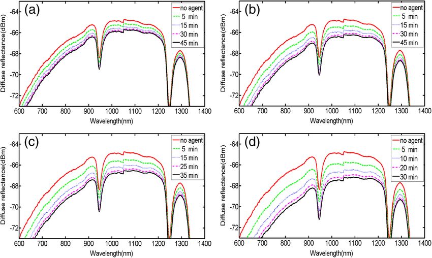
as Ref. 26. was applied with different solutions. Figures 2(a), 2(b), 2(c), and
2(d) correspond to the results of anhydrous glycerol and 30,
40, and 50% liquid paraffin glycerol mixtures, respectively.
2.3 Evaluation of the Reduction of Diffuse Reflection The curves in each figure were obtained at different time inter-
Reflection spectrum collected by the spectrum analyzer system vals. It can be seen that the results from all four agents have
can be directly used to evaluate the diffuse reflectance. Some similar trends qualitatively; that is, the diffuse reflection
parameters have been used to evaluate it quantitatively.22,29 decreases gradually with time elapsing over the whole
The optical clearing effect of OCAs can be revealed by the wavelength range investigated. In addition, we can see the
reduction of diffuse reflectance from the finger skin. Intensity reduced extent is different among these agents. From Fig. 2,
we also find there is better effect at short wavelengths than
that at long wavelengths, which is similar to the results of
many agents.16,22,24
In order to compare the diffuse reflectance decrease, the data
of samples without and with the agents treatment were extracted
where the spectrum analyzer detected the minimal diffuse reflec-
tance at the wavelength 1054.4 nm (ΔR), respectively. The stat-
istical results of all 12 volunteers are shown in Fig. 3, where the
negative percentage represents the decrease in reflectance com-
pared to the control. The average diffuse reflectance decreased
∼19.8, 28.8, 33.8, and 43.5% for anhydrous glycerol, 30,
40, and 50% liquid paraffin glycerol mixtures, respectively.
Three mixed solutions deliver more effective capability of
decrease of diffuse reflectance than glycerol alone does. It indi-
cates the trend that the more liquid paraffin is added, the larger
decrease of diffuse reflectance is caused, which is similar to the
effect of DMSO as shown in Ref. 22.
Two examples of the reduction of diffuse reflectance at
Fig. 1 Sketch map of the experiment system. 1054.4 nm with time elapsing of anhydrous glycerol, 30, 40,
Journal of Biomedical Optics 105001-2 October 2013 • Vol. 18(10)
Downloaded From: https://www.spiedigitallibrary.org/journals/Journal-of-Biomedical-Optics on 04 Nov 2021
Terms of Use: https://www.spiedigitallibrary.org/terms-of-useChen, Liang, and Zhang: Study on reflection of human skin with liquid paraffin. . .
Fig. 2 Changes of diffuse reflectance for human finger skin before and after application of different agents over the range of 600 nm - 1400 nm. (a), (b),
(c) and (d) are anhydrous glycerol, 30%, 40% and 50% liquid paraffin glycerol mixtures, respectively. The different curves in each figure are cor-
responding to different time intervals from top to bottom, (a) no agent, 5, 15, 30, and 45 min, (b) no agent, 5, 15, 30, and 45 min, (c) no agent, 5, 15, 25,
and 35 min, and (d) no agent, 5, 10, 20, and 30 min.
scattering characteristics of biotissues are reversible after apply-
ing liquid paraffin glycerol mixture, which is an essential char-
acteristic of OCAs. For all these solutions, 50% liquid paraffin
glycerol mixture has the largest decreasing speed. However, the
optimal reduction of diffuse reflection of anhydrous glycerol is
not reached within 45 min.
In addition, we can see from Fig. 4 that the reduction of
diffuse reflectance is larger than 20% after 15 min mixture
treatment, and this can be maintained for more than 25 min.
We find 50% concentration has the largest reduced effect in
Figs. 3 and 4. According to Ref. 26, 30% concentration has the
best optical clearing effect under the surface of 700 μm. It has
been known that blood flows in blood containing layers (living
Fig. 3 Average reduction of diffuse reflectance (ΔR) at 1054.4 nm for epidermis, dermis) will wash away a part of agent periodically
anhydrous glycerol and 30%, 40%, and 50% liquid paraffin glycerol for in vivo samples after the penetration of the osmotic agents.
mixtures, respectively. Most researchers think the mechanism of the internal biotissue
enhancement includes the match of refractive indices and tissue
dehydration due to the osmotic properties of OCAs. The gross
and 50% liquid paraffin glycerol mixtures are given in Fig. 4. volume of the mixture is the same for 30 to 50% mixture in this
They are shown with red, blue, black, and green curves, respec- study and Ref. 26. After excluding other possibilities, as a pos-
tively. It can be seen from Fig. 4 that all the agents make the sible explanation, we think that under the synergistic effect of
diffuse reflectance decrease. Thirty to fifty percentage liquid the liquid paraffin more glycerol for 30% mixture can penetrate
paraffin glycerol mixtures have much better effect than that into the tissue and reach the deeper tissue.
of anhydrous glycerol. Furthermore, the mixtures improve the Although there are numerous studies about optical clearing
speed of the reduction of diffuse reflectance, which is accom- techniques, the mechanism of optical clearing is still not com-
panied by the increase of liquid paraffin. It further proves that pletely clear; so evaluating an OCA thoroughly from different
OCAs combining hydrophilic agents with lipophilic agents aspects is necessary before it can be used in clinic. Based on the
improve the speed of percutaneous penetration over the whole results of this manuscript and Ref. 26, we think the optical clear-
spectrum investigated. ing effect should be evaluated by combining the reduction of
It can be seen from Fig. 4 that the curves of diffuse reflection diffuse reflection from the surface (such as the reflectance spec-
within 45 min after applying 30 to 50% liquid paraffin glycerol trum) with the improvement of the returned light from the
mixtures have the common trend of first decreasing and then deep biotissue (such as OCT images). In addition, influence
increasing, and have a maximum reduction of diffuse reflection, of interaction of osmotic agents and in vivo skin surface on
although they have slight difference for different persons light diffuse reflection and penetration should be further studied
[Figs. 4(a) and 4(b)]. This phenomenon further proves the in the future.
Journal of Biomedical Optics 105001-3 October 2013 • Vol. 18(10)
Downloaded From: https://www.spiedigitallibrary.org/journals/Journal-of-Biomedical-Optics on 04 Nov 2021
Terms of Use: https://www.spiedigitallibrary.org/terms-of-useChen, Liang, and Zhang: Study on reflection of human skin with liquid paraffin. . .
Fig. 4 Two examples of the reduction of diffuse reflectance at 1054.4 nm with time elapsing of anhydrous glycerol and 30%, 40%, and 50% liquid
paraffin glycerol mixtures.
4 Conclusion 4. V. V. Tuchin, Tissue Optics: Light Scattering Methods and Instruments
for Medical Diagnosis, 2nd ed., SPIE, Bellingham, Washington
The synergistic effect of reduction of diffuse reflection on (2007).
human finger induced by different concentrations of liquid par- 5. E. A. Genina, A. N. Bashkatov, and V. V. Tuchin, “Tissue
affin combined with glycerol was investigated by spectroscopy. optical immersion clearing,” Expert Rev. Med. Devices 7(6), 825–
From the experimental results, 30 to 50% mixed solutions are 842 (2010).
6. V. V. Tuchin, X. Xu, and R. K. Wang, “Dynamic optical coherence
more effective and efficient than anhydrous glycerol over visible
tomography in studies of optical clearing, sedimentation and aggrega-
and near-infrared (600 to 1400 nm) wave band. The speed of the tion of immersed blood,” Appl. Opt. 41(1), 258–271 (2002).
reduction of diffuse reflectance is accelerated by adding liquid 7. V. V. Tuchin et al., “Light propagation in tissues with controlled optical
paraffin over the whole spectrum investigated. In addition, there properties,” J. Biomed. Opt. 2(4), 401–417 (1997).
is better effect at short wavelengths than at long wavelengths. 8. A. N. Bashkatov et al., “Optical clearing of skin tissue produced by
application of glucose solution: in vivo study,” Proc. SPIE 6163,
Acknowledgments 616313 (2006).
9. E. A. Genina et al., “Optical clearing of the eye sclera in vivo caused
The authors acknowledge the support from the National by glucose,” Quantum Electron. 36(12), 1119–1124 (2006).
Natural Science Foundation of China (Grant No. 11374167) 10. G. Vargas et al., “Use of an agent to reduce scattering in skin,” Lasers
and the Tianjin Foundation of Natural Science Surg. Med. 24(2), 133–141 (1999).
(No. 09JCZDJC18300). The authors thank Dr. Yan Li for help- 11. H. Cheng et al., “Hyperosmotic chemical agent’s effect on in vivo
cerebral blood flow revealed by laser speckle,” Appl. Opt. 43(31),
ful discussions. She is from the Department of Dermatology,
5772–5777 (2004).
Tianjin Medical University General Hospital. 12. J. W. Fluhr, R. Darlenski, and C. Surber, “Glycerol and the skin: holistic
approach to its origin and functions,” Br. J. Dermatol. 159(1), 23–34
(2008).
References 13. C. G. Rylander et al., “Dehydration mechanism of optical clearing in
1. R. K. Wang and V. V. Tuchin, “Optical tissue clearing to enhance tissue,” J. Biomed. Opt. 11(4), 041117 (2006).
imaging performance for OCT,” Chapter 28 in Optical Coherence 14. A. T. Yeh et al., “Reversible dissociation of collagen in tissues,”
Tomography: Technology and Applications, W. Drexler and J. G. J. Invest. Dermatol. 121(6), 1332–1335 (2003).
Fujimoto, Eds., pp. 855–886, Springer, Berlin Heidelberg (2008). 15. J. Hirshburg et al., “Collagen solubility correlates with skin optical
2. V. V. Tuchin, “Optical clearing of tissues and blood using the immersion clearing,” J. Biomed. Opt. 11(4), 040501 (2006).
method,” J. Phys. D: Appl. Phys. 38(15), 2497–2518 (2005). 16. X. Xu and Q. Zhu, “Feasibility of sonophoretic delivery for effective
3. L. V. Wang and H. Wu, Biomedical Optics: Principles and Imaging, skin optical clearing,” IEEE Trans. Biomed. Eng. 55(4), 1432–1437
John Wiley & Sons Inc., Hoboken, New Jersey (2007). (2008).
Journal of Biomedical Optics 105001-4 October 2013 • Vol. 18(10)
Downloaded From: https://www.spiedigitallibrary.org/journals/Journal-of-Biomedical-Optics on 04 Nov 2021
Terms of Use: https://www.spiedigitallibrary.org/terms-of-useChen, Liang, and Zhang: Study on reflection of human skin with liquid paraffin. . .
17. M. H. Khan et al., “Optical clearing of in vivo human skin: implications 23. M. Brezinski et al., “Index matching to improve optical coherence
for light-based diagnostic imaging and therapeutics,” Lasers Surg. Med. tomography imaging through blood,” Circulation 103(15), 1999–2003
34(2), 83–85 (2004). (2001).
18. X. Wen et al.,” In vivo skin optical clearing by glycerol solutions: 24. D. Zhu et al., “Imaging dermal blood flow through the intact rat skin
mechanism,” J. Biophoton. 3(1–2), 44–52 (2010). with an optical clearing method,” J. Biomed. Opt. 15(2), 026008 (2010).
19. J. Jiang and R. K. Wang, “Comparing the synergistic effects 25. X. Wen et al., “Enhanced optical clearing of skin in vivo and optical
of oleic acid and dimethyl sulfoxide as vehicles for optical coherence tomography in-depth imaging,” J. Biomed. Opt. 17(6),
clearing of skin tissue in vitro,” Phys. Med. Biol. 49(23), 5283–5294 066022 (2012).
(2004). 26. J. Wang et al., “Evaluation of optical clearing with the combined liquid
20. X. Xu and Q. Zhu, “Evaluation of skin optical clearing enhancement paraffin and glycerol mixture,” Biomed. Opt. Express 2(8), 2329–2338
with azone as a penetration enhancer,” Opt. Commun. 279(1), 223– (2011).
228 (2007). 27. J. W. Wilson et al., “Optical clearing of archive-compatible paraffin
21. X. Xu and R. K. Wang, “The role of water desorption on optical clearing embedded tissue for multiphoton microscopy,” Biomed. Opt. Express
of biotissue: studied with near infrared reflectance spectroscopy,” Med. 3(11), 2752–2760 (2012).
Phys. 30(6), 1246–1253 (2003). 28. H. Shan et al., “Study on application of optical clearing technique in
22. X. Xu and R. K. Wang, “Synergistic effect of hyperosmotic agents of skin diseases,” J. Biomed. Opt. 17(11), 115003 (2012).
dimethyl sulfoxide and glycerol on optical clearing of gastric tissue 29. H. Zhong et al., “Synergistic effect of ultrasound and thiazone-PEG 400
studied with near infrared spectroscopy,” Phys. Med. Biol. 49(3), on human skin optical clearing in vivo,” Photochem. Photobiol. 86(3),
457–468 (2004). 732–737 (2010).
Journal of Biomedical Optics 105001-5 October 2013 • Vol. 18(10)
Downloaded From: https://www.spiedigitallibrary.org/journals/Journal-of-Biomedical-Optics on 04 Nov 2021
Terms of Use: https://www.spiedigitallibrary.org/terms-of-useYou can also read