Partial uterine prolapse and ovarian cysts in two Djungarian hamsters
←
→
Page content transcription
If your browser does not render page correctly, please read the page content below
Case Report                                                         Veterinarni Medicina, 66, 2021 (01): 40–44

                                                                     https://doi.org/10.17221/144/2020-VETMED

Partial uterine prolapse and ovarian cysts
in two Djungarian hamsters
Hatice Esra Colakoglu1*, Murat Onur Yazlik1, Arzu Esen1,
Arda Selin Tunc2
1
  Small Animal Hospital, Department of Obstetrics and Gynecology, Faculty of Veterinary
  Medicine, Ankara University, Ankara, Turkey
2
  Department of Pathology, Faculty of Veterinary Medicine, Ankara University, Ankara, Turkey
*Corresponding author: canatan@ankara.edu.tr

Citation: Colakoglu HE, Yazlik MO, Esen A, Tunc AS (2021): Partial uterine prolapse and ovarian cysts in two Djungarian
hamsters. Vet Med-Czech 66, 40–44.

Abstract: A 2-year-old multiparous (Case 1) and a 2.5-year-old nulliparous (Case 2) Djungarian hamster each
presented with a history of a prolapsed mass from the vulva. A partial uterine prolapse was diagnosed in both
cases, according to the clinical and diagnostic examinations. The prolapsed mass was replaced in each hamster,
and an ovariohysterectomy was performed. The histopathological examination of the removed tissues revealed
a cyst and papillary hyperplasia in the ovary. This first case report, to our knowledge, demonstrates the possibility
of a uterine prolapse with a cyst and papillary hyperplasia in the ovary and how to surgically manage this condi-
tion. The report could also contribute to having a better understanding of the occurrence of a uterine prolapse
without parturition in hamsters.

Keywords: cysts; ovariohysterectomy; prolapse; uterus

   A uterine prolapse is a protrusion of one or two          a mouse (Chawla et al. 2019). There are no case
uterus horns through the opening of the vulva.               reports about a uterine prolapse and the clinical
If only one of the horns is prolapsed, the condition         management of a uterine prolapse in hamsters re-
is called a partial uterine prolapse. The occurrence         gardless of parturition.
of a uterine prolapse can be seen in women and                 Cystic ovaries have been described as being a com-
most domestic animals (Jelovsek et al. 2007; Deroy           mon occurrence in all small rodents older than
et al. 2015). The exact cause of a uterine prolapse          2 years, especially in hamsters (Martorell 2017).
is still unknown. Many risk factors have a role              The aetiology of ovarian cysts is not clear in small
in the occurrence of a uterine prolapse; genetic             rodents. Cystic ovaries are often associated with
predisposition, aberrant connective tissue, obesity,         concurrent diseases in the reproductive tract
advancing age, parturition influencing the forma-            (Keller et al. 1987; Paterson 2006).
tion of uterine prolapse in women like in animals              There is no information about the occurrence of
(Dietz 2008; Miesner and Anderson 2008). Uterine             a uterine prolapse, which has not been associated
prolapse has rarely been described in exotic small           with parturition. This case herein describes the
mammals, and only referred to in guinea pigs                 clinical presentation of a uterine prolapse in two
(Richardson 2000; Bennet 2012), rabbits (Rosell              Djungarian hamsters with ovarian cysts and the
and de la Fuente 2016; Di Graloma et al. 2019) and           successful treatment with an ovariohysterectomy.

40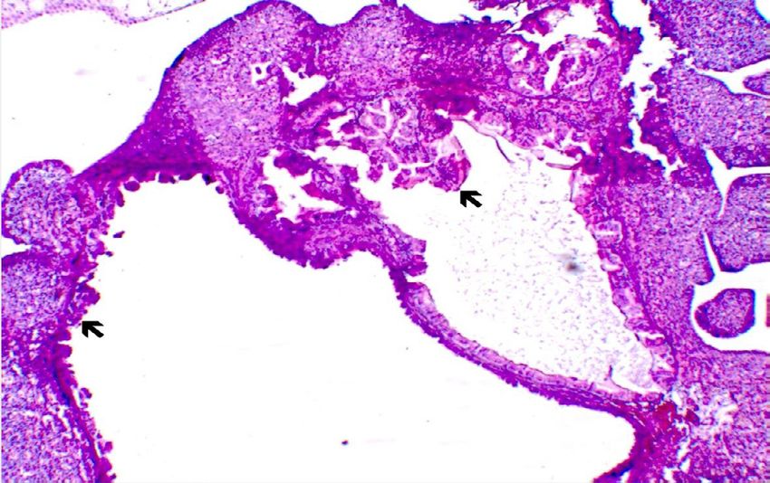
Case Report Veterinarni Medicina, 66, 2021 (01): 40–44
https://doi.org/10.17221/144/2020-VETMED
Case summary
Figure 1
CASE 1
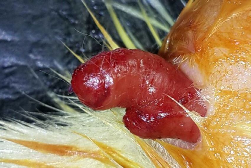
A 2-year old multiparous Djungarian hamster was
presented to a small animal clinic with a history
of mass prolapses from the vulva (Figure 1). The fe-
male was being kept together with a male. The owner
did not report inappetence or weakness. The ham-
ster gave birth 2 months previously and there was
no history of abortion.
The owners observed that the hamster had come
into heat a few days ago. The mass was noticed Figure 1. Prolapsed mass of Case 1
2 days prior to the visit to the clinic. During the
routine clinical examination, the activity, body
condition and general health status were judged Figure 2
as good. The tissue, which protruded from the vul-
va was diagnosed as a uterine horn. Initially, the
swollen mass was kept under pressure gently with
hyperosmotic solutions (30% dextrose; 20% manni-
tol) to decrease the oedema and reduce the volume
of the mass. This procedure was planned for the
replacement of the prolapsed mass. However, this
type of intervention was not successful and subse-
quently a decision to solve the problem surgically
was made.
General anaesthesia was induced and maintained
with isoflurane administered in 100% oxygen. The
hamster was placed in the dorsal position. The wall
of the abdomen was routinely prepared for the sur-
gery and a midline laparotomy was performed.
At first, the prolapsed uterine horn was replaced
to its normal anatomical position (Figure 2), then
an ovariohysterectomy was performed per the own-
er’s request (Figure 3). Figure 2. Replacing the prolapsed tissue from Case 1. Pro-
The abdomen was closed according to a routine lapsed horn (arrow)
procedure. Preventively, enrofloxacin was admin-
istered 10 mg/kg twice a day for 5 days.
Figure 3
CASE 2
A 2.5 year-old Djungarian hamster was pre-
sented to our small animal clinic with a history
of a mass with haemorrhages protruding from the
vulva. The owner reported that the mass was found
4 days previously. There were no signs of inappe-
tence or weakness. The owner also reported that
the hamster was nulliparous and had come into
heat before the mass prolapse. The mass was iden-
tified as a uterine horn. On the horn of the uterus, Figure 3. Both ovaries and horns of the uterus from Case 1
41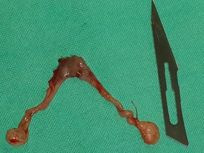
Case Report                                                         Veterinarni Medicina, 66, 2021 (01): 40–44

                                                                    https://doi.org/10.17221/144/2020-VETMED

surface erosions, haemorrhagic areas and cannibal-           made. An ovariohysterectomy was performed, simi-
ism symptoms were seen (Figure 4). Because of the            lar to the procedure as in Case 1. The abdominal
injury to the prolapsed tissue and haemorrhages,             wound was closed according to standard proce-
the decision to perform surgery was immediately              dures. Enrofloxacin 10 mg/kg b.i.d. was adminis-
                                                             tered for 5 days.
  Figure 4                                                      In the post-operative hospitalisation process of the
                                                             hamsters, signs of pain, such as an abnormal pos-
                                                             ture, decreased activity and appetite, abnormal ag-
                                                             gression, elevated respiratory rate (NRC 2009) were
                                                             not present and all the sutures were removed on the
                                                             fifth day after surgery.
                                                                The histopathological examination of the tissues
                                                             revealed that ovaries in both cases had cystic struc-
                                                             tures, papillary hyperplasia (Figures 5 and 6) and
                                                             subacute steatitis. There were no pathological le-
                                                             sions detected on the uterine tissue.

Figure 4. Necrotic and haemorrhagic areas on the uter-       DISCUSSION AND CONCLUSION
ine horn (black arrow) and tail (white arrow) from Case 2
                                                               This case report tackles the random occurrence
Figure 5                                                     of a uterine prolapse with cysts and papillary hy-
                                                             perplasia in the ovary. A uterine prolapse more
                                                             commonly occurs in women, cows, pigs, and cats
                                                             (Jelovsek et al. 2007; Miesner and Anderson 2008;
                                                             Deroy et al. 2015). It is rarely reported in exotic ani-
                                                             mals such as guinea pigs (Richardson 2000; Bennet
                                                             2012), rabbits (Di Girolamo et al. 2019) and mice
                                                             (Chawla et al. 2019). However, there are no data
                                                             in literature about the occurrence of a uterine pro-
                                                             lapse in hamsters.
                                                               The main cause of the uterine prolapse is the
                                                             relaxation of the pelvic ligaments and muscles,
                                                             and, as a result, the inability to support the uterus.
                                                             A uterine prolapse is occasionally observed in ani-
Figure 5. Papillary hyperplastic area from Case 1 (arrow).   mals with dystocia and parturition. The predispos-
H&E; ×100                                                    ing factors could include high levels of oestrogen
                                                             and relaxin, being overweight, having an altered
 Figure 6                                                    micro- and macro-mineral metabolism, constipa-
                                                             tion, ovarian cysts or a genetic predisposition (Dietz
                                                             2008; Reid 2011; Yotov et al. 2013; Deroy et al. 2015).
                                                             In the cases reported hereof, there was no recent
                                                             parturition in the two cases and, therefore, the uter-
                                                             ine prolapse could not be associated with parturi-
                                                             tion. Because of the similarities between human and
                                                             rodent anatomy, rodents are accepted as a model
                                                             in studies of uterine prolapse (Couri et al. 2012).
                                                             In human medicine, the main causes of uterine pro-
                                                             lapse have been described as the weakness of the pel-
Figure 6. Cystic and papillary hyperplastic areas from       vic floor diaphragm and the loss of integrity of the
Case 2 (arrows). H&E; ×40                                    uterosacral and cardinal ligament complex (Reid

42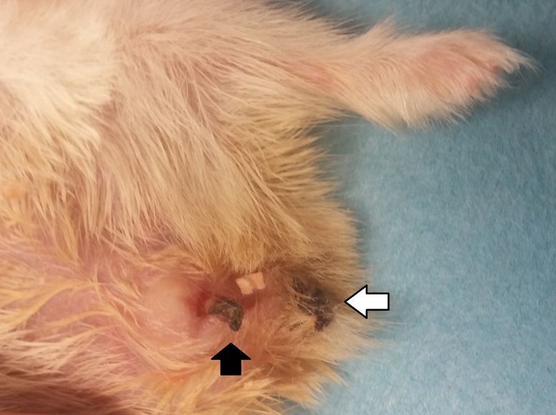
Case Report                                                      Veterinarni Medicina, 66, 2021 (01): 40–44

                                                                  https://doi.org/10.17221/144/2020-VETMED

2011). A uterine prolapse is presumed to be a con-        Barthold 2007; Girling 2013; Martorell 2017). The
nective tissue disorder and changes in the collagen       aetiology of ovarian cysts is unknown, but a cer-
tissue have a role in prolapse formation (Rinne and       tain role may be played by oestrogenic substances
Kirkinen 1999). Oestrogen also plays a role in the        in hay. Serous cysts (cystic rete ovarii) are one of the
collagen metabolism. Oestrogen stimulates the col-        types of cysts seen in exotic animals. These cysts
lagen degradation by increasing the proteinase ac-        are incapable of steroidogenesis. Cysts are usually
tivity such as matrix metalloproteinase 2 (MMP-2)         asymptomatic, however, as they grow in size, sym-
(Jackson et al. 2002). MMP-2 would lead to a loss         metric alopecia, abdominal distension and infertil-
in strength of the fibrous collagen and the reduc-        ity may be observed (Martorell 2017). Cystic ovaries
tion of its mechanical strength and increase the risk     are often associated with concurrent diseases in the
of a prolapse (Jackson et al. 1996; Ma et al. 2012).      reproductive tract such as cystic endometrial hy-
  Based on the owners’ observations, we know that         perplasia, mucometra or endometritis (Keller et al.
both hamsters came into heat days before the uter-        1987; Paterson 2006; Pilny 2014).
ine prolapse. Exposure to oestrogen might be ef-             A definitive diagnosis requires an ovariectomy/
fective in decreasing the tension of the structural       ovariohysterectomy along with a histopathologic
contents. Decreasing the tension triggers a uterine       examination of the affected tissues (Sayers and
prolapse like a domino effect.                            Smith 2010).
  In both cases, the oestrogen during the oestrous           In both cases, the owners stated that both ham-
cycle might have contributed to the development           sters did not experience infertility and showed oes-
of the uterine prolapse.                                  trus before the prolapse. The cysts were detected
  Additionally, Dietz (2008) reported a progres-          accidentally during the surgery. It is thought that
sive relaxation of the pelvic support of the uter-        these cysts may be inactive rete cysts and associ-
us and vagina with an advancing age in women.             ated with the uterine pathology as mentioned in the
Therefore, this relaxation may lead to a clinically       literature.
important uterine prolapse in susceptible women              The fact that a uterine prolapse has a multifacto-
as well (Dietz 2008). The average life of hamsters        rial aetiopathogenesis, further research, especially
is 3 years, and both hamsters in the study were           from the genetics point of view, need to be under-
of an advanced age.                                       taken to understand the exact cause.
  Because of the similarities between humans and             In conclusion, although it is a rare case in veteri-
rodents, it could be stated that cause of the uter-       nary literature, a uterine prolapse may appear with
ine prolapse may be related to the relaxation of the      cystic and papillary hyperplasia in hamsters regard-
pelvic ligaments and muscles depending on the ad-         less of parturition.
vanced age in both hamsters.
  A uterine prolapse is a life-threatening situation.
Treatment approaches vary depending on the breed          Conflict of interest
and tissue injury. The most appropriate treatment
procedure for a uterine prolapse in hamsters is a sur-      The authors declare no conflict of interest.
gical approach. Because of the anatomy, structure
and size of the genital tissues, there was no possibil-
ity to push the prolapsed tissue back and to place        REFERENCES
a suture in the vulva. Also, connective tissue disor-
ders and the uterine tissue function loss might be ir-    Abramowitch SD, Feola A, Jallah Z, Moalli PA. Tissue me-
reversible (Abramowitch et al. 2009). Accordingly,          chanics, animal models, and pelvic organ prolapse: A re-
an ovariohysterectomy was performed with the idea           view. Eur J Obstet Gynecol Reprod Biol. 2009 May;Suppl 1:
that the traumatic tissue will not be functional, and       S146-58.
to prevent the recurrence of a uterine prolapse.          Bennett RA. Soft tissue surgery. In: Quesenberry KE, Car-
  The histopathological examination of the ovar-            penter JW, editors. Ferrets, rabbits, and rodents: Clinical
ian tissue revealed cysts and papillary hyperplasia         medicine and surgery. 3rd ed. St. Louis, MO, USA: Else-
in both cases. Cystic ovaries have been described           vier; 2012. p. 326-38.
as being a common occurrence in all small rodents         Chawla S, Mahara K, Bathrachalam C. Successful treatment
older than 2 years, especially in hamsters (Percy and       of postparturient pelvic prolapse in mouse (Mus muscu-

43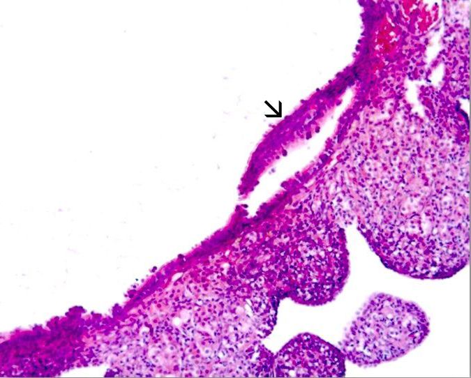
Case Report                                                              Veterinarni Medicina, 66, 2021 (01): 40–44

                                                                         https://doi.org/10.17221/144/2020-VETMED

  lus) using a novel hydropropulsion technique. J Exotic Pet     Martorell J. Reproductive disorders in pet rodents. Vet Clin
  Med. 2019 April;29:79-82.                                        Exot Anim. 2017 May;20(2):589-608.
Couri BM, Lenis AT, Borazjani A, Paraiso MF, Damaser             Miesner MD, Anderson DE. Management of uterine and
  MS. Animal models of female pelvic organ prolapse: Les-          vaginal prolapse in the bovine. Vet Clin North Am Food
  sons learned. Expert Rev Obstet Gynecol. 2012 May 1;7(3):        Anim Pract. 2008 Jul;24(2):409-19.
  249-60.                                                        NRC – National Research Council. National Research Coun-
Deroy C, Bismuth C, Carozzo C. Management of a com-plete           cil (US) committee on recognition and alleviation of
  uterine prolapse in a cat. JFMS Open Rep. 2015 Jun 1;            pain in laboratory animals. Recognition and alleviation
  1(1): [4].                                                       of pain in laboratory animals. Washington (DC): National
Dietz HP. The aetiology of prolapse. Int Urogynecol J Pelvic       Academies Press (US); 2009. 177 p.
  Floor Dysfunct. 2008 Oct;19(10):1323-9.                        Paterson S. Dermatology of mammals. In: Paterson S, edi-
Di Girolamo N, D’Ovidio D, Del Duca V, Donnelly TM,                tor. Skin diseases of exotic pets. 1st ed. Iowa, USA: Black-
  Montani A, Selleri P. Surgical resolution of uterine pro-        well Publishing Professional; 2006. p. 242-4.
  lapse in three pet rabbits. J Small Anim Pract. 2019 Dec 7.    Percy DH, Barthold SW. Rats. In: Percy DH, Barthold SW,
  Forthcoming.                                                     editors. Pathology of laboratory rodents and rabbits. 3rd ed.
Girling SJ. Common diseases of small mammals. In: Girl-            Iowa, USA: Wiley-Blackwell Publishing; 2007. p. 125-77.
  gling S, editor. Veterinary nursing of exotics pets. 2nd ed.   Richardson VCG. The reproductive system. In: Richardson
  New Jersey, USA: Willey Online Library; 2013. p. 59-90.          VCG, editor. Diseases of domestic guinea pigs. 2 nd ed.
Jackson SR, Eckford SD, Abrams P, Avery NC, Tarlton JF,            Malden, USA: Blackwell Publishing; 2000. p. 14-38.
  Bailey AJ. Changes in metabolism of collagen in genitou-       Pilny A. Ovarian cystic disease in guinea pigs. Vet Clin Exot
  rinary prolapse. The Lancet. 1996 June 15;347(9016):             Anim. 2014 Jan;17(1):69-75.
  1658-61.                                                       Rinne KM, Kirkinen PP. What predisposes young women
Jackson S, James M, Abrams P. The effect of oestradiol on          to genital prolapse?. Eur J Obstet Gynecol Reprod Biol.
  vaginal collagen metabolism in postmenopausal women              1999 May;84(1):23-5.
  with genuine stress incontinence. BJOG. 2002 Mar;109(3):       Reid F. Uterine prolapse-preservation or excision? Obstet
  339-44.                                                          Gynaecol Reprod Med. 2011 June;21(6):176-9.
Jelovsek JE, Maher C, Barber MD. Pelvic organ prolapse.          Rosell JM, de la Fuente LF. Causes of mortality in breeding
  The Lancet. 2007 Mar 24;369(9566):1027-38.                       rabbits. Prev Vet Med. 2016 May 1;127:56-63.
Keller LS, Griffith JW, Lang CM. Reproductive failure as-        Sayers I, Smith S. Mice, rats, hamsters and gerbils. In: Mer-
  sociated with cystic rete ovarii in guinea pigs. Vet Pathol.     edith A, Johnson-Delaney C, editors. BSAVA manual of
  1987 July 1;24(4):335-9.                                         exotic pets. 5th ed. Gloucester, England: BSAVA; 2010.
Ma Y, Guess M, Datar A, Hennesey A, Cardenas I, Johnson J,         p. 1-27.
  Connell KA. Knockdown of Hoxa11 in vivo in the utero-          Yotov ST, Atanasov A, Antonov A, Karadaev M. Post oestral
  sacral ligament and uterus of mice results in altered col-       vaginal prolapse in a non-pregnant heifer (a case report).
  lagen and matrix metalloproteinase activity. Biol Reprod.        Trakia J Sci. 2013;11(1):95-101.
  2012 Apr 5;86(4):100.
                                                                                                      Received: July 9, 2020
                                                                                                 Accepted: October 22, 2020

44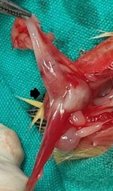
You can also read