Simplified method to perform CLARITY imaging
←
→
Page content transcription
If your browser does not render page correctly, please read the page content below
Poguzhelskaya et al. Molecular Neurodegeneration 2014, 9:19
http://www.molecularneurodegeneration.com/content/9/1/19

 METHODOLOGY                                                                                                                                    Open Access

Simplified method to perform CLARITY imaging
Ekaterina Poguzhelskaya1,2†, Dmitry Artamonov1,2†, Anastasia Bolshakova1,2, Olga Vlasova1,2
and Ilya Bezprozvanny1,2,3*

  Abstract
  Background: Imaging methods are used widely to understand structure of brain and other biological objects.
  However, sample penetration by light microscopy is limited due to light scattering by the tissue. A number of
  methods have been recently developed to solve this problem. In one approach (SeeDB) simple procedure for
  clarifying brain samples for imaging was described. However, this method is not compatible with immunostaining
  approach as SeeDB-prepared tissue is not permeable to the antibodies. Another technique for clearing brain tissue
  (CLARITY) was optimized for immunochemistry, but this method technically much more demanding than SeeDB.
  Results: Here we report optimized protocol for imaging of brain samples (CLARITY2). We have simplified and
  shortened the original protocol. Following hydrogel fixation, we cut brain tissue to 1–1.5 mm thick coronal slices.
  This additional step enabled us to accelerate and simplify clearing, staining and imaging steps when compared
  to the original protocol. We validated the modified protocol in imaging experiments with brains from line M Thy1-GFP
  mouse and in immunostaining experiments with antibodies against postsynaptic protein PSD-95 and striatal-specific
  protein DARPP32.
  Conclusions: The original CLARITY protocol was optimized and simplified. Application of the modified CLARITY2 protocol
  could be useful for a broad range of scientists working in neurobiology and developmental biology.
  Keywords: CLARITY, See deep brain, Neuronal structure, 3D brain tissue reconstruction, Neuroimaging, Confocal,
  Two-photon

Background                                                                            The SeeDB procedure takes only 1 week, requires
Understanding structural organization of the brain is                                 minimum reagents and efforts and does not transform
critical for modern neuroscience studies. A number of                                 the sample. Prepared SeeDB samples are transparent to
advanced imaging methods have been developed for                                      visible light but not permeable for macromolecules or
studies of brain structure. However, due to light scatter-                            antibodies. Thus, SeeDB procedure is not compatible
ing the penetration of light into brain tissue is limited.                            with immunostaining. Other methods have been re-
For confocal microscopy the depth of imaging is limited                               ported that use chemical solvents with high refractive-
to 30 micron, for two-photon microscopy it is no more                                 index to reduce light scattering [3-5]. Similar to SeeDB,
than 600–800 microns [1]. Imaging at such depths also                                 these methods are not compatible with immunostaining
require use of strong excitation power, causing local                                 applications.
overheating of the sample. To alleviate the problem of                                  The most recently developed method is CLARITY [6].
light scattering, recently several procedures have been                               This method is based on transforming the intact brain
developed for obtaining optically transparent brain sam-                              tissue to the nanoporous hydrogel formed by cross-
ple for imaging. The simple and fast protocol called See                              linked three-dimensional network of hydrophilic poly-
Deep Brain (SeeDB) method was initially published [2].                                mers. The lipid phase of the brain is solubilized in
                                                                                      detergent and removed by electrophoresis. As a result,
* Correspondence: ilya.bezprozvanny@utsouthwestern.edu                                optically transparent brain sample is obtained for im-
†
 Equal contributors
1
 Department of Medical Physics, St.Petersburg State Polytechnical University,         aging studies. The nanoporous hydrogel is permeable
St Petersburg 195251, Russia                                                          to the antibodies and other macromolecules, which makes
2
 Laboratory of Molecular Neurodegeneration, St.Petersburg State                       CLARITY compatible with immunostaining applications.
Polytechnical University, St Petersburg 195251, Russia
Full list of author information is available at the end of the article                However, several steps of published CLARITY protocol [6]

                                         © 2014 Poguzhelskaya et al.; licensee BioMed Central Ltd. This is an Open Access article distributed under the terms of the
                                         Creative Commons Attribution License (http://creativecommons.org/licenses/by/4.0), which permits unrestricted use,
                                         distribution, and reproduction in any medium, provided the original work is properly credited. The Creative Commons Public
                                         Domain Dedication waiver (http://creativecommons.org/publicdomain/zero/1.0/) applies to the data made available in this
                                         article, unless otherwise stated.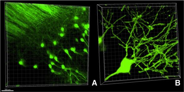
Poguzhelskaya et al. Molecular Neurodegeneration 2014, 9:19                                                                           Page 2 of 5
http://www.molecularneurodegeneration.com/content/9/1/19

are technically difficult. In particular, electrophoretic clear-          based on our own experience and experience of other la-
ing of the sample is a time consuming and challenging step                boratories (forum.claritytechniques.org) the clearing step
in the CLARITY procedure.                                                 remains challenging and takes substantial amount of time
                                                                          and effort. We reasoned that clearing whole brain is com-
New in method                                                             plicated and slow process, but for most applications it is
Here we describe a simplified version of the CLARITY                      not necessary to keep whole brain intact. Using vibratome
procedure (CLARITY2). The initial hydrogel fixation pro-                  (World Precisions Instruments) we cut the hydrogen-fixed
cedure is identical to the original protocol [6]. Following               mouse brains to 1–1.5 mm thick coronal slices (Figure 1A).
hydrogel fixation, we cut brain tissue in 1–1.5 mm thick                  These slices were put into 50 ml conical tubes filled with
coronal slices using vibratome. This additional step en-                  10 ml of clearing solution for 7 days (37°C, shaking at
abled us to accelerate and simplify clearing, staining and                21 rpm, fresh solution changes every 3 days). We discov-
imaging steps when compared to the original protocol.                     ered that this simple step resulted in efficient clearing of
Using CLARITY2 procedure, we have been able to obtain                     the slices (Figure 1A). In order to speed up clearing
high quality confocal and two-photon images of neuronal                   procedure standard protein electrophoresis chamber
structures from Thy1-GFP transgenic mice [7] and follow-                  (for example from BioRad) can also be utilized. If elec-
ing immunostaining of brain samples with antibodies                       trophoresis chamber is used, then the slices clearing
against synaptic protein PSD-95 and striatal-specific pro-                procedure takes 2–3 days when compared to 7 days
tein DARRP-32.                                                            with passive clearing. The quality of images obtained
                                                                          with passive and electrophoresis-based approach was
Results and discussion                                                    similar, and we present images from passively cleared
The adult wild type mice (FVB strain) was used to                         samples in this paper.
develop the protocol. The mouse brain was obtained                           In our pilot experiments the cleared slices were im-
and embedded into hydrogel by following previously                        munostained with mouse monoclonal antibodies against
described protocol [6] without any modifications (see                     post-synaptic density protein PSD-95 and rabbit monoclo-
Methods for details). The next step in the published                      nal antibodies against striatal specific protein DARPP32.
protocol is clearing from lipid/detergent micelles. The                   Original CLARITY procedure required 2 weeks incubation
original procedure [6] requires use of electrophoretic                    with primary and secondary antibodies to enable sufficient
clearing chamber (ETC.) for the purpose of clearing the                   penetration of antibodies into the sample [6]. Coronal slices
sample. This step is technically difficult and numerous vari-             in our experiments could be efficiently stained after 2 days
ations of ETC. chamber design have been described since                   incubation with primary antibodies and overnight incuba-
initial publication (forum.claritytechniques.org). However,               tion with secondary antibodies. The stained structures were

 Figure 1 Simplified CLARITY clearing procedure. (A) Hydrogel-embedded coronal slices before (bottom) and after (top) passive clearing
 procedure. (B) Coronal slices on microscope cover glass prepared for imaging. (C) Confocal images of striatal region co-stained with DARPP32
 (green) and PSD-95 (red) antibodies. (D) Confocal images of hippocampus region stained with PSD-95 antibodies (red). Scale bar is 150 microns
 on panels C-D.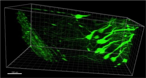
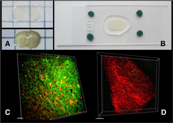
Poguzhelskaya et al. Molecular Neurodegeneration 2014, 9:19                                                                 Page 3 of 5
http://www.molecularneurodegeneration.com/content/9/1/19

visualized by Alexa-488-conjugated anti-rabbit and Alexa-            immunostaining experiments. Using 20x objectives we
594-conjugated anti-mouse secondary antibodies. For                  have been able to image up to depth of 250 microns
imaging experiments coronal slices were positioned on                using confocal microscopy (Figure 1, 2 and 3). When
microscope slides in PBST solution and covered by a                  the samples were imaged with two-photon microscope
cover slip (Figure 1B). Imaging in flat slices (Figure 1B)           (Thorlabs, 20x Olympus objective), the depth of pene-
did not require use of FocusClear solution needed for                tration was increased up to 1–1.5 mm, that is spanning
image reconstruction at depths more than 2 mm [6].                   whole thickness of the slice. The 3D reconstruction of
Using confocal microscope (Thorlabs) equipped with                   neuronal shapes based on two-photon imaging data is
20x Olympus objectives we have been able to easily                   shown for line M GFP-positive neurons on Figure 4
visualize the DARRP-32-positive and PSD-95-positive                  and Additional file 1.
(Figure 1B) structures in the brain samples. The ex-                    In conclusion, here we describe modified and simpli-
ample of striatal region co-stained for PSD-95 (red)                 fied version of CLARITY protocol (CLARITY2). Follow-
and DARPP-32 (green) is shown on Figure 1C. The                      ing imbedding into the hydrogel, the brain samples were
hippocampal region stained for PSD-95 (red) is shown                 cut into 1–1.5 mm thick coronal slices by vibratome. As
on Figure 1D. We attempted to compare the quality of                 a result, clearing, immunostaining and imaging proce-
images obtained with original [6] and modified procedure,            dures have been accelerated and simplified. Clearing
but run into technical difficulties with ETC-clearing step           could be performed by passive incubation of slices in the
when using original protocol (data not shown). As a result,          clearing solution for 1 week and did not require a special
we have not been able to obtain cleared brain samples using          ETC. chamber. Active clearing could be also done with
original procedure.                                                  brain slices using standard protein electrophoresis cham-
   To demonstrate versatility of the modified method we              ber to shorten the clearing procedure to 2–3 days in
also processed the sections obtained from Thy-GFP mice               duration. Antibody staining and imaging steps were also
(line M) brains [7]. In these experiments 1–1.5 mm thick             simplified and accelerated when compared to the ori-
coronal sections from line M mice were passively clarified           ginal protocol. We achieved 250 micron penetration into
as described above. The images of GFP-positive hippocam-             the thickness of the slice using confocal microscope and
pal neurons were collected using confocal microscope                 have been able to image whole thickness of the slice
equipped with 20x (Figure 2A) or 60x (Figure 2B) Olympus             (1.5 mm) using two-photon microscopy. Imaging in flat
objectives.                                                          slices could be performed on microscope slides and did
   In the next series of experiments we used anti-PSD95              not require use of expensive FocusClear solution needed
mouse monoclonal antibodies and Alexa-594-conjugated                 for image reconstruction at depths more than 2 mm [6].
anti-mouse secondary antibodies to stain coronal sections            We hope that described CLARITY2 procedure will be
from line M brains. Obtained confocal images were used               useful for mapping neuronal circuits in mice brain and
for 3D reconstruction of neuronal structures (Figure 3A              for imaging post-mortem human brain samples. When
and B). On these images individual GFP-positive neurons              compared to original CLARITY procedure [6] analysis
(green) are surrounded by PSD-95-positive synaptic struc-            of images obtained using CLARITY2 procedure may re-
tures (red).                                                         quire digital “stitching” of images from adjacent coronal
   The results shown on Figures 1, 2 and 3 validate                  sections, but this can be done relatively easily when ne-
the modified procedure for confocal imaging and                      cessary using image analysis software.

 Figure 2 Confocal images of hippocampal neurons from line M Thy1-GFP mouse. (A) Neuronal structures of hippocampus, 20x objective.
 Scale bar is 300 microns. (B) 3D reconstruction of individual GFP-positive neuron, 60x objective.
Poguzhelskaya et al. Molecular Neurodegeneration 2014, 9:19 Page 4 of 5
http://www.molecularneurodegeneration.com/content/9/1/19
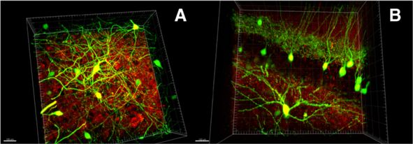
Figure 3 Confocal images of striatal and hippocampal neurons from line M Thy1-GFP mouse. (A, B). 3D reconstruction of GFP-positive
neurons (green) and PSD95 staining (red) is shown. 20x objective was used. Different brain regions are shown on panels (A) and (B). Scale bar is
100 microns on panels A and B.
Methods temperature (20°C). To avoid skin contact or inhalation,
Mice prepare solutions in a fume hood wearing gloves while
Wild type FVB/NJ (Stock Number: 001800) or line M preparing both of them.
mice Tg(Thy1-EGFP)MJrs/J (Stock Number: 007788) [7]
were used. Both lines were obtained from the Jackson Mice perfusion
Laboratory and the breeding colonies were established Thaw the 40 ml of hydrogel at room temperature.
in the animal facility located in the Laboratory of Gently mix the content, avoid air bubbles. Keep the tube
Molecular Neurodegeneration in St Petersburg State with hydrogel on ice. Provide animal anesthesia – in our
Polytechnical University. All mouse experiments have experiments mice were anesthetized with 300 ul of
been performed according to the procedures approved Urethane (250 mg/ml). Transcardially perfuse the mouse
by the local animal control authorities. with 20 ml of ice-cold solution of hydrogel with perfu-
sion rate of 10 ml per minute. Quickly extract the brain
Hydrogel and clearing solutions and place it in a conical tube containing remaining
Both solutions were prepared by following protocol de- 20 ml of hydrogel. Wrap tube in aluminum foil if the
scribed in the original article [6]. Briefly, to prepare sample contains fluorescent substance (such as GFP) to
hydrogel solution combine and mix 40 ml of acrylamide avoid photobleaching. Place the tube in a refrigerator
(40%), 10 ml of bis-acrylamide (2%), 1 g of VA-044 initi- (4°C) for 1–2 days to ensure full penetration of the sam-
ator (10% wt), 40 ml of 310 PBS, 100 ml of 16% PFA and ple by hydrogel.
210 ml of dH2O. Hydrogel solution is stored at −20°C, all
manipulations with hydrogel are performed on ice. For Hydrogel tissue embedding
clearing solution combine and mix 123.66 g boric acid, Tissue embedding was performed according to the
400 g sodium dodecyl sulphate, and 9 l dH2O. Add protocol described in the original article [6]. Briefly,
dH2O to 10 l and add NaOH until the pH has reached 50 mL conical tube containing hydrogel-soaked brain
8.5. This solution can be made, stored and used at room sample was positioned to the dessication chamber in a
Figure 4 Two-photon imaging of hippocampal neurons from line M Thy1-GFP mouse. Single frame from 3D neuronal image reconstruction
(Additional file 1). 20x objective was used. Scale bar is 150 micron.Poguzhelskaya et al. Molecular Neurodegeneration 2014, 9:19 Page 5 of 5
http://www.molecularneurodegeneration.com/content/9/1/19
fume hood. The dessication chamber is filled with the ni- Confocal and 2 –photon imaging systems (ThorLabs)
trogen gas from the tank. Switch the desiccation chamber equipped with 20× and 60× Olympus objectives (w.d.
valve from nitrogen gas flow to the vacuum pump. Keep 2.0 mm) were used in our imaging experiments. Obtained
the vacuum on for 10 minutes, then switch the vacuum off images were analyzed using Imaris v7.4.2. Software.
and slowly fill the dessication chamber with the nitrogen
gas. Open the dessication chamber and tightly close the top Additional file
of the sample tube. Minimize exposure of the sample to air
as oxygen impedes formation of hydrogel. Submerge the Additional file 1: 3D reconstruction of hippocampal neurons form line
M Thy1-GFP mice based on two-photon imaging data (20 x objective).
tube in 37°C incubator on the rotator. Incubate for 3 hours
or until solution has polymerized. In a fume hood, extract
Competing interests
the hydrogel-embedded brain sample from the gel. The authors declare that they have no competing interests.
Tissue clearing Authors’ contributions
EP and DA performed the experiments. EP, DA, AB analyzed data. EP, DA,
Coronal slices (1–1.5 mm in thickness) were cut from OV and IB designed research. EP, DA, AB, OV, IB wrote the paper and prepare
hydrogel-embedded brain using World Precisions In- figures for publication. All authors read and approved the final manuscript.
struments vibratome (NVSLM1 Motorized Advance
Authors’ information
Vibroslice). The slices are numbered, placed in 50 ml Ekaterina Poguzhelskaya at poguzhelskaya@mail.ru is a contact for technical
plastic tubes and incubated in 10 ml clearing solution questions on the procedures described in this paper.
for 7–10 days at a temperature of 37C with shaking
Acknowledgements
21 rpm. Fresh clearing solution was exchanged every We would like to thank the members of Laboratory of Molecular
3 days. The clearing of tissue was evaluated by eye. In Neurodegeneration in St Petersburg State Polytechnical University for their
most experiments 7 days was enough for complete clearing, help and support in the course of these studies. This work was supported by
the contract with the Russian Ministry of Science 11.G34.31.0056 (I.B) and NIH
but for some samples additional 3 days of clearing was grants R01NS074376 and R01NS080152 (I.B.).
needed. The standard Bio-Rad chamber for Western blot-
ting (Mini-Protean Tetra Cell system # 165–8000) could be Author details
1
Department of Medical Physics, St.Petersburg State Polytechnical University,
used to speed up a clearing to 2–3 days in needed. If St Petersburg 195251, Russia. 2Laboratory of Molecular Neurodegeneration,
electrophoretic clearing is used, the chamber is placed to a St.Petersburg State Polytechnical University, St Petersburg 195251, Russia.
3
thermostat at 37°C, and the voltage is set for 12 V. Department of Physiology, University of Texas Southwestern Medical Center
at Dallas, Dallas, TX, USA.
Following passive (7 days) or electrophoretic (2–3
days) clearing the slices are washed from clearing solu- Received: 27 March 2014 Accepted: 20 May 2014
tion in PBST (0.1% Triton X- 100 in PBS) for 2 days. Published: 26 May 2014
The slices are now clarified. References
1. Helmchen F, Denk W: Deep tissue two-photon microscopy. Nat Methods
Immunochemistry 2005, 2:932–940.
2. Ke MT, Fujimoto S, Imai T: SeeDB: a simple and morphology-preserving
The cleared brain slices are placed in PBST. The primary optical clearing agent for neuronal circuit reconstruction. Nat Neurosci
antibody is added at 1:1000 dilution for 2 days, then slices 2013, 16:1154–1161.
are washed with PBST 5 times for 30 minutes. The second- 3. Dodt HU, Leischner U, Schierloh A, Jährling N, Mauch CP, Deininger K,
Deussing JM, Eder M, Zieglgänsberger W, Becker K: Ultramicroscopy:
ary antibody is added at 1:1000 overnight in PBST, and the three-dimensional visualization of neuronal networks in the whole
slices washed once for 30 min in PBST. The antibodies mouse brain. Nat Methods 2007, 4:331–336.
used in our experiments were: mouse monoclonal 4. Staudt T, Lang MC, Medda R, Engelhardt J, Hell SW: 2,2’-thiodiethanol:
a new water soluble mounting medium for high resolution optical
anti-PSD-95 (Pierce, MA1-045), rabbit monoclonal microscopy. Microsc Res Tech 2007, 70:1–9.
anti-DARPP32 (Cell Signaling 19A3), Alexa Fluor488 Goat 5. Gonzalez-Bellido PT, Wardill TJ: Labeling and confocal imaging of neurons
anti-rabbit (Life Technologies, A-11008), Alexa Fluor594 in thick invertebrate tissue samples. Cold Spring Harb Protoc 2012,
2012:969–983.
Goat anti-mouse (Life Technologies, A-11005). 6. Chung K, Wallace J, Kim SY, Kalyanasundaram S, Andalman AS, Davidson TJ,
Mirzabekov JJ, Zalocusky KA, Mattis J, Denisin AK, Pak S, Bernstein H,
Imaging Ramakrishnan C, Grosenick L, Gradinaru V, Deisseroth K: Structural and
molecular interrogation of intact biological systems. Nature 2013,
The slices are removed from PBST and put on a glass 497:332–337.
slide. The slices are covered with the cover glass, which 7. Feng G, Mellor RH, Bernstein M, Keller-Peck C, Nguyen QT, Wallace M,
is cemented to the glass slide using plastiline (Figure 1B). Nerbonne JM, Lichtman JW, Sanes JR: Imaging neuronal subsets in transgenic
mice expressing multiple spectral variants of GFP. Neuron 2000, 28:41–51.
The coronal slices are put on the microscope slide in the
small drop of liquid (PBST) to avoid drying and to pre- doi:10.1186/1750-1326-9-19
vent formation of air bubbles. The plastiline is mounted Cite this article as: Poguzhelskaya et al.: Simplified method to perform
CLARITY imaging. Molecular Neurodegeneration 2014 9:19.
around the sample and the sample is covered with a
cover slip. Be very careful to avoid trapped air bubbles.You can also read