A pediatric pheochromocytoma presented as a multiorgan failure syndrome and brief discussion of the related medical literature - Hellenic ...
←
→
Page content transcription
If your browser does not render page correctly, please read the page content below
Original Case Report

A pediatric pheochromocytoma presented as a multi-
organ failure syndrome and brief discussion of the
related medical literature

                                      Abstract
                                      A 9 years old boy presented to our hospital complaining of night sweat and weight loss (5kg) for 3 months
                                      followed by cough, nausea, vomiting and malaise for two weeks. During that time he was found to have
Wen Li1 MD,                           continuous hypertension. The clinical, electrocardiogram (ECG), renal scintigraphy and biologic ndings
Chao Ma2 MD, PhD,                     suggested multi organ-failure syndrome. Computed tomography (CT) and ultrasound revealed a right
Lingyu Zhang3 MD,                     adrenal tumor. Vanillymandelic acid (VMA) in the 24 hours urine sample was not elevated. Pheoc-
Jinming Yu4 PhD                       hromocytoma was suspected given his hypertension, ultrasound and CT ndings. Pre-operative stabi-
                                      lization of his blood pressure was achieved over the following 4 weeks, after treatment with alpha- and
                                      beta-blockers, sodium nitroprusside and diuretics. Subsequently, right adrenalectomy was successfully
                                      performed. Histological examination showed that the tumor was a pheochromocytoma. Technetium-
1. School of Medicine. Jinan, China   99m-ethylene dicysteine (99mTc-EC) renal scintigraphy con rmed severe kidney function impairment. In
Department of Nuclear Medicine,       conclusion: Our pediatric case of pheochromocytoma was rst presented, with cardiac, renal and hepatic
Qianfo Shan Hospital A liated to      failure which prompted us to the diagnosis of pheochromocytoma .
Shandong University, Jinan,
250014, China                         Hell J Nucl Med 2016; 19(2): 159-163     Epub ahead of print: 22 June2016      Published online: 2 August 2016
2. Nuclear Medicine, A liated
Xinhua Hospital, School of
Medicine Shanghai Jiaotong
University, Shanghai, China
3. Department of Nuclear Medicine,    Introduction
Qianfo Shan Hospital A liated to
Shandong University, China
4. Department of Radiation
Oncology, Shandong
Cancer Hospital a liated to
Shandong University, Jinan, China
                                      P       heochromocytomas (PCC) derive from chroma n cells of the sympathetic
                                              nervous system and produce excessive amounts of catecholamines, which are
                                              responsible for hypertensive surges, palpitations, headache, and diaphoresis
                                      commonly found with these tumors [1]. Pheochromocytoma in the pediatric popula-
                                      tion is rare with an incidence of 2 per million [2]. Etiopathogenesis, management, and
Keywords: - Pediatric pheochromo-
                                      outcome of pediatric PCC is still obscure because of limited number of cases studied [3-
cytoma                                5]. Appropriate evaluation and management are essential for a favorable outcome. Des-
-Multi-organ failure                  pite its low incidence, PCC is the most frequent endocrine tumor in childhood [2, 6], usu-
-Clinical presentations               ally appearing at the age of 8 or 9 years. Almost 10% of the cases will have a family history
-Laboratory ndings                    because of the association of PCC to multiple endocrine neoplasm syndromes [7]. Phe-
                                      ochromocytoma is a potentially lethal cause of childhood hypertension. Pheochromo-
- Renal scintigraphy
                                      cytoma with multi-organ failure syndrome has been reported in adults [1, 8, 9] but not in
                                      children. We report a pediatric PCC with multi-organ failure as an initial presentation
                                      which was successfully treated by surgery.

Corresponding author:
Jinming Yu PhD
Department of Radiation               Case Report
Oncology,
Shandong Cancer Hospital and
Institute, Jinan, Shandong, China
Tel: +86-13806406293                  A 9 years old boy presented to our hospital complaining of night sweating and weight
E-mail: sdyujinming@126.com
                                      loss (5kg) during the last 3 months followed by cough, nausea, vomiting and malaise for
                                      the last two weeks. He had no family history. He reported bronchitis treated for 4 days
                                      with a nonsteroidal anti-in ammatory agent but had no improvement for his cough and
Rece ved:                             night sweat. On examination, he had no fever and no abdominal pain. His vital signs in-
 26 April 2016                        cluded a heart rate of 115 beats per minute, blood pressure of 130/90mmHg, respiratory
Accepted revised:
                                      rate of 20 breaths per minute, and blood oxygen saturation of 98%. His examination was
 20 May 2016
                                      unremarkable for cardiac abnormalities. His lungs clinical examination was normal and
                                      his lower extremities were nonedematous.

159
 93      Hellenic Journal of Nuclear Medicine • May-August 2016                                                              www.nuclmed.gr
Original Case Report
Initial laboratory values revealed elevated: creatinine, 1%-3% [1]. The causative factors for hypertension include re-
urea nitrogen, troponin and B-type natriuretic peptide (pro-
BNP) levels (Table 1). Immunological markers including anti-
glomerular basement membrane antibody, anti-double-
stranded DNA antibody and antinuclear antibody were ne-
gative. Electrocardiogram (ECG) showed a sinusal tachy-
cardia, T waves inverted in leads II, III, V5 and aVF, QT
prolongation. Echocardiography, M type, revealed enlar-
gement of the left atrium and left ventricle (LV) with hypo-
kinesia and low ejection fraction (EF) of 40%. Blood pressure
and LVEF are listed in Table 2. Chest radiogram showed pat-
chy in ltrations in left lung. The clinical, ECG and biologic n-
dings suggested multiorgan-failure syndrome: cardiac fai-
lure (increased troponin and proBNP, decreased LVEF), renal
Figure 1. Abdominal ultrasound revealed a 45mm×34mm solid lesion at the right
failure (creatinine, 166.5 mol/L and markedly decreased
adrenal gland.
e ective renal plasma ow (ERPF) by renal dynamic scinti-
graphy), hepatic cytolysis (alanine aminotransferase,
134IU/L) and intravascular disseminated coagulation ( brin
degradation products 7.59mg/L, thrombin time 30%). The
24h urine sample of VMA was not elevated. Despite multiple
organ failure, the hemodynamic state was stable. An abdo-
minal ultrasound (US) revealed a 45mm×34mm solid lesion
at the right adrenal gland and di use hyperecho in both
kidneys (Figure 1). Plain and enhanced computerized tomo-
graphy (CT) showed a 42×33cm round mass at the right
adrenal gland with inhomogenous enhancement (Figure
2a-2b). These ndings were compatible with a PCC. Plain
and enhanced CT also showed decreased density on both
kidneys with inhomogenous enhancement especially on
the left kidney (Figure 2c-2d). Pheochromocytoma was
suspected given his hypertension, the US and CT imaging
99m
ndings. Renal dynamic imaging using Tc-EC showed
reduced and inhomogenous radioactivity in the right kid-
ney and atrophy of the left kidney (Figure 3a) with markedly Figure 2. Plain and enhanced computerized tomography con rmed a 42×33cm
decreased ERPF (Figure 3b). round mass at the right adrenal gland with inhomogenous enhancement (Figure
The patients was preoperativly treated with teicoplanin 2a-2b); Decreased density at both kidneys with inhomogenous enhancement
for lung infection, with supportive liver protection therapy, especially in the left kidney were also shown by plain and enhanced CT (Figure 2c-
and for heart failure with milrinone, alpha-blockers (phento- 2d).
lamine, doxazosin), a beta-blocker (propranolol), sodium
nitroprusside, and diuresis medication including pirono-
lactone and hydrochlorothiazide. Before operation, blood
pressure was stabilized during 4 weeks.
Subsequently, right adrenalectomy was successfully per-
formed. Histological examination showed that the tumor
was a PCC (Figure 4). Pathology features did not suggest of
malignancy. There were no complications related to surgery.
After right adrenalectomy, the blood pressure was restored
to normal (Table 2), and patient's symptoms of severe head-
aches was reduced. Furthermore, his renal function im-
proved (Table 1).
Discussion
Hypertension is increasingly recognized as a public health Figure 3. 99mTc-EC renal dynamic imaging (120 seconds per frame) showed reduced
issue for children and adolescents. The prevalence of and inhomogeneous radioactivity in the right kidney and atrophy in the left kidney
hypertension in children is estimated to be approximately (Figure 3a) which had a markedly decreased ERPF (Figure 3b).
www.nuclmed.gr Hellenic Journal of Nuclear Medicine • May-August 2016 160
93Original Case Report
Table 1. Laboratory values throughout the patient's hospital course
Month/day 2/24 2/27 2/29 3/1 3/3 3/5 3/7 3/11 3/17
Troponin
7.52 6.36 2.684 1.720 0.802 0.211 0.130 0.028
(Original Case Report
rasound and CT could not suggest renal insu ciency but
showed abnormal echo and density in both kidneys in our
case. Renal scintigraphy can identify renal function im-
pairment. In adults, most cases of cardiomyopathy with PCC
result in partial or complete remission after treatment. The
majority of cases of dilated cardiomyopathy improve within
6 weeks to 16 months to improve [23]. Our patient had
slightly enlarged left atrium and ventricles, hypokinesia and
40% LVEF on presentation, with improvement to 49% when
discharged. Our patient's renal failure also improved. Our
pediatric case of PCC with cardiac, renal and hepatic failure
is the rst one reported according to our knowledge and
emphasizes the importance of early diagnosis [9].
Pheochromocytoma tumors may overproduce norepine-
phrine and/or epinephrine [24]. The diagnosis of PCC is
based on serum and urinary levels of catecholamines or
their metabolites [2]. The combination of urinary metane-
phrines and vanillylmandelic acid (VMA) was reported to
have a diagnostic sensitivity of 98% in detecting PCC [14, 25,
26]. Large tumors had the highest levels of urinary VMA [27].
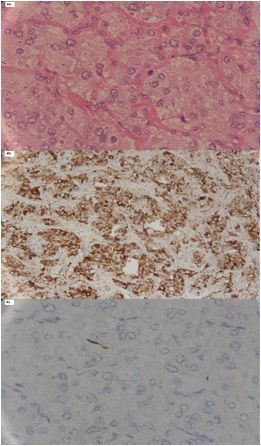
Figure 4. HE staining (X400) showed that the pheochromocytoma cells were Plasma free and urinary metanephrines and also total meta-
arranged in an alveolar manner forming nests (Figure 4a). Immunohistochemistry nephrines were performed equivalently for diagnosing PCC
revealed positive CgA (Figure 4b) and S100 in tumor cells (Figure 4c). in 46 adult cases without renal insu ciency [24]. Urinary
VMA was reported to be elevated in 53%, and epinephrine
and norepinephrine in 42% in a total of 19 pediatric patients
nal artery stenosis and coarctation of the aorta, hyperthy- with PCC [28]. Patients with chronic renal failure can not
roidism, adrenogenital syndrome, Conn's and Cushing's often take the 24 hours urine collection test which makes it
syndromes, brain tumors, and frequently essential hyper- di cult to diagnose PCC in patients with end stage renal
tension [7]. Pheochromocytoma is a potentially lethal cause disease [29]. In our case the VMA in the 24 hours collected
of childhood hypertension and accounts for about 1% of urine was not elevated which may be due to renal
pediatric hypertensive patients [3]. Pheochromocytoma is insu ciency.
common in preadolescent boys and teenage girls [2,10-14]. In addition, ultrasound (US), computed tomography (CT),
Most of the tumors are right sided (n=6/16) as also in our magnetic resonance imaging (MRI), and iodine-123-
123
case. Sporadic cases of PCC in children account in 14/16 of metaiodobenzylguanidine ( I-MIBG) scintigraphy are
patients [14] as in our case. The typical triad of symptoms in useful for the localization of PCC [14]. Magnetic resonance
PCC consists of episodic headache, sweating, and palpi- imaging is currently the best tool for tumor localization [13].
tations, but the clinical presentation in children may be The accuracy of these techniques is enhanced considerably
di erent [2]. Pheochromocytoma may be completely asym- when a morphologic study (CT or MRI) is combined with a
123
ptomatic as showed in a case by Eren (2015) [2] . Our case functional study such as I-MIBG scintigraphy [30]. Planar
123/131
had no abdominal palpitations. In children PCC is di erent I-MIBG or single photon emission tomography (SPET)/
from adults. More often is multifocal and extra-adrenal CT scans were used in both children and adults speci cally
[13,15]. Hypertension in children may be continuous rather for the detection of atypically located PCC, for distant
than paroxysmal [2] , as was in our case. metastases, and for recurrences [25, 26, 30, 31]. Other
Multi-organ failure syndrome due to PCC with renal failure researchers showed that somatostatin receptors imaging
using gallium-68 (68Ga)-DOTA(0)-tyr(3)-octreotate ( Ga-
68
[16], acute myocardial infarction, cardiac arrhythmias, and
heart failure [1, 9], pulmonary hemorrhage, and cyclic hypo- DOTATATE) was better for the localization of sporadic
tension [1, 17-19], shock, rhabdomyolysis, hepatic cytolysis, metastatic PCC as compared to uorine-18- uoro-2-deoxy-
18
acute pancreatitis, and intravascular disseminated coagu- D-glucose ( F-FDG) PET/CT, and CT/MRI [32].
lation [8] has been reported in adults. In adults, PCC related When a PCC is suspected in a child, careful and expedi-
shock may be due to extensive intravascular volume deple- tious management is essential to prevent complications or
tion, hypocalcemia, and desensitization of adrenergic re- even death. Pre- intra- and postoperative medical manage-
ceptors [20]. The mechanism of renal injury is presumed to ment is as important as the surgical procedure [14, 15].
be intense vasoconstriction induced by catecholamines, Malignancy in PCC is very di cult to diagnose histologically,
leading to muscle ischemia [20, 21]. In children, PCC with so the biologic behavior of the tumor may determine its
rhabdomyolysis is an unusual manifestation [22]. Elevated classi cation. Pediatric malignant PCC actually is very rare
serum BUN, creatinine and renal dynamic imaging using ranging from 8.3% to 13.1% which is lower than that in
99m
Tc-EC in our case, con rmed severe kidney function adults (ranging from 32% and 42%) [14]. Tumor size may be
impairment. However, conventional imaging including ult- an indicator of malignant potential. It has been reported that
www.nuclmed.gr Hellenic Journal of Nuclear Medicine • May-August 2016 162
93Original Case Report
benign tumors had a mean diameter of 45±4mm as in our luation, diagnosis, and treatment. World J Urol 1999; 17: 35-9.
case, whereas malignant tumors, 86±13mm [6]. 12. Waguespack SG, Rich T, Grubbs E et al. A current review of the
etiology, diagnosis, and treatment of pediatric pheochromo-
Recurrence of PCC is very important from the clinical
cytoma and paraganglioma. J Clin Endocrinol Metab 2010; 95:
point of view. Children who experience recurrence will usu- 2023-37.
ally develop recurrent symptoms within a year of diagnosis. 13. Ross JH. Pheochromocytoma. Special considerations in children.
The presence of extra-adrenal cells in tissues where chro- Urol Clin North Am 2000; 27: 393-402.
ma n cells are not normally found indicates metastatic 14. Ciftci AO, Tanyel FC, Senocak ME, Buyukpamukcu N. Pheochro-
disease. The incidence of PCC recurrence varies from 10% to mocytoma in children. J Pediatr Surg 2001; 36: 447-52.
40% [26, 33]. There were no speci c clinical or laboratory 15. Reddy VS, O'Neill JA Jr, Holcomb GW 3rd et al. Twenty- ve-year
surgical experience with pheochromocytoma in children. Am
data in our patient to predict recurrence [14]. Interestingly, a Surg 2000; 66: 1085-91; discussion 1092.
rare case of adrenal insu ciency following unilateral adre- 16. Fujiwara M, Imachi H, Murao K et al. Improvement in renal dys-
nalectomy of a PCC was reported in a 10 years old child function and symptoms after laparoscopic adrenalectomy in a
which may be explained that PCC had dual-hormone (also patient with pheochromocytoma complicated by renal dysfun-
adrenocorticotropic hormone ACTH) secreting capacity, ction. Endocrine 2009; 35: 57-62.
and that the child had unrecognized subclinical Cushing's 17. Dinckal MH, Davutoglu V, Soydinc S et al. Phaeochromocytoma-
induced myocarditis mimicking acute myocardial infarction. Int J
syndrome pre-operatively [34]. Our case had normal ACTH
Clin Pract 2003; 57: 842-3.
and no indications of Cushing's syndrome. 18. Attar MN, Moulik PK, Salem GD et al. Phaeochromocytoma
In conclusion, our patient exhibited a rare kind of PCC presenting as dilated cardiomyopathy. Int J Clin Pract 2003; 57:
complicated by renal, cardiac and hepatic failure, which was 547-8.
successfully treated by laparoscopic surgery. Technetium- 19. Kimura Y, Ozawa H, Igarashi M et al. A pheochromocytoma cau-
99m
Tc-EC renal dynamic imaging not only revealed kidney sing limited coagulopathy with hemoptysis. Tokai J Exp Clin Med
atrophy, but also severe kidney impairment better than US 2005; 30: 35-9.
20. Hamrin B. Sustained hypotension and shock due to an adrenaline-
or contrast enhanced CT. This case, according to our secreting phaeochromocytoma. Lancet 1962; 2: 123-4.
knowledge, is rst reported and emphasizes the importance 21. Imperadore F, Azzolini M, Piscioli F et al. A rare cause of cardiogenic
to diagnose PCC in case of an unexplained multi-organ shock: catecholamine cardiomyopathy of pheochromocytoma.
failure syndrome so as to reduce mortality. Ital Heart J 2002; 3: 375-8.
22. Celik H, Celik O, Guldiken S et al. Pheochromocytoma presenting
Acknowledgment with rhabdomyolysis and acute renal failure: a case report. Ren Fail
2014; 36: 104-7.
This work was supported by the Development of Science
23. Lin PC, Hsu JT, Chung CM, Chang ST. Pheochromocytoma un-
and Technology Project of Shandong Province (2013G- derlying hypertension, stroke, and dilated cardiomyopathy. Tex
GB14067) Heart Inst J 2007; 34: 244-6.
24. Grouzmann E, Drouard-Troalen L, Baudin E et al. Diagnostic
accuracy of free and total metanephrines in plasma and fractio-
Bibliography nated metanephrines in urine of patients with pheochromo-
1. Park M, Hryniewicz K, Setaro JF. Pheochromocytoma presenting cytoma. Eur J Endocrinol 2010; 162: 951-60.
with myocardial infarction, cardiomyopathy, renal failure, 25. Lucon AM, Pereira MA, Mendonca BB et al. Pheochromocytoma:
pulmonary hemorrhage, and cyclic hypotension: case report and study of 50 cases. J Urol 1997; 157: 1208-12.
review of unusual presentations of pheochromocytoma. J Clin 26. Bravo EL. Evolving concepts in the pathophysiology, diagnosis, and
Hypertens (Greenwich) 2009; 11: 74-80. treatment of pheochromocytoma. Endocr Rev 1994; 15: 356-68.
2. Eren E, Saglam H, Caliskan Y et al. Pediatric patients with pheochro- 27. Sawin RS. Functioning adrenal neoplasms. Semin Pediatr Surg 1997;
mocytoma: Experience of a tertiary health center. Pediatr Int 2015; 6: 156-63.
57: 875-9. 28. Bhansali A, Rajput R, Behra A et al. Childhood sporadic pheo-
3. Pickard JL, Ross G Jr., Silver D. Coexisting extraadrenal pheochro- chromocytoma: clinical pro le and outcome in 19 patients. J
mocytoma and renal artery stenosis: a case report and review of Pediatr Endocrinol Metab 2006; 19: 749-56.
the pathophysiology. J Pediatr Surg 1995; 30: 1613-5. 29. Maeda T, Kozakai N, Nishiyama T et al. [Pheochromocytoma with
4. Ein SH, Shandling B, Wesson D et al. Recurrent pheochromo- chronic renal failure: a case report]. Hinyokika Kiyo 2009; 55: 409-12.
cytomas in children. J Pediatr Surg 1990; 25: 1063-5. 30. Pattou FN, Combemale FP, Poirette JF et al. Questionability of the
5. Ein SH, Weitzman S, Thorner P et al. Pediatric malignant pheo- bene ts of routine laparotomy as the surgical approach for pheo-
chromocytoma. J Pediatr Surg 1994; 29: 1197-201. chromocytomas and abdominal paragangliomas. Surgery 1996;
6. Pham TH, Moir C, Thompson GB et al. Pheochromocytoma and 120: 1006-11; discussion 1012.
paraganglioma in children: a review of medical and surgical 31. Sharma P, Dhull VS, Jeph S et al. Can hybrid SPECT-CT overcome the
management at a tertiary care center. Pediatrics 2006; 118: 1109-17. limitations associated with poor imaging properties of 131I-MIBG?:
7. Newman KD, Ponsky T. The diagnosis and management of endo- Comparison with planar scintigraphy and SPECT in pheochro-
crine tumors causing hypertension in children. Ann N Y Acad Sci mocytoma. Clin Nucl Med 2013; 38: e346-53.
2002; 970: 155-8. 32. Janssen I, Chen CC, Taieb D et al. 68Ga-DOTATATE PET/CT in the
8. Herbland A, Bui N, Rullier A et al. Multiple organ failure as initial pre- Localization of Head and Neck Paragangliomas Compared with
sentation of pheochromytoma. Am J Emerg Med 2005; 23: 565-6. Other Functional Imaging Modalities and CT/MRI. J Nucl Med 2016;
9. Tamdy A, Oukerraj L, Khatri D et al. Acute myocardial infarction re- 57: 186-91.
vealing a pheochromocytoma: a case report. Ann Cardiol Angeiol 33. Marshall DG, Ein SH. Two boys with four pheochromocytomas
(Paris) 2010; 59: 97-9. each. J Pediatr Surg 1986; 21: 815-7.
10. Kaufman BH, Telander RL, van Heerden JA et al. Pheochromocytoma 34. Sjoeholm A, Li C, Leem C et al. Adrenal insu ciency in a child
in the pediatric age group: current status. J Pediatr Surg 1983; 18: following unilateral excision of a dual-hormone secreting phae-
879-84. ochromocytoma. Endocrinol Diabetes Metab Case Rep 2015;
11. Walther MM, Keiser HR, Linehan WM. Pheochromocytoma: eva- 2015: 150041.
163
93 Hellenic Journal of Nuclear Medicine • May-August 2016 www.nuclmed.grYou can also read