Cancer Association of South Africa (CANSA)
←
→
Page content transcription
If your browser does not render page correctly, please read the page content below

Cancer Association of South Africa (CANSA)

                                                                         Fact Sheet
                                                                             on
                                                                     Cancer of the Spleen

Introduction
The spleen is an organ found in virtually all
vertebrates. Similar in structure to a large lymph
node, it acts primarily as a blood filter.

                                        [Picture Credit: Spleen]

The spleen plays important roles in regard to red
blood cells (also referred to as erythrocytes) and the
immune system. It removes old red blood cells and
holds a reserve of blood, which can be valuable in case
of haemorrhagic shock, and also recycles iron. As a part of the mononuclear phagocyte system, it
metabolises haemoglobin removed from senescent erythrocytes. The globin portion of haemoglobin
is degraded to its constitutive amino acids, and the haeme portion is metabolised to bilirubin, which
is removed in the liver.

The spleen also synthesises antibodies in its white pulp and removes antibody-coated bacteria and
antibody-coated blood cells by way of blood and lymph node circulation. A study published in 2009
using mice found that the spleen contains, in its reserve, half of the body's monocytes within the red
pulp. These monocytes, upon moving to injured tissue (such as the heart), turn into dendritic cells and
macrophages while promoting tissue healing. The spleen is a centre of activity of the mononuclear
phagocyte system and can be considered analogous to a large lymph node, as its absence causes a
predisposition to certain infections.

In humans, the spleen is brownish in colour and is located in the left upper quadrant of the abdomen.

Cancer of the Spleen
Cancer of the spleen is a malignancy of white blood cells involving tumour deposits in the spleen.
There are a number of different types of spleen cancers including lymphoma, non-Hodgkin's
lymphoma and some types of T-cell lymphomas.

Most splenic cancer do not start in the spleen, and those that do, are almost always lymphomas.
Lymphoma is a type of blood cancer that develops in the lymphatic system. It is more common for a
Researched and Authored by Prof Michael C Herbst
[D Litt et Phil (Health Studies); D N Ed; M Art et Scien; B A Cur; Dip Occupational Health; Dip Genetic Counselling; Dip
Audiometry and Noise Measurement; Diagnostic Radiographer; Medical Ethicist]
Approved by Ms Elize Joubert, Chief Executive Officer [BA Social Work (cum laude); MA Social Work]
February 2021                                                                                                    Page 1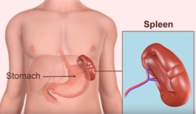
lymphoma to start in another part of the lymphatic system and invade the spleen than it is for
lymphoma to start in the spleen itself.

Lymphoma is probably the most common splenic malignancy and is usually a manifestation of
generalised lymphoma. Primary splenic lymphoma is rare. Most of the primary splenic lymphomas are
non-Hodgkin lymphomas (marginal zone cell lymphoma). The most common finding is splenomegaly,
but it may be absent in up to 30% of lymphoma patients.

Ultrasound may depict a solitary lesion or slightly ill-defined inhomogeneous hypoechoic lesions.
Another pattern is a general diffuse inhomogeneity with minute hypoechoic lesions less than 1 cm in
size.

Staging of lymphomas on CT can be limited as only 45%–70% of lymphomas show diffuse splenic
infiltration or tumour foci less than 1 cm in diameter so that the diagnosis of lymphoma can sometimes
only be made microscopically. The focal lesions with diameter from 1 to 10 cm are typically of low
attenuation and rarely enhance so may be better demonstrated on post-contrast scans.

MRI findings are non-specific and similar to those of metastases from other primary tumours.
Typically, lymphomas are hypointense or nearly isointense on T1-weighted images and hyperintense
on T2-weighted images. Injection of contrast medium may improve detection of splenic lymphoma.

Although the spleen is the most vascular organ in the body, it is an infrequent site for metastatic
disease (3.4% of metastatic carcinoma). Explanations proposed for the relative paucity of splenic
metastases have included:

•   the sharp angle made by the splenic artery which makes it difficult for tumour emboli to enter the
    spleen
•   the rhythmic contractile nature of the spleen which squeezes out the tumour emboli
•   the absence of afferent lymphatics to carry metastatic tumour to the spleen; and
•   anti-tumour activity due to a high concentration of lymphoid tissue in the spleen.

Apart from these factors, the frequency of splenic metastases may have been underestimated as they
are often asymptomatic and occur late in the disease. Splenic metastases are most commonly found
in malignant melanoma, lung, breast or ovarian carcinomas.

On ultrasound, they can show various degrees of echogenicity, but are usually hypoechoic.

On CT, splenic metastases typically appear as hypodense lesions which may be solid or cystic and with
inhomogeneous contrast enhancement indicating a mixture of vascularisation or necrosis.

On MRI, metastases are predominantly hypointense on T1-weighted images and hyperintense on T2-
weighted images, with occasionally inhomogeneous contrast enhancement. MRI is more accurate for
the detection of splenic metastases which are necrotic or haemorrhagic.

Shimono, J., Miyoshi, H., Arakawa, F., Yamada, K., Sugio, T., Miyawaki, K., Eto, T., Miyagishima, T.,
Kato, K., Nagafuji, K., Akashi, K., Teshima, T. & Ohshima, K. 2019.
“The hepatitis C virus (HCV) is a single-stranded RNA virus which is thought to be involved in the onset
of B cell lymphoma. HCV-positive diffuse large B cell lymphoma (DLBCL) has been reported to clinically
manifest in extranodal lesions (e.g., in the liver, spleen, and stomach). Here, we investigated HCV-
Researched and Authored by Prof Michael C Herbst
[D Litt et Phil (Health Studies); D N Ed; M Art et Scien; B A Cur; Dip Occupational Health; Dip Genetic Counselling; Dip
Audiometry and Noise Measurement; Diagnostic Radiographer; Medical Ethicist]
Approved by Ms Elize Joubert, Chief Executive Officer [BA Social Work (cum laude); MA Social Work]
February 2021                                                                                                    Page 2
positive and -negative primary splenic DLBCL (p-spDLBCL) and non-primary splenic DLBCL (ordinary DLBCL). Furthermore, to examine HCV lymphomagenesis, RNA in situ hybridization (ISH), RT-PCR (reverse-transcription polymerase chain reaction), and NS3 immunostaining of HCV viral nonstructural proteins were performed. HCV-positive p-spDLBCL patients presented fewer B symptoms (asymptomatic) and better performance status, with elevated presence of splenic macronodular lesions and more germinal center B cell (GCB) sub-group cases than HCV-negative p-spDLBCL patients. However, HCV-positive ordinary DLBCL patients were found to have more non-GCB sub-group cases than HCV-negative ordinary DLBCL patients. HCV-positive DLBCL patients showed 20.6% (7/34) NS3 positivity, 16.7% (1/6) HCV-RNA in situ positivity, and 22.2% (2/9) detection of HCV-RNA in tumor tissue by RT-PCR. Splenic samples were found to have a higher frequency of HCV detection than lymph node samples, thus suggesting that HCV may be closely related to lymphomagenesis, especially in splenic lymphoma.” Incidence of Cancer of the Spleen in South Africa The National Cancer Registry (2017) does not provide any information regarding the incidence of cancer of the spleen. Padilla, O., Tam, W. & Geyer, J.T. 2020. “Hematopoietic neoplasms involving the spleen are uncommon, but T cell neoplasms involving the spleen are extremely rare. The rarity of splenic involvement by T cell neoplasms has resulted in a limited body of literature describing their splenic characteristics. As a result, our purpose in this review article is to provide and summarize some of the characteristics seen by different T cell neoplasms that may involve the spleen.” Signs and Symptoms of Cancer of the Spleen The following are common signs and symptoms of cancer of the spleen: • Abdominal pain or fullness, especially in the upper abdomen • Bone and joint pain • Easy bleeding or bruising • Fatigue • Fever and chills • Frequent infections • Night sweats • Swollen lymph nodes • Unexplained weight loss (when not trying to lose weight) Serious Symptoms that Might Indicate a Life-threatening Condition in Cancer of the Spleen In some cases of cancer of the spleen, complications can arise that are life threatening. Immediate expert medical assistance should be sought: • Bluish colouration of the lips or fingernails • Change in level of consciousness or alertness, such as passing out or unresponsiveness Researched and Authored by Prof Michael C Herbst [D Litt et Phil (Health Studies); D N Ed; M Art et Scien; B A Cur; Dip Occupational Health; Dip Genetic Counselling; Dip Audiometry and Noise Measurement; Diagnostic Radiographer; Medical Ethicist] Approved by Ms Elize Joubert, Chief Executive Officer [BA Social Work (cum laude); MA Social Work] February 2021 Page 3
•   Change in mental status or sudden behaviour change, such as confusion, delirium, lethargy,
    hallucinations and delusions
•   Chest pain
•   Chest tightness
•   Chest pressure
•   Heart palpitations
•   High fever (higher than 38oC)
•   Rapid heart rate (tachycardia)
•   Respiratory or breathing problems, such as shortness of breath, difficulty breathing, laboured
    breathing, wheezing
•   Severe abdominal pain.

Causes of Cancer of the Spleen
A number of factors increase the risk of developing leukaemia and lymphoma, cancers that may
involve in the spleen. Not all individuals with risk factors will develop cancer of the spleen. Risk
factors include:
• Advanced age (although cancer of the spleen can occur at all ages)
• A compromised immune system due to such conditions as HIV/Aids
• A history of having used corticosteroids
• A history of having used medication during organ transplant
• History of previous cancer of cancer treatment, e.g. lymphoma or leukaemia
• Exposure to heavy metals
• Exposure to radiation

Treatment of Cancer of the Spleen
Treatment for cancer of the spleen cancer will depend on the type of cancer and how much it has
spread. The removal of the spleen is a possible treatment.

Spleen removal surgery is called a splenectomy. A splenectomy is a procedure usually done in cases
such as: trauma, blood disorders (idiopathic thrombocytopenia purpura (ITP), thalassaemia,
haemolytic anaemia, sickle cell anaemia), cancer (lymphoma, Hodgkin disease, leukaemia), and
hypersplenism to name a few.

Spleen removal is typically a minimally invasive laparoscopic surgery, meaning that surgeons make
several small incisions and use special surgical tools and a small camera to conduct the surgery. In
certain cases, a surgeon may opt for one large incision, instead.

One can live without a spleen because other organs, such as the liver and lymph nodes, can take over
the duties of the spleen. Nevertheless, removing the spleen can have serious consequences. One will
be more at risk to develop infections. Often, doctors recommend getting vaccines, including a
pneumococcus vaccine, Haemophilus B vaccine, Meningococcal vaccine, and yearly flu vaccine after a
splenectomy. It is important to see a doctor at the first sign of infection if one does not have a spleen.

Researched and Authored by Prof Michael C Herbst
[D Litt et Phil (Health Studies); D N Ed; M Art et Scien; B A Cur; Dip Occupational Health; Dip Genetic Counselling; Dip
Audiometry and Noise Measurement; Diagnostic Radiographer; Medical Ethicist]
Approved by Ms Elize Joubert, Chief Executive Officer [BA Social Work (cum laude); MA Social Work]
February 2021                                                                                                    Page 4
Fallah, J. & Olszewski, A.J. 2019.
OBJECTIVES: To examine the use of splenectomy, chemotherapy, and subsequent overall survival
(OS) in contemporary patients with splenic lymphomas.
METHODS: We analyzed records of 6450 patients with various splenic lymphomas recorded in the
National Cancer Data Base (2004-2013). Survival was compared using Mantel-Byer test to account for
guarantee-time bias, stratified by age, sex, comorbidities, and lymphoma stage.
RESULTS: Splenectomy rate was overall 58%, and varied from 49% in splenic marginal zone (SMZL) to
77% in follicular lymphoma (FL). It significantly decreased across all histologies over time (overall from
69% in 2004, to 44% in 2013). Thirty-day mortality after splenectomy was 4%. Chemotherapy use
varied from 40% in FL to 76% in diffuse large B-cell lymphoma (DLBCL), but increased significantly
only for SMZL and T-cell lymphomas over time. Overall, 57% of splenectomies were performed as
diagnostic procedures, which was significantly less common in academic hospitals (p < 0.0001).
Following a diagnostic splenectomy, chemotherapy was not administered to 29% of patients with
DLBCL, 49% with mantle cell, and 42% with T-cell lymphomas. Median OS ranged from 12.4 years for
FL to 1.0 year for T-cell lymphomas. We found no association between performance of splenectomy
and OS across all histologies. Patients with DLBCL who did not receive chemotherapy after a
diagnostic splenectomy had significantly worse OS (p = 0.001). The association between post-
splenectomy chemotherapy and OS was not observed in FL or SMZL.
CONCLUSION: many splenic lymphomas may be treated without surgery, but a high proportion of
diagnostic splenectomies indicates an ongoing need for less invasive diagnostic modalities.
Yoshizawa, J., Kubo, N., Ishizone, S., Karasawa, F. & Nakayama, A. 2017.
BACKGROUND: Solitary metastasis of a malignancy to the spleen is rare, particularly for
gastric cancer. Only a few case reports have documented isolated splenic metastasis from early
gastric cancer. We describe a case of splenic metastasis from early gastric cancer.
CASE PRESENTATION: A 60-year-old man underwent a distal gastrectomy for early gastric cancer. It
infiltrated the submucosa with pathological nodal involvement (pT1bN2M0, stage IIB). One year after
the gastrectomy, an abdominal computed tomography scan showed a low-density lesion, 17 mm in
diameter, at the upper pole of the spleen. Positron emission tomography/computed tomography
showed focal accumulation of fluorine-18 fluorodeoxyglucose in the spleen without extrasplenic
tumor dissemination or metastasis. We diagnosed splenicmetastasis of gastric cancer, and performed
a splenectomy. Histological examination confirmed moderately differentiated tubular
adenocarcinoma and poorly differentiated adenocarcinoma (solid type) that was consistent with the
features of the primary gastric cancer. The splenic tumor was pathologically and
immunohistochemically diagnosed as a metastasis from the gastric carcinoma. More than 18 months
after the splenectomy, the patient has had no evidence of recurrent gastric cancer.
CONCLUSION: When solitary metastasis to the spleen is suspected during the postoperative follow-
up of a patient with gastric cancer, a splenectomy is a potentially effective treatment.
About Clinical Trials
Clinical trials are research studies that involve people. They are conducted under controlled
conditions. Only about 10% of all drugs started in human clinical trials become an approved drug.
Clinical trials include:
• Trials to test effectiveness of new treatments
• Trials to test new ways of using current treatments
• Tests new interventions that may lower the risk of developing certain types of cancers
Researched and Authored by Prof Michael C Herbst
[D Litt et Phil (Health Studies); D N Ed; M Art et Scien; B A Cur; Dip Occupational Health; Dip Genetic Counselling; Dip
Audiometry and Noise Measurement; Diagnostic Radiographer; Medical Ethicist]
Approved by Ms Elize Joubert, Chief Executive Officer [BA Social Work (cum laude); MA Social Work]
February 2021 Page 5• Tests to find new ways of screening for cancer The South African National Clinical Trials Register provides the public with updated information on clinical trials on human participants being conducted in South Africa. The Register provides information on the purpose of the clinical trial; who can participate, where the trial is located, and contact details. For additional information, please visit: www.sanctr.gov.za/ Medical Disclaimer This Fact Sheet is intended to provide general information only and, as such, should not be considered as a substitute for advice, medically or otherwise, covering any specific situation. Users should seek appropriate advice before taking or refraining from taking any action in reliance on any information contained in this Fact Sheet. So far as permissible by law, the Cancer Association of South Africa (CANSA) does not accept any liability to any person (or his/her dependants/estate/heirs) relating to the use of any information contained in this Fact Sheet. Whilst the Cancer Association of South Africa (CANSA) has taken every precaution in compiling this Fact Sheet, neither it, nor any contributor(s) to this Fact Sheet can be held responsible for any action (or the lack thereof) taken by any person or organisation wherever they shall be based, as a result, direct or otherwise, of information contained in, or accessed through, this Fact Sheet. Sources and References Consulted or Utilised Fallah, J. & Olszewski, A.J. 2019. Diagnostic and therapeutic splenectomy for splenic lymphomas: analysis of the National Cancer Data Base. Hematology. 2019 Dec;24(1):378-386. Giovagnoni, A., Giorgi, C. & Goteri, F. 2005. Tumours of the spleen. Cancer Imaging, 5(1): 73-77. Doi: 10.1102/1470- 7330.2005.0002. Health Grades http://www.healthgrades.com/conditions/spleen-cancer National Cancer Institute. http://www.cancer.gov/clinicaltrials/learningabout/what-are-clinical-trials Padilla, O., Tam, W. & Geyer, J.T. 2020. T-cell neoplasms in the spleen. Semin Diagn Pathol. 2020 Oct 9;S0740- 2570(20)30089-7. Right Diagnosis http://www.rightdiagnosis.com/s/spleen_cancer/intro.htm Shimono, J., Miyoshi, H., Arakawa, F., Yamada, K., Sugio, T., Miyawaki, K., Eto, T., Miyagishima, T., Kato, K., Nagafuji, K., Akashi, K., Teshima, T. & Ohshima, K. 2019. Clinicopathological features of HCV-positive splenic diffuse large B-cell lymphoma. Ann Hematol. 2019 May;98(5):1197-1207. doi: 10.1007/s00277-019-03628-8. Epub 2019 Feb 7. Live Science http://www.livescience.com/44725-spleen.html Spleen http://medmum.com/spleen-pain/ Researched and Authored by Prof Michael C Herbst [D Litt et Phil (Health Studies); D N Ed; M Art et Scien; B A Cur; Dip Occupational Health; Dip Genetic Counselling; Dip Audiometry and Noise Measurement; Diagnostic Radiographer; Medical Ethicist] Approved by Ms Elize Joubert, Chief Executive Officer [BA Social Work (cum laude); MA Social Work] February 2021 Page 6
Wikipedia
https://en.wikipedia.org/wiki/Spleen

Yoshizawa, J., Kubo, N., Ishizone, S., Karasawa, F. & Nakayama, A. 2017. Curative resection by splenectomy for solitary
splenic metastasis from early gastric cancer: a case report and literature review. BMC Cancer. 2017 Jun 20;17(1):436. doi:
10.1186/s12885-017-3434-y.

Researched and Authored by Prof Michael C Herbst
[D Litt et Phil (Health Studies); D N Ed; M Art et Scien; B A Cur; Dip Occupational Health; Dip Genetic Counselling; Dip
Audiometry and Noise Measurement; Diagnostic Radiographer; Medical Ethicist]
Approved by Ms Elize Joubert, Chief Executive Officer [BA Social Work (cum laude); MA Social Work]
February 2021                                                                                                    Page 7
You can also read