Pathogenicity of viral nervous necrosis virus

Page created by Jay Green
 
CONTINUE READING
Pathogenicity of viral nervous necrosis virus
Iranian Journal of Fisheries Sciences                    13(1)168-177                           2014

                                                                                       Pathogenicity of viral nervous necrosis virus
                                                                                                for Guppy fish, Poecilia reticulata
Downloaded from jifro.ir at 10:43 +0430 on Wednesday May 29th 2019

                                                                      Nazari, A.1‫ ;٭‬Hassan, M. D.2; Bovo, G.3; Zorriehzahra, M. J.4; Azmi,T. I.2
                                                                                                               and Arshad, S.S.2

                                                                                           Received: August 2012                         Accepted: September 2012
                                                                     Abstract
                                                                          The pathogenicity of a Nervous Necrosis Virus isolate obtained from naturally infected Golden
                                                                     grey mullet (Liza auratus) suffering serious mortalities in Iranian coastline water of the Caspian Sea
                                                                     was investigated for first time. An experimental infection has been performed using three groups, two
                                                                     experimental groups and one control group of Guppy (Poecilia reticulata) with mean weight
                                                                     0.47±0.09 g, at temperature 25ºC. The infectious dosage (50 ml) with TCID50/ml= 10 4.25for 2 hours in
                                                                     group 1and 4 hours in group 2 developed the disease with immersion method. Clear clinical signs
                                                                     associated with significant mortality were observed since 15 dpi. Cumulative mortalities rose to 100%
                                                                     at 30 dpi. While in the control group no mortality was recorded.
                                                                         Virus was re-isolated on SSN-1 cell line that showing typical CPE developed after inoculation
                                                                     with tissues filtrate from dead fish. Histopathological examination of exposed fish, showed clear
                                                                     vacuolization in the granular layer of the retina and cerebellum. TEM micrographs revealed
                                                                     intracytoplasmic vacuoles in the retina of infected Guppy. IHC revealed the presence of viral antigens
                                                                     in the brain and retina.
                                                                         These results confirmed the pathogenicity of the NNV isolate obtained from Golden grey mullet
                                                                     suffering high mortality with regard to suggest that the same agent isolated from golden grey mullet is
                                                                     very likely the cause of the mortality observed in the same species.

                                                                     Keywords: VNN, Golden grey mullet, Caspian Sea

                                                                     1-Department of Biology, Falavarjan Branch, Isalmic Azad University, Esfahan, Iran.
                                                                     2-Faculty of Veterinary Medicine, University of Putra. No. 43400 Serdang, Selangor, Malaysia.
                                                                     3-Fish Pathology Department ,OIE Reference Lab for Encephalopathy and Retinopathy, Institute Zooprofilattico
                                                                      Sperimentale delle Venezie, Viale dell'Università, 10- 35020 Legnaro, Padova- Italy.
                                                                     4-Iranian Fisheries Research Organization, P.O.Box: 13185-116, Tehran, Iran
                                                                     *Corresponded author‫׳‬s email: alireza_15869@yahoo.com
169       Nazari et al., Pathogenicity of viral nervous necrosis virus for ...

                                                                     Introduction
                                                                           Betanodaviruses are the etiological agents            The Guppy was selected as a VNN susceptible
                                                                     of the disease known as viral nervous necrosis              species because of the difficulties to adapt
                                                                     or viral encephalopathy and retinopathy.                    grey mullet to the aquarium environment. In
Downloaded from jifro.ir at 10:43 +0430 on Wednesday May 29th 2019

                                                                     Piscine nodaviruses belong to the genus                     the present study, susceptibility of Guppy to
                                                                     Betanodavirus, within the family Nodaviridae                piscine nodavirus for confirmation of isolated
                                                                     (Schneemann et al., 2005). Betanodaviruses                  VNN viral particles from Grey mullet by
                                                                     are small (25-30 nm in diameter) RNA viruses                experimentally infection was explained.
                                                                     with an icosahedral morphology consisting of
                                                                     a single coat protein and a bisegmented protein             Materials and methods
                                                                     RNA1 and RNA2 (Iwamoto et al., 1999). The                         Ninety adult male and female guppies
                                                                     disease may affect more than forty fish species             with mean weight 0.47±0.09 g were obtained
                                                                     worldwide particular during the larval and                  from Inland Water Aquaculture Research
                                                                     juvenile stage. Adult fish may also be affected             Center in Bandar Anzali affiliated to IFRO.
                                                                     in some species (Nakai, 2007). Affected fish                The fishes were acclimatized in clean aquaria
                                                                     exhibit    neurological     disorders    such     as        for two weeks before the challenge trials and
                                                                     abnormal swimming behavior, loss of appetite,               fed with commercial ornamental fish pellet
                                                                     lethargy, enlargement of swim bladder and                   twice daily. The temperature was maintained at
                                                                     darkness in some species of fish (Bovo and                  25ºC and the aquaria were aerated. During the
                                                                     Florio, 2008). Histopathologically, the disease             two weeks, mortality wasn’t observed.
                                                                     is characterized by vacuolation of the grey                       The challenge virus used in this study
                                                                     matter of the brain, spinal cord and the                    was    obtained      from   SSN-1cell     culture
                                                                     granular layers of the retina (Munday et al.,               inoculated with brain and eye homogenized
                                                                     2002).                                                      tissues originating from naturally infected
                                                                        Grey mullet is an important commercial                   Grey mullet. The cell monolayer with marked
                                                                     fish species in northern Iran. Its capture rate             CPE was scraped from the flasks, and fresh
                                                                     declined from 6446 MT in 2002 to 2780 MT in                 EMEM media (SIGMA) was added. Then, the
                                                                     2009. Epizootic affecting Golden Grey mullet                scraped cells and media were dispensed into
                                                                     in Iranian coastline water of Caspian Sea were              centrifuge tube and centrifuged at 1500 rpm,
                                                                     recognized since 2004 (Zorriehzahra et al.,                 for 10 min at 4ºC. The cell pellet was collected
                                                                     2005). Moribund fish revealed abnormal                      and resuspended in EMEM medium. This new
                                                                     swimming behavior, belly up at rest, bilateral              supernatant was freeze-thawed twice at -70ºC
                                                                     exophthalmia,      hemorrhage     in    the     skin.       to break the cells and permit the virus release.
                                                                     Histopathological      lesions     consisted      of        The supernatant was centrifuged again at 4000
                                                                     vacuolation in the CNS and retina. By RT-                   rpm for 20 min at 4ºC to separate the cell
                                                                     PCR, VNN was detected in wild fish living in                associated viral particles from the cell debris.
                                                                     the Caspian Sea (Zorriehzahra et al., 2005).                Titration of the virus was performed in SSN-1
Iranian Journal of Fisheries Sciences13(1) 2014                    170

                                                                     cells in 96 well-plates and the TCID50 /mL-1                    Brain and eyes tissues were pooled and
                                                                     calculated according to Reed and Muench                   homogenized in EMEM supplemented with
                                                                     (1938).                                                   200 IU mL-1 penicillin (Gibco), 200 µg mL-1
                                                                          Three      experimental          groups    each      streptomycin (Gibco), 20 µg mL-1 gentamycin
Downloaded from jifro.ir at 10:43 +0430 on Wednesday May 29th 2019

                                                                     consisting of 30 specimens was established,               (Gibco) and 2 µg mL-1 fungizone (Gibco) in a
                                                                     i.e. test group 1, test group 2 and control               proportion of 1:10 sample weight, volume.
                                                                     group. Fish were immerged in 6L of water                  Homogenized          samples   were       incubated
                                                                     added with 50ml of infected supernatant                   overnight at 4ºC, centrifuged and the resultant
                                                                                      4.25
                                                                     (TCID 50/ml= 10         ) respectively for 2 hours in     supernatants kept at 4ºC until used. The virus-
                                                                     test group 1 and 4 hours in test group 2. The             containing supernatants were diluted 1:100 and
                                                                     control group was immerged for 4 hours in 6 L             inoculated in SSN-1 cells in flask. After 7 days
                                                                     of water containing 50ml cell culture media               of incubation at 25ºC, CPE was observed.
                                                                     (EMEM). All the aquaria were aerated                            Five   µm       paraffin-embedded          tissue
                                                                     accordingly.                                              sections were de-waxed and rehydrated in two
                                                                          After the challenge, fish were transferred           xylene baths and an ethanol series (100, 70),
                                                                     into 60 L aquaria and fed twice daily. The                and rinsed in distilled water. The sections were
                                                                     temperature was maintained at 25ºC. Clinical              then treated with H2O2 in methanol. Non-
                                                                     signs and mortalities were recorded for a                 specific antibody binding sites were blocked
                                                                     period of 30 days. After 15 dpi when clinical             with normal goat serum. Then, the primary
                                                                     signs and mortality appeared, moribund fish               antibody (Anti RGNNV Noda monoclonal Ab,
                                                                     were sampled and submitted to laboratory                  AQUATIC DIAGNOSTIC LTD, UK) was
                                                                     investigations. The whole head were fixed in              added and incubated at room temperature for
                                                                     Bouin      fixative        for      histopathological     60 min. After rinsing in TBS (5 min), the
                                                                     examination    and       parallel     samples   were      secondary antibody (goat anti- mouse IgG
                                                                     prepared for virus isolation and fixed in 4%              biotin conjugate, SIGMA ALDRICH CO,
                                                                     glutaraldehyde for TEM.                                   USA) was added and incubated at 22 °C for 30
                                                                          Samples were fixed in aqueous Bouin’s                min. After washing, 3, 3΄-diaminobenzidine
                                                                     fluid processed by an               automatic tissue      (DAB) was added, and the reaction was
                                                                     processor in Pathology Lab, Diagnostic Center             allowed to develop for 5 min. The sections
                                                                     of Iranian Veterinary Organization, Pajouhesh             were washed in distilled water, counter stained
                                                                     Blvd, Karaj, Iran and embedded in paraffin                with Hematoxylin and mounted for light
                                                                     wax. Five µm sections were deparaffinized,                microscopy examination.
                                                                     rehydrated and then stained with H&E for                       Brain   and     eye   were   fixed     in     4%
                                                                     histopathological examination. Some tissue                glutaraldehyde and then post–fixed in 1%
                                                                     sections        were             retained         for     osmium tetroxide. Ultra thin sections were
                                                                     Immunohistochemical examination.                          stained with uranyl acetate/lead citrate and
171                            Nazari et al., Pathogenicity of viral nervous necrosis virus for ...

                                                                     examined                             with     a       PHILIPS-400           electron           infection. In the group 1, the cumulative
                                                                     microscope.                                                                                    mortality was significantly higher from day 17
                                                                                                                                                                    onward      (p
Iranian Journal of Fisheries Sciences13(1) 2014                  172
Downloaded from jifro.ir at 10:43 +0430 on Wednesday May 29th 2019

                                                                     Figure 2: Cell culture isolation of NNV from infected brain tissue of Guppy. Marked CPE
                                                                              (arrows) appeared in SSN-1 cell culture after 5days of 3rd passage. Unstained. X 200.

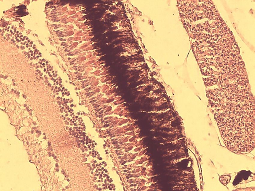
                                                                          The eyes and brains of infected Guppy             In the eye, the ganglion cell layer of the retina
                                                                     showed vacuolated cells. In the brain, vacuoles        was vacuolated (Fig.4). The results exhibited a
                                                                     were seen in the stratum griseum ventricular           very light reaction between MAb and viral
                                                                     layer and stratum album central layer of               antigens in the brain tissue (Fig. 5).The eye
                                                                     mesencephalon region, as well as in the septal         samples revealed intracytoplasmic vacuoles in
                                                                     area, ventral and dorsal olfactory area in             the retina cells (Fig. 6).
                                                                     olfactory bulb of telencephalon region (Fig. 3).

                                                                                                                        S

                                                                                                         V
                                                                                                B

                                                                     Figure 3: Vacuolation in the olfactory bulb of telencephalon region in brain tissue of infected
                                                                                Guppy at 15 dpi (arrows). S: Septal area. V: Ventral olfactory area. H&E. X 400.
173    Nazari et al., Pathogenicity of viral nervous necrosis virus for ...
Downloaded from jifro.ir at 10:43 +0430 on Wednesday May 29th 2019

                                                                     Figure 4: Light microscopic image of retina section of infected Guppy at 15 dpi. Vacuolation was
                                                                              seen in ganglion cell layer (arrows). H&E. X 400.

                                                                     Figure 5: Brain tissue section from test group1 Guppy at 15 dpi stained with IHC dye. Golden
                                                                              brown color (arrow) indicated light reaction between MAb and inter-cellular viral
                                                                              antigens. X 1000.

                                                                                                                  A1                               A2
Iranian Journal of Fisheries Sciences13(1) 2014                 174

                                                                     Figure 6: VNN bathed challenge Guppy, Poecilia reticulata 15 dpi. A1 & A2. Electron
                                                                                   micrograph showing the vacuoles in the cytoplasm of the retina cell, (arrows). Scale
                                                                                   bar ═ 0.2 µm.
                                                                     Discussion
Downloaded from jifro.ir at 10:43 +0430 on Wednesday May 29th 2019

                                                                          The experimental infection in Guppy was             lasted for 30 days. In both studies up to 100%
                                                                     performed to evaluate the VNN strain isolated            mortality were observed, but in Hegde’s
                                                                     from naturally infected Grey mullet. In the              report, absence of clinical signs was detected
                                                                     current study, clinical signs, typical CPE and           while in our study typical signs were
                                                                     changes in the retina and brine were observed            associated to mortality.
                                                                     in infected Guppy. In the present study,                       In experimental infections, clinical signs
                                                                     significant differences in viral pathogenicity           may vary according to differences on the time
                                                                     were observed between 2 groups when the                  of transmission, age of fish and route of
                                                                     times of challenge were varied.                          infection (Aranguren et al., 2002). Other
                                                                          In the current study, the sequence of               observations    indicated   that   pattern   of
                                                                     mortalities     indicated     the    similarity    of    pathogenicity may differ when different fish
                                                                     mortalities in other fish species affected with          species and virus strains or isolate are
                                                                     NNV (Aranguren et al., 2002). In naturally               compared (Mladineo, 2003).
                                                                     infected Guppy listlessness, emaciation and                     Furthermore, immature immune system
                                                                     mortality were observed (Hegde et al., 2003).            could also lead to higher mortality in short
                                                                     In   other     species,   although     the    highest    period in the fish larvae and juvenile as
                                                                     mortalities were observed in larvae and                  compared to lower mortality but longer time of
                                                                     juveniles     (Yoshikoshi     and    Inoue,    1990;     death in adult fish (Aranguren et al., 2002). On
                                                                     Glazebrook et al., 1990; Breuil et al., 1991;            the other hand, the viral exposure dose plays
                                                                     Mori et al., 1991; Renault et al., 1991), larger         an important role in the pathogenicity of the
                                                                     fish showed also developed clinical signs and            infection. The researchers indicated that the
                                                                     suffered from mortalities (Fukuda et al., 1996;          incubation period before appearance of clinical
                                                                     Skliris and Richards, 1999).                             signs following artificial infection may vary
                                                                          The clinical signs observed in this study           according to species and depends on dose of
                                                                     were slightly different from the experimental            virus inoculums (Grotmol et al., 1999).
                                                                     infection reported in a previous investigation                Histopathology investigations revealed
                                                                     (Hegde et al, 2003). In that previous study,             vacuolation changes in the eye and brain.
                                                                     0.1g Guppy was challenged with NNV isolated              Similar signs were reported in naturally
                                                                     from Guppy and Epinephelus tauvina and                   infected Grey mullet, which was the source of
                                                                     mortality lasted for 15 days, while, in our trial,       the virus in this study (Zorriehzahra et al.,
                                                                     the weight of the challenged Guppy were                  2005) as well as in other marine and
                                                                     0.47±0 and they have been exposed to a NNV               freshwater fish species (Nguyen et al., 1996;
                                                                     isolated from Golden grey mullet and mortality           Peducasse et al., 1999). Likewise, this
175        Nazari et al., Pathogenicity of viral nervous necrosis virus for ...

                                                                     observation was similar to first report of                   vacuolation in the retina was also reported by
                                                                     natural infection of Guppy larvae to VNN                     Tanaka et al. (2004) with EM micrographs
                                                                     (Hagde et al., 2003).                                        previously.
                                                                           The observed signs, in particular the                      The IHC performed both in brain and eyes
Downloaded from jifro.ir at 10:43 +0430 on Wednesday May 29th 2019

                                                                     erratic swimming behavior, could be referred                 sections showed very few positive foci and
                                                                     to cell vacuolation and necrotic changes                     light reaction between specific MAb and viral
                                                                     caused by the virus in the brain. As              other      antigens scattered in the target tissues. This
                                                                     studies noted, the brain and eye were main                   result could be due to a low sensitivity of Mabs
                                                                     target tissues for NNV in most of infected fish              employed in our test against the GMNNV
                                                                     species . In Figure 3, small vacuoles were seen              strain or low density of NNV invasive particles
                                                                     in the stratum griseum periventriculare of optic             in brain. This hypothesis earlier mentioned by
                                                                     tectum in mesencephalon. Vacuolated cells in                 Zorriehzahra et al. (2005) could be supported
                                                                     this area have been reported in sevenband                    by the existence pathogenicity of viral strains
                                                                     grouper,        Epinephelus         septemfasciatus          obtained      from    Golden      grey     mullet
                                                                     previously      (Tanaka     et   al.,    2004).    The       characterized by different pathogenicity. Low
                                                                     vacuolated cells shown in Fig. 3 were located                vacuolation in the retina presumably indicated
                                                                     in the ventral olfactory area and septal area of             that optic nerves and retina were infected after
                                                                     cerebral hemisphere or olfactory bulb of                     brain infection as described by Mladineo
                                                                     telencephalon. Both areas contained the mass                 (2003) using Immunolabeling. In the current
                                                                     of nerve cells which transmitted olfactory                   study, horizontal transmission was employed
                                                                     impulses to other centers (Hibiya, 1982). The                for pathogenicity of NNV isolated from
                                                                     many small nerve cells in olfactory bulb form                Golden grey mullet to Guppy fish. Thus, it
                                                                     a granule cell layer (Tanaka et al., 2004). The              could concluded that isolated virus from
                                                                     telencephalon      on     the    other    hand     was       Golden grey mullet in the current study is
                                                                     responsible        for     olfaction,       memory,          similar to isolate virus by Zorriehzahra et al.
                                                                     reproductive and feeding behavior (Roberts,                  (2005).
                                                                     2000). The abnormal swimming observed in
                                                                     experimentally infected Guppy could be                       References
                                                                     associated with vacuolar changes and necrosis                Aranguren, R., Tafalla, C., Novoa, B. and
                                                                     of brain cells in the above mentioned area. The                 Figueras, A., 2002. Experimental transmission
                                                                     retina showed vacuolation in inner ganglion                     of encephalopathy and retinopathy induced by
                                                                     cell layer. Similar necrotized area in NNV                      nodavirus to sea bream Sparus aurata l., using
                                                                     infection was described by Tanaka et al.                        different infection models. Journal of Fish
                                                                     (2004).                                                         Diseases, 25, 317-324.
                                                                           TEM micrographs from retina layer in the               Bovo, G. and Florio, D., 2008. Viral diseases of
                                                                     eye of infected Guppy revealed the presence of                  cultured marine fish. In Fish Diseases, vol. 1,
                                                                     intracytoplasmic vacuolation. Intracytoplasmic                  Eiras, J.C., H. Segner, T. Wahli, and B. G.
Iranian Journal of Fisheries Sciences13(1) 2014                          176

                                                                        Kapoor, (Eds.), Science Publishers, Enfield,             Hibiya, T., 1982.      An atlas of fish histology.
                                                                        NH: 186-230.                                                Normal and pathological features. Tokyo,
                                                                     Breuil, G., Bonami, J. R., Pepin, J. F. and                    Kosonsha Ltd, 147P.
                                                                        Pichot, Y., 1991. Viral infection        (piconalike     Húsgarõ, S., Grotmol, S., Hjeltnes, B. K.,
                                                                                ) associated with mass mortalities in
Downloaded from jifro.ir at 10:43 +0430 on Wednesday May 29th 2019

                                                                        virus                                                       Rodseth, O. M. and Biering, E., 2001.
                                                                        hatchery-reared        sea-bass      (dicentrarchus         Immune response to a recombinant capsid
                                                                        labrax   )larvae an juveniles. Aquaculture, 97,             protein of striped jack nervous necrosis virus
                                                                        109-116.                                                    (SJNNV) in turbot scophthalmus maximus and
                                                                     Chi, S. C., Shieh, J. R. and Lin, S. J., 2003.                 Atlantic halibut hippoglossus hippoglossus, and
                                                                        Genetic       and     antigenic      analysis       of      evolution of a vaccine against SJNNV. Diseases
                                                                        betanodaviruses       isolated      from       aquatic      of Aquatic Organisms, 45, 33-44.
                                                                        organisms in Taiwan. Disease of Aquatic                  Iwamoto, T., Mori, K., Arimoto, M. and Nakai,
                                                                        Organisms, 55, 221-228.                                     T., 1999. High permissivity of the fish cell line
                                                                     Fukuda, Y., Neuyen, H. D., Furuhashi, M., and                  SSN-1 for piscine nodaviruses. Diseases of
                                                                        Nakai, T. 1996. Mass mortality of cultured                  Aquatic Organisms, 39, 37-47.
                                                                        sevenband             grouper,           epinephelus     Mladineo, I., 2003. The immunohistochemical
                                                                        septemfasciatus, associated with viral nervous              study of nodavirus changes in larval, juvenile
                                                                        necrosis. Fish Pathology, 31, 165-170.                      and adult sea bass tissue. Journal of Applied
                                                                     Glazebrook, J. S., Heaaman, M. P., and de Beer,                Ichthyology, 19, 366-370.
                                                                        S.   W.     1990.   Picona-like     viral    particles   Mori, K., Nakai, T., Negahara, M., Muroga, K.,
                                                                        associated with mass mortalities in larval                  Mckuchi, T., and Kanno, T. 1991. A viral
                                                                        barramundi, lates calcarifer bloch. Journal of              disease in hatchery-reared larvae and juveniles
                                                                        Fish Diseases, 13, 245-249.                                 of redspotted grouper. Fish Pathology, 26, 209-
                                                                     Grotmol, S., Bergh, O., and Totland, G. K. 1999.               210.
                                                                        Transmission of viral encephalopathy and                 Munday, B. L., Kwang, J. and Moody, N., 2002.
                                                                                     (
                                                                        retinopathy VER) to yolk-sac larvae of the                  Betanodavirus infection of teleost fish: A
                                                                        Atlantic halibut Hippoglossus hippoglossus:                 review. Journal of Fish Diseases, 25, 127-142.
                                                                        occurrence of nodavirus in various organs and a          Nakai, T., 2007. Viral Nervous Necrosis (VNN):
                                                                        possible route of infection. Diseases of Aquatic            Global status of outbreaks, diagnosis, research
                                                                        Organisms, 39, 95-106.                                      and     surveillance.   SEAFDEC         International
                                                                     Hegde. A., Teh, H. C. Lam, T. J. and Sin, Y. M.,               Workshop on Emerging Fish Diseases in Asia.
                                                                        2003.     Nodavirus     infection   in      freshwater      Bangkok, Thailand, SEAFDEC.
                                                                        ornamental fish, Guppy, Poicelia reticulata-             Nguyen, H. D., Nakai, T. and Muroga, K., 1996.
                                                                        comparative characterization and pathogenicity              Progression of striped jack nervous necrosis
                                                                        studies. Archives of Virology, 148, 575-586.                virus    (SJNNV)        in   adult     striped    jack
                                                                                                                                    pseudocaranx      dentex     larvae.   Diseases     of
                                                                                                                                    Aquatic Organisms, 24, 99-105.
177      Nazari et al., Pathogenicity of viral nervous necrosis virus for ...

                                                                     Peducasse, S., Castric, R., Thiery, R., Jeffroy, J.,           Dicentrachus labrex infected in different ways.
                                                                        Le ven, A. and Baudin-Laurencin, F., 1999.                  Disease of Aquatic Organisms, 36, 11-20.
                                                                        Comparative studies of viral encephalopathy              Reed, L.L. and Munch, H., 1938. A simple
                                                                        and    retinopathy     in   juvenile     sea      bass      method of estimating fifty percent endpoints.
Downloaded from jifro.ir at 10:43 +0430 on Wednesday May 29th 2019

                                                                       American Journal of Hygiene, 27, 493-497.                    diseased   sevenband      grouper     Epinephelus
                                                                     Renault, T., Haffner, P. H., Baudin, L.F., Breuil,             septemfasciatus. Fish Pathology, 33, 31-36.
                                                                        G. and Bonami, J.R., 1991. Mass mortalities in           Tanaka, S., Takagi, M. and Miyazaki, T., 2004.
                                                                        hatchery-reared sea bass Lates calcarifer larvae            Histopathological studies on viral nervous
                                                                        associated with the presence in the brain and               necrosis of sevenband grouper, Eepinephelus
                                                                        retina of virus-like particles. Bull Eur Ass Fish           septemfasciatus thunberg, at the grow-out stage.
                                                                        Pathol, 11, 68-73.                                          Journal of Fish Diseases, 27, 385-399.
                                                                     Roberts, R., 2000. Fish pathology (3 Ed). London,           Yoshikoshi, K. and Inoue, K., 1990. Viral nervous
                                                                        W.B.Saunders, 472P.                                         necrosis in hatchery- reared larvae and juveniles
                                                                     Schneemann A., Ball L. A., Delsert C., Johnson                 of Japanese parrotfish Oplegnathus fasciatus
                                                                        J.E. Nishizawa T., Family Nodaviridae. In                   (Temminck   &Schlegel).     Journal     of    Fish
                                                                        Virus Taxonomy. 8th Report ICTV. Edited by:                 Diseases, 13, 69-77.
                                                                        Fauquet    CM,       Mayo   MA,        Maniloff     J,   Zorriehzahra, M. J., Nakai, T., Sharifpour, I.,
                                                                        Desselberger U, Ball LA. Elsevier Academic                  Kaw gomez, D., Chi, S.C. and Soltani, M.,
                                                                        Press, San Diego, CA, 2005, 865-872.l                       2005. Mortality of wild Golden grey mullet Liza
                                                                     Skliris, G.P. and Richards, R. H., 1999. Induction             auratus in Iranian waters of the Caspian Sea
                                                                        of nodavirus disease in seabass Dicentrarchus               associated with viral nervous necrosis-like
                                                                        labrax, using different infection models. Virus             agent. Iranian Journal of Fisheries Science,
                                                                        Research, 63, 85-93.                                        4(2), 43-58.
                                                                     Tanaka, A., Aori, H. and Nakai, T., 1998.
                                                                        Pathogenecity of the nodavirus detected from
You can also read