Fact sheet Mycobacterium ulcerans - (Buruli ulcer) disease
←
→
Page content transcription
If your browser does not render page correctly, please read the page content below
Mycobacterium ulcerans
            (Buruli ulcer) disease
                                              Fact sheet

Mycobacterium ulcerans disease - referred to globally as Buruli ulcer (BU), also as ‘Bairnsdale ulcer’ in Victoria
and ‘Daintree ulcer’ in Queensland - is recognised as one of the world’s neglected tropical diseases. It is
primarily considered a human disease and infection in humans is characterised by progressive, painless
destruction and necrosis of the skin, leading to the formation of ulcers. Australia is the only developed
country with significant local transmission of M. ulcerans disease in humans and the only country to report
the disease in wild animals. The mode of transmission and environmental reservoir are not completely
understood, but research in Australia suggests a potential role for small mammals, particularly possums, in
the ecology of this pathogen.

Mycobacterium ulcerans [MU] (Family Mycobacteriaceae) is a slow growing mycobacterium, closely related to
M. marinum. The bacterium produces a destructive toxin, mycolactone, which causes tissue damage and
inhibits the local immune response.

The bacterium has been reported a range of both domestic and wildlife mammalian species in endemic areas
of Australia. Laboratory-confirmed cases have been diagnosed in humans, koalas (Phascolarctos cinereus)
(McOrist et al. 1985), common ringtail possums (Pseudocheirus peregrinus), a common brushtail possum
(Trichosurus vulpecula), a mountain brushtail possum (T. cunninghami), a long-footed potoroo (Potorous
longipes), a black rat (Rattus rattus)1 as well as horses, dogs, a cat and an alpaca. The organism has been
found in the faeces of ringtail and brushtail possums and northern brown bandicoots (Isoodon macrourus) in
endemic areas of Australia (Fyfe et al. 2010; Carson et al. 2014; O'Brien et al. 2014; Röltgen et al. 2017; Singh
et al. 2019).

1   National wildlife health information system (eWHIS) data.
Human disease occurs in over 30 countries worldwide. Foci of infection have been reported in Australia (see
below), Africa (e.g. Benin, Cote d’Ivoire and Ghana), Asia and the Western Pacific (e.g. Japan and Papua New
Guinea), and the Americas (e.g. French Guiana). Australia is the only country to report the disease in wildlife.

There are at least three long-standing and recognised endemic areas for BU in humans in Australia: East
Gippsland near Bairnsdale, Vic; Far North Qld (FNQ) between Mossman and the Daintree River, and the
Capricorn Coast of Qld. Since the 1990s, new endemic areas have emerged on the Bellarine and Mornington
Peninsulas of Vic, not far from Melbourne. In 2019, two new potential transmission areas were identified in
Victoria, Airey’s Inlet (Surf Coast) and the Geelong suburb of Belmont (VicHealth 2019). Case numbers in
humans have increased significantly in Vic [340 reported human cases in Victoria in 2018; 297 in 2019 (Vic
Government 2020)], as have the number of human cases associated with severe disease (Tai et al. 2018;
VicHealth 2019). In 2020 a clinical case was reported in a ringtail possum with lesions in Essendon, a northern
suburb of Melbourne, with the organism confirmed by PCR, culture and isolation. There are anecdotal reports
of other similar unconfirmed cases in ringtail possums in the same area (Ban et al. 2020). Human cases were
detected in Essendon, Moonee Ponds and Brunswick West, first reported in early 2021. These are the first
non-coastal locations in Victoria recognised as potential risk areas for MU infection (Vic Health 2021).

Laboratory-confirmed cases of MU disease in wildlife have only been reported in Vic. All reported cases in
wildlife have been identified in locations where human cases have also occurred. There have been no
confirmed cases of MU disease in wildlife in Qld, NT or other states. In NSW, in reptiles, there have been
confirmed infections with a different subspecies of MU, not associated with human disease (J Fyfe pers.
comm. September 2018).

A study found 9/42 ringtail and 1/21 brushtail possums examined at an endemic Victorian site had clinical BU
lesions. Of these cases, 82% (9/11) of ringtail possums also had MU positive faeces, compared to 16% (5/31)
of ringtail possums without BU lesions, at the same location (Fyfe et al. 2010).

Globally, the geographic distribution of human BU case clusters is always highly focal (Carson et al. 2014).

The epidemiology of MU disease in Australia is not well understood and appears to be changing in south-east
Australia, with increasing numbers of human cases and expansion into new geographic areas (Tai et al. 2018).
The reasons for the change in epidemiology are not known. Most recent Australian studies have focused on
endemic and emerging-endemic areas in Victoria. More information is available at
www2.health.vic.gov.au/public-health/infectious-diseases/beating-buruli.

The incubation period is unknown in animals, but in humans it may be as long as several months (median 4.5
months) (Trubiano et al. 2013). Humans come into contact with the pathogen only in specific geographic
areas, which are frequently near water bodies – either along coastal areas or inland near slow flowing rivers,
swamps and lakes. The mode of transmission is unknown, but it is considered an environmental pathogen and
there is no evidence to indicate that the disease is transmitted from person-to-person (or animal-to-animal,
or animal-to-human). Exposure to the pathogen is thought to occur via contaminated water, soil or

                        WHA Fact sheet: Mycobacterium ulcerans disease | February 2021 | 2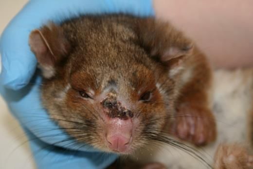
vegetation in specific geographic areas, and terrestrial mammals and or mosquitos may play an
epidemiological role.
It is possible that there are different modes of transmission for different geographic and epidemiological
scenarios (Singh et al. 2018) and that there are differences in virulence across geographically separate strains.
Evidence suggests transmission may occur after exposure times of less than two hours (Trubiano et al. 2013).
The long incubation period in humans and frequently, an extended period to diagnosis have hindered an
understanding of the epidemiology.
Clinical cases have been reported in a range of marsupial species from endemic areas of Vic (Fyfe et al. 2010;
O'Brien et al. 2014). There appears to be a significant disease burden in ringtail possums in some areas and
adult animals appear to be most commonly affected. Ringtail possums appear more likely to have persistent
lesions and to suffer from systemic disease associated with BU than other infected marsupials (O'Brien et al.
2014).
Research has investigated a potential role for a mammalian reservoir and/ or amplifying host. In Vic, common
ringtail and common brushtail possums from endemic areas, both with and without clinical disease, have
been shown to shed MU in their faeces (Fyfe et al. 2010; Carson et al. 2014; O'Brien et al. 2014). Focal areas
of high prevalence of MU in possum faeces corresponded to focal areas of human cases (Fyfe et al. 2010;
Carson et al. 2014). These findings could reflect a common environmental source of infection or may suggest
an epidemiologically important role for possums. One hypothesis is that possums or other terrestrial
mammals may act as an environmental reservoir of MU in SE Australia; they may ingest the organism from
the environment, amplify it and shed it in the faeces. There is no evidence of direct transmission from animals
to humans.
The epidemiological model in FNQ appears to be different to SE Australia, with outbreaks occurring
sporadically, possibly associated with heavy rainfall and associated environmental changes. Research in these
areas found MU in northern brown bandicoot faeces, indicating a possible role for this wildlife species in the
ecology of MU disease (Röltgen et al. 2017).
Research increasingly suggests a role for mosquitoes or other biting vectors in the transmission of M. ulcerans
from the environment to humans (Johnson et al. 2007; Quek et al. 2007; Johnson and Lavender 2009; Wallace
et al. 2017; Johnson 2019), however it is likely that there are several routes of infection, including possibly
other puncturing injuries (Wallace et al. 2017).
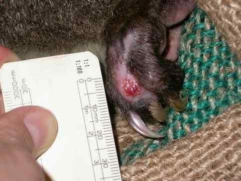
In wildlife, the most common clinical presentation is an ulcer or lesion, usually on the animal’s nose, ear, paw
or tail (Fig. 1). Behavioural signs may include lethargy, limited movement and, in the case of koalas, resting
on the ground. Non-healing ulcers are typical also in humans and other domestic animals.
WHA Fact sheet: Mycobacterium ulcerans disease | February 2021 | 3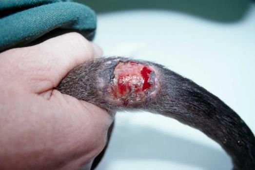
A                                                 B

Figure 1. A. Long-footed potoroo with large lesion at the base of the tail. B. Ringtail possum with lesion on the nose.

Diagnostic criteria include:
       •   animal lives in an endemic area
       •   animal has an ulcer with no other apparent cause, usually on an extremity, and often with
           undermined edges
       •   acid-fast bacilli demonstrated in diagnostic specimens.

Most cases of M. ulcerans in wildlife exhibit cutaneous ulcerative lesions, sub-cutaneous nodules or swelling
of paws, limbs or digits. There may be an undermined skin ulcer, with necrotic fat frequently visible at the
base (Fig. 1). There may be minimal inflammatory response. Systemic infection is also known to occur.
Necropsy examinations of a long-footed potoroo, koalas and ringtail possums have revealed the presence of
M. ulcerans in internal organs as well as lesions. The bacterium produces mycolactone toxin, which causes
tissue damage and hinders the local immune response.

Traumatic skin ulcers, other mycobacterial and infectious skin ulcers.

       •   swabs (dry or in transport medium)
       •   fresh tissue
       •   paraffin-embedded fixed tissue sections.

       •   direct smear examination for acid fast bacilli (AFB)
       •   culture for M. ulcerans
       •   polymerase chain reaction (PCR)
       •   histopathology.
PCR is the most rapid, sensitive and specific method for the diagnosis of M. ulcerans disease (Fyfe et al. 2007).
This test is performed at the Victorian Infectious Diseases Reference Laboratory (address: 792 Elizabeth St,
Melbourne 3000, phone: 03 9342 9379, www.vidrl.org.au). It is advisable to contact the laboratory prior to
sending a specimen for testing.

                          WHA Fact sheet: Mycobacterium ulcerans disease | February 2021 | 4
Various medical and surgical methods have been used to treat MU disease in domestic animals, including
antibiotic treatment, surgical excision and cryosurgery (Elsner et al. 2008; van Zyl et al. 2010; O'Brien et al.
2011). In wildlife, the infection has been known to resolve without treatment, as in the case of a mountain
brushtail possum from Bellbird Creek (Fig. 2) and a brushtail possum from Point Lonsdale (unpublished). In
severe cases, animals have been euthanased. Human treatment is not addressed in this fact sheet.
A B
Figure 2. Mountain brushtail possum with lesion (A) at diagnosis and (B) later without treatment.
While the mode of transmission of MU is unknown, effective strategies for prevention and control of the
disease remain to be devised. Referral of diseased animals for treatment or euthanasia by veterinarians
experienced in the diagnosis of this condition is recommended.
There is no targeted surveillance program for MU disease in wildlife. A greater awareness of the disease
among veterinarians and wildlife carers, particularly in endemic areas such as Raymond Island, Phillip Island
and the Bellarine and Mornington Peninsulas, as well as active case finding as part of research, has led to
increased reporting of cases in wildlife and domestic animals.
The WHO Collaborating Centre for MU - based at the Victorian Infectious Diseases Reference Laboratory
(VIDRL) in Melbourne - maintains records of all known human and animal cases of MU disease in Australia. In
Victoria, the Department of Health publishes statistics on notifiable infectious diseases (in humans), including
MU infection, on its website www2.health.vic.gov.au.
Wildlife disease surveillance in Australia is coordinated by Wildlife Health Australia. The National Wildlife
Health Information System (eWHIS) captures information from a variety of sources including Australian
government agencies, zoo and wildlife parks, wildlife carers, universities and members of the public.
Coordinators in each of Australia's States and Territories report monthly on significant wildlife cases identified
in their jurisdictions. NOTE: access to information contained within the National Wildlife Health Information
System dataset is by application. See the WHA website for more information:
www.wildlifehealthaustralia.com.au/ProgramsProjects/eWHISWildlifeHealthInformationSystem.aspx#requests.
There are almost 30 cases reported in eWHIS from free-living marsupials from endemic areas of Victoria,
mostly in ringtail possums.
WHA Fact sheet: Mycobacterium ulcerans disease | February 2021 | 5The WHO has identified six main priorities for research into BU: mode of transmission; development of simple
diagnostic tests; drug treatments and new treatment modalities; development of vaccines; social and
economic studies; and studies to determine the incidence and prevalence.
In Australia, much research has focussed on determining the mode of transmission and environmental source
of MU. Further work is needed to gain a better understanding of the potential role of ringtail and/or brushtail
possums, if there are any epidemiological interactions between these two species, and whether the relative
population density of ringtail and brushtail possums influences endemicity and/or emergence of BU in
humans. Possum faecal surveys may provide a useful public health monitoring tool and may act as sentinels
for incidence in humans.
Work is required to better understand the prevalence of MU lesions in possums in human endemic areas, the
susceptibility of different Australian marsupial species to MU infection, the possibility of gut amplification in
marsupial species and whether MU disease poses a risk to populations of possums or other marsupial species.
Genomic studies could help to explore whether different pathogen strains are associated with different
virulence, and whether strains are evolving and changing in virulence over time (Tai et al. 2018).
Mycobacterium ulcerans infection is primarily a human disease and is notifiable in the state of Victoria. The
source of infection in humans remains unknown and there are no confirmed cases of transmission from
wildlife to humans. All age groups of humans are at risk, however severe disease is more common in older
patients (Tai et al. 2018).
There is much to be learned about MU disease. Further research is needed to better understand the possible
epidemiological role of possums or other terrestrial mammals. The current interest in important but less well
known tropical diseases and the ‘One Health’ approach will hopefully continue to support increased
awareness of the disease and the resources needed to develop tools for diagnosis, treatment, prevention and
control.
Ban S, Cox-Witton K, Grillo T (2020) Mycobacterium ulcerans in a ringtail possum in a new location. Animal
Health Surveillance Quarterly 25, 13-17.
Carson C, Lavender CJ, Handasyde KA, O'Brien CR, Hewitt N et al. (2014) Potential wildlife sentinels for
monitoring the endemic spread of human buruli ulcer in South-East australia. PLOS Neglected Tropical
Diseases 8, e2668.
Elsner L, Wayne J, O'Brien CR, McCowan C, Malik R et al. (2008) Localised Mycobacterium ulcerans infection in
a cat in Australia. Journal of Feline Medicine & Surgery 10, 407-12.
Fyfe JA, Lavender CJ, Handasyde KA, Legione AR, O'Brien CR et al. (2010) A Major Role for Mammals in the
Ecology of Mycobacterium ulcerans. PLOS Neglected Tropical Diseases 4, e791.
WHA Fact sheet: Mycobacterium ulcerans disease | February 2021 | 6Fyfe JA, Lavender CJ, Johnson PD, Globan M, Sievers A et al. (2007) Development and application of two
multiplex real-time PCR assays for the detection of Mycobacterium ulcerans in clinical and environmental
samples. Applied and Environmental Microbiology 73, 4733-4740.
Johnson P (2019) Buruli ulcer in Australia. In 'Buruli Ulcer.' (Eds G Pluschke, K Röltgen.) pp. 61-76. (Springer:
Tokyo).
Johnson P, Azuolas J, Lavender CJ, Wishart E, Stinear TP et al. (2007) Mycobacterium ulcerans in mosquitoes
captured during an outbreak of Buruli ulcer, southeastern Australia. Emerging Infectious Diseases 13, 1653-
1660.
Johnson P, Lavender C (2009) Correlation between Buruli ulcer and vector-borne notifiable diseases, Victoria,
Australia. Emerging Infectious Diseases 15, 614-615.
McOrist S, Jerrett IV, Anderson M, Hayman J (1985) Cutaneous and respiratory tract infection with
Mycobacterium ulcerans in two koalas (Phascolarctos cinereus). Journal of Wildlife Diseases 21, 171-173.
O'Brien C, Handasyde KA, Hibble J, Lavender CJ, Legione AR et al. (2014) Clinical, microbiological and
pathological findings of Mycobacterium ulcerans infection in three Australian Possum species. PLOS Neglected
Tropical Diseases 8, e2666.
O'Brien C, McMillan E, Harris O, O'Brien DP, Lavender CJ et al. (2011) Localised Mycobacterium ulcerans
infection in four dogs. Australian Veterinary Journal 89, 506-10.
Quek TY, Athan E, Henry MJ, Pasco JA, Redden-Hoare J et al. (2007) Risk factors for Mycobacterium ulcerans
infection, southeastern Australia. Emerging Infectious Diseases 13, 1661-6.
Röltgen K, Pluschke G, Johnson PD, Fyfe J (2017) Mycobacterium ulcerans DNA in Bandicoot Excreta in Buruli
Ulcer–Endemic Area, Northern Queensland, Australia. Emerging Infectious Diseases 23, 2042.
Singh A, McBride W, Govan B, Pearson M (2018) Potential Animal Reservoir of Mycobacterium ulcerans: A
Systematic Review. Tropical Medicine and Infectious Disease 3, 56.
Singh A, William McBride J, Govan B, Pearson M (2019) Survey of local fauna from endemic areas of Northern
Queensland, Australia for the presence of Mycobacterium ulcerans. International Journal of Mycobacteriology
8, 48-52.
Tai AY, Athan E, Friedman ND, Hughes A, Walton A et al. (2018) Increased Severity and Spread of
Mycobacterium ulcerans, Southeastern Australia. Emerging Infectious Diseases 24, 58.
Trubiano JA, Lavender CJ, Fyfe JA, Bittmann S, Johnson PD (2013) The incubation period of Buruli ulcer
(Mycobacterium ulcerans infection). PLOS Neglected Tropical Diseases 7, e2463.
van Zyl A, Daniel J, Wayne J, McCowan C, Malik R et al. (2010) Mycobacterium ulcerans infections in two
horses in south-eastern Australia. Australia Veterinary Journal 88, 101-106.
Vic Government (2020) Surveillance of notifiable conditions in Australia. Melbourne. Available at
www.health.vic.gov.au/ideas/downloads/daily_reports/rptVS_SNIDSVictorianSummary_GR.pdf.
Vic Health (2021) 'Buruli ulcer bacteria identified in inner west Melbourne.' Available at
https://www2.health.vic.gov.au/about/news-and-events/healthalerts/buruli-ulcer-bacteria [Accessed 25 Feb
2021].
WHA Fact sheet: Mycobacterium ulcerans disease | February 2021 | 7VicHealth (2019) Possible new transmission areas for Buruli ulcer in Victoria. Victoria. Available at
https://www2.health.vic.gov.au/about/news-and-events/healthalerts/increased-incidence-of-buruli-ulcer-in-
victoria-april-2018 [Accessed 1 Nov 2019].
Wallace JR, Mangas KM, Porter JL, Marcsisin R, Pidot SJ et al. (2017) Mycobacterium ulcerans low infectious
dose and mechanical transmission support insect bites and puncturing injuries in the spread of Buruli ulcer.
PLOS Neglected Tropical Diseases 11, e0005553.
We are extremely grateful to the many people who had input into this fact sheet and would specifically like to
thank the Victorian Infectious Diseases Reference Laboratory (VIDRL), Christina McCowan, Caroline Lavender,
Janet Fyfe and John Hayman who have a special interest in this condition in Australia.
Updated: February 2021
We are interested in hearing from anyone with information on this condition in Australia, including laboratory
reports, historical datasets or survey results that could be added to the National Wildlife Health Information
System. If you can help, please contact us at admin@wildlifehealthaustralia.com.au
Wildlife Health Australia would be very grateful for any feedback on this fact sheet. Please provide detailed
comments or suggestions to admin@wildlifehealthaustralia.com.au We would also like to hear from you if
you have a particular area of expertise and would like to produce a fact sheet (or sheets) for the network (or
update current sheets). A small amount of funding is available to facilitate this.
This fact sheet is managed by Wildlife Health Australia for information purposes only. Information contained
in it is drawn from a variety of sources external to Wildlife Health Australia. Although reasonable care was
taken in its preparation, Wildlife Health Australia does not guarantee or warrant the accuracy, reliability,
completeness, or currency of the information or its usefulness in achieving any purpose. It should not be
relied on in place of professional veterinary or medical consultation. To the fullest extent permitted by law,
Wildlife Health Australia will not be liable for any loss, damage, cost or expense incurred in or arising by
reason of any person relying on information in this fact sheet. Persons should accordingly make and rely on
their own assessments and enquiries to verify the accuracy of the information provided.
WHA Fact sheet: Mycobacterium ulcerans disease | February 2021 | 8You can also read