DETECTION OF ANTIBODIES AGAINST LUMPY SKIN DISEASE VIRUS BY VIRUS NEUTRALIZATION TEST AND ELISA METHODS - Semantic Scholar
←
→
Page content transcription
If your browser does not render page correctly, please read the page content below
Acta Veterinaria-Beograd 2019, 69 (1), 47-60
                                                                   UDK: 636.2.09:616.98:578.821
Research article                                                   DOI: 10.2478/acve-2019-0003

DETECTION OF ANTIBODIES AGAINST LUMPY SKIN
DISEASE VIRUS BY VIRUS NEUTRALIZATION TEST
AND ELISA METHODS

SAMOJLOVIĆ Milena1, POLAČEK Vladimir1, GURJANOV Vladimir2,
LUPULOVIĆ Diana1, LAZIĆ Gospava1, PETROVIĆ Tamaš1, LAZIĆ Sava1*

Scientific Veterinary Institute „Novi Sad“, Novi Sad, Republic of Serbia;
1

Veterina DOO, PIK Bečej, Bečej, Republic of Serbia
2

(Received 13 July 2018, Accepted 21 February 2019)

        Infection of cattle with lumpy skin disease virus (LSDV) is very important from the
        aspect of livestock production. Although it can cause significant economic losses,
        available serological assays are still not sufficiently efficient and reliable. A 3-day VNT
        was performed using Madin-Darby bovine kidney (MDBK) cell line and LSDV isolated
        from clinically infected cow to improve serological diagnostics of lumpy skin disease
        (LSD).
        In total, 325 cattle sera samples were tested in order to compare the performances of
        VNT and ELISA. Tested samples originated from 125 cows before the occurrence of
        LSD in the Republic of Serbia and 200 tested samples originated from vaccinated cows.
        Sera samples from vaccinated cows were collected starting from the vaccination day to
        4 months after vaccination. In 7 different time intervals after vaccination sampling was
        carried out in 20 cows originating from one herd and in 3 different time intervals in 20
        cows originating from a different herd each time of sampling.
        Out of 200 samples from vaccinated cows, antibodies against LSDV were detected in
        68 (34%) samples by VNT, and in 60 (30%) samples by ELISA. No positive finding was
        detected by VNT in samples collected before the occurrence of LSD in Serbia, while
        one positive finding was detected in the same samples by ELISA. The first presence
        of antibodies in vaccinated cattle was detected by both tests 20 days after vaccination,
        and the largest number of animals with antibodies against LSDV was detected 30 days
        after vaccination.
        Comparing the results obtained by VNT and ELISA, it was calculated that kappa index
        was 0.913. The specificity of VNT and ELISA was 100% and 99.2%, respectively. VNT
        is simpler to perform compared to the recommended virus neutralization test by the
        OIE and can improve LSD serological diagnostics with additional sensitivity testing.
        Key words: antibodies, ELISA, Lumpy skin disease virus, Virus neutralization test,
        specificity, sensitivity

*Corresponding author: e-mail: lazic@niv.ns.ac.rs

                                                                                                47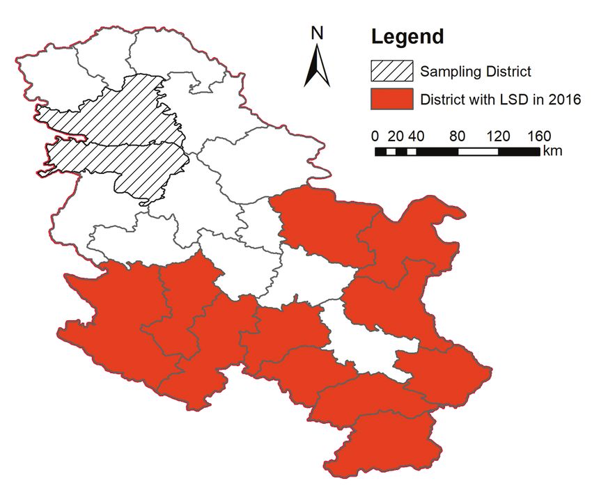
Acta Veterinaria-Beograd 2019, 69 (1), 47-60

                                      INTRODUCTION

Lumpy Skin Disease (LSD) is a contagious disease of cattle and buffaloes, although
the disease was found in some wildlife species (impalas and gazelles). The disease
is characterized by changes on the skin and mucous membranes in the form of
inflammatory necrotic spots, which can cause severe general health disorders, significant
decrease in milk production and growth, abortions and sterility [1]. Significant changes
in hematological and biochemical parameters were observed in infected animals [2].
LSD is on the OIE list of notifiable diseases, because of its great economic impact
and huge economic losses it can cause [3]. However, Carn and Kitching [4] found that
not all experimentally infected cattle would display clinical signs of the disease, so
this must be taken into consideration when determining measures for preventing the
spread of the disease.
The causative agent of LSD is a virus belonging to the family Poxviridae, subfamily
Capripoxviridae, genus Capripox [5]. Beside lumpy skin disease virus (LSDV), genus
Capripox consists of sheeppox virus (SPV) and goatpox virus (GPV). Viruses within
this genus share 97% of nucleotide identity are serologically indistinguishable and
cross-protective, so they are used in vaccines for the prevention of these diseases [6-8].
The first case of LSD in cattle in the Republic of Serbia was reported on June 5,
2016. A total of 225 cases of the disease and 267 infected cattle were registered.
The last case of the disease in the Republic of Serbia was registered on October 1,
2016. [9]. The virus was detected in the samples from skin nodules, and afterwards
isolated [10]. The isolated LSDV named SERBIA/Bujanovac/2016 was sequenced
in the length of the complete genome and submitted into NCBI GenBank under the
number KY702007. Sequencing of the genome revealed that LSDV isolated in Serbia
consists of 150661 nucleotides and that it shares 99.95% of nucleotide identity with
the Neethling Warmbaths LW strain, which was isolated in South Africa in 1999 [11].
According to the recommendation of the World Organization for Animal Health
(OIE) detection of antibodies against LSDV can be performed by virus neutralization
test, agar gel immunodiffusion test, indirect fluorescence test, ELISA as well as by
Western blot [12]. Virus neutralization test is “the gold standard” and is considered
the most specific method for the detection of antibodies against LSDV, but it is
not sensitive enough to identify each animal that was in contact with the virus [13].
Immunodiffusion and immunofluorescence tests for the detection of antibodies
against LSDV, are less specific, while Western blot is specific and sensitive enough,
but complicated to set up and very expensive for routine use, while ELISA requires
standardization of the coated antigen in the microtiter plates [12,14]. On the other
hand, main advantages of ELISA are its cost, speed and possibility to run a large
number of samples on one plate [15]. Based on the current knowledge on the detection
of antibodies against LSDV, OIE indicates that virus neutralization test is suitable
for determining status of the herd and individual animals to LSDV infection. Also,
according to the obtained results of current studies, virus neutralization test can be

48
Samojlović et al.: Detection of antibodies against Lumpy skin disease virus by Virus neutralization test and ELISA methods
used for the confirmation of the clinical status of infected animals, determining the
eradication policy and prevalence of LSD, or immune status of vaccinated cattle in
the heard. However, it was determined that cattle with mild or asymptomatic infection
will not always develop antibody levels detectable with VNT, as well as that vaccinated
cattle will sometimes develop low levels of antibodies below the detection limit [9].
As recommended by OIE, key elements of VNT Protocol include using Capripoxvirus
of standard titer of 100 TCID/50, 1:5 to 1:500 dilution of test sera, Vero or LT
(lamb testis) cells, or other cells susceptible to Capripoxvirus on which the virus was
titrated. Testing is done in flat-bottomed 96-well microtiter plates for cell culture.
Microtiter plates are incubated for 9 days at 37°C. Samples are considered positive if
neutralization index is ≥ 1:5 [12].
Effectors of cell mediated immunity play a dominant role in the development of
protection against LSD, however antibodies, as effectors of the humoral immunity,
are important indicators of immune reactivity in both vaccinated and infected animals
[3,14,16,17]. Antibodies against LSDV in vaccinated animals appear 10 days after
vaccination and remain detectable in the majority of vaccinated animals 30 days after
vaccination [18].
VNT and ELISA have been used for the detection of antibodies against LSDV in
different studies. Hedger and Hamblin [19] found antibodies in wild animals by VNT.
Babiuk et al. [20,21] detected antibodies in experimentally infected animals with
LSDV by comparative testing of samples by VNT and ELISA. Similarly, Bowden
et al. [14] performed comparative testing of samples by VNT and ELISA to detect
antibodies against LSDV in experimentally infected animals. Walid et al. [22] detected
antibodies against LSDV in clinically infected cows, fevered cows, and in-contact
cows. Furthermore, Tilahun et al. [23] used VNT to follow seroconversion in cows
vaccinated against LSD.
The aim of this paper was to present the results of detection of antibodies against
LSDV obtained by comparative testing of cattle sera samples by virus neutralization
test (VNT) and ELISA, in parallel. In addition to that, the aim of this paper was to
present the compatibility of the obtained results, as well as the results of specificity and
sensitivity of VNT and commercial ELISA (ELISA) for the detection of antibodies
against LSDV.
MATERIAL AND METHODS
Sera samples of cows from Južnobački and Srem Districts were used in this study
(Figure 1). In this area LSD was not reported during the LSD outbreak in Serbia
in 2016 and vaccination against LSD started during July 2016. Tested cows were
of different breed (Black and Red Holstein, Simmental breed, and crosses of these
breeds) and age (2-6 years).
49Acta Veterinaria-Beograd 2019, 69 (1), 47-60
Figure 1. Republic of Serbia – Districts with LSD in 2016 and Sampling Districts
Blood sera samples from the sera bank of
Scientific Veterinary Institute “Novi Sad”
From the sera bank of the Scientific Veterinary Institute “Novi Sad” 125 cattle sera
samples were randomly chosen. These samples were collected during 2015 and until
June 1, 2016, i.e. before LSD outbreak in Serbia. From the year 2015, 94 samples were
tested, and from the year 2016, 31 samples were tested. Tested cattle originated from
52 different farms.
Blood sera samples from vaccinated cows
Vaccination of cattle against LSD in the Republic of Serbia was conducted with
„OBP Lumpy Skin Disease“ vaccine (Onderstepoort, Biological Products). Samples
were collected from randomly selected vaccinated cows originating from four herds.
In the 20 cows originating from one herd, sampling was carried out in 7 different
time intervals after vaccination. A total of 140 sera samples from the same 20 cows
of one herd were collected on days 0, 10, 20, 30, 45, 60, and 75 after vaccination.
Sampling was also carried out in 3 different time intervals after vaccination in 20
cows originating from a different herd each time of sampling. In this way, 60 more
samples were collected on days 90, 105 and 120 after vaccination. Hence, testing was
performed in a total of 200 sera samples collected from vaccinated cows.
50Samojlović et al.: Detection of antibodies against Lumpy skin disease virus by Virus neutralization test and ELISA methods
Commercial ELISA set kit
A commercial ELISA kit „ID Screen® Capripox Double Antigen Multi-species“,
manufactured by „IDvet, (France), Lot: B54, was used to detect specific antibodies
against LDSV. Testing was carried out according to manufacturer’s instruction.
The results of the test are defined based on the calculated S/P ratio (Formula 1). If
the S/P ratio is higher than 30%, tested sample is defined as positive on the presence
of antibodies against LSDV.
Formula 1. Calculation of S/P ratio
Virus neutralization test
Virus neutralization test was performed using LSD virus isolate (SERBIA/Bujanovac/
2016). The LSDV was isolated from skin nodules of a clinically infected cow during
LSD outbreak in Serbia. The virus isolate was cultivated and titrated on MDBK cell
line (ATCC, CCL-22). The titer of virus isolate used in VNT was 6,0 log10 TCID/50
after the fourth passage.
VNT was performed in commercial flat-bottomed 96-well microtiter plates for cell
culture manufactured by „Sarstedt“, (Lot: 2079082) using cell culture medium Eagle
MEM with Hepes buffer, manufactured by „Sigma“ (Lot: SLBQ2618V), with 10%
fetal bovine serum, manufactured by Capricorn (Lot: CP16-1377).
Virus neutralization test procedure
VNT procedure:
–– 50 µL of cell culture medium with 10% fetal bovine serum was added to all wells
of the microtiter plate, except virus control, cell control and virus back titration
wells. Control sera and test sera were then titrated in a volume of 50 µL using
twofold dilutions from 1:2 to 1:256
–– 50 µL of appropriate dilution of stock virus was added to every well containing
control and test sera
–– Microtiter plates were incubated for one hour at 37°C and after incubation
100 µL of MDBK cell suspension of concentration 185000 cell/ml was added
to every well containing control and test sera
–– Microtiter plates were incubated for three days (72 hours) in an incubator at 37°C,
after that, plates were read microscopically for the presence of cytopathogenic
effect (CPE) of LSD virus
VNT contained the following controls:
–– Positive serum control (antibody titer value 1:8 – 1:32)
51Acta Veterinaria-Beograd 2019, 69 (1), 47-60
–– Negative serum control (fetal bovine serum)
–– Controll of MDBK cells in 8 wells (Figure 2A)
–– Control of working dilution of stock virus of 100 TCID/50 in 8 wells
(Figure 2B)
–– A back titration of the working dilution of stock virus using eight wells per
tenfold dilution (10, 1 and 0 TCID/50 in 50µL) (Figures 2C and 2D showing
cytopathogenic effect of 10 and 1 TCID/50 of diluted virus).
Figure 2. The control of MDBK cell line (A), the controls of LSDV of 100 TCID/50 (B),
LSDV of 10 TCID/50 (C) and LSDV of 1 TCID/50 (D)
Reading of the results with verification – criteria
–– Virus titer in back titration (3 serial tenfold dilutions of working virus) was
within 30 and 300 TCID/50
–– No cytomorphological changes were observed in cell line control wells
–– Antibody titer of positive control serum was 1:8 – 1:32,
–– Antibody titer of negative control serum wasSamojlović et al.: Detection of antibodies against Lumpy skin disease virus by Virus neutralization test and ELISA methods
–– Determined antibody titer of 1:2 or higher was considered to be a positive
finding for the presence of antibodies against LSDV.
Statistical analysis
For the statistical analysis of obtained results in this study kappa test was used according
to: http://epitools.ausvet.com.au/content.php?page=Compare2Tests, and specificity
and sensitivity of the compared methods were calculated according to Thrusfield [24].
RESULTS
Results of detection of antibodies against LSDV by VNT and ELISA
The results of antibody detection against LSDV in 125 samples from the sera bank of
the Scientific Veterinary Institute “Novi Sad” and 200 samples from vaccinated cattle,
obtained by both VNT and ELISA, are presented in Table 1.
Table 1. Results of antibody detection against LSDV by VNT and ELISA
Results of VNT Results of ELISA
Number
Tested Number Number Positive Number Number Positive
of tested
samples of positive of negative samples of positive of negative samples
samples
samples samples (%) samples samples (%)
Samples from the
125 0 125 0 1* 124 0.8
sera bank
Samples collected
from vaccinated 200 68 132 34 60 140 30
cattle
TOTAL 325 68 257 20.92 61 264 18.77
*S/P ratio value = 69.19%
In total 68 out of 325 sera (20.92%) were positive by VNT, whereas 61 (18.77%)
sera reacted positive by ELISA. No positive finding of antibodies against LSDV was
detected by VNT in samples from the sera bank of the Scientific Veterinary Institute
“Novi Sad”, while by ELISA positive antibody finding was detected in one sample
from the sera bank. In 200 sera samples from vaccinated cattle, positive finding was
determined in 68 samples (34%) by VNT and in 60 (30%) samples by ELISA (Table 1).
Antibodies against LSDV became detectable 20 days after vaccination. Antibodies
were detected in 6 (30%) cows by VNT, and with antibody titer values in the range
from 1:8 to 1:32. By ELISA, antibodies were detected in 2 (10%) cows, with S/P values
50.73% and 63.80%. The largest number of cows with positive antibody finding was
detected 30 days after vaccination. Antibodies were found in 15 (75%) cows by VNT
with antibody titer values from 1:8 to 1:64, and in 13 (65%) cows by ELISA, with S/P
values from 29.97% to 170.47%. Analyzing the results obtained by both tests, in the
53Acta Veterinaria-Beograd 2019, 69 (1), 47-60
same vaccinated cows, it was found that the number of animals with antibodies against
LSDV decreased nearly 50% on days 45, 60 and 75 after vaccination compared to day
30 after vaccination. On days 45 and 60 after vaccination, there was no significant
reduction in antibody titer values, nor in the S/P values, which were in the range
from 1:16 to 1:64 and 141.14% to 146.35%, respectively. On day 75 after vaccination,
antibody titer values were slightly reduced, and were from 1:2 to 1:32, while S/P values
were from 53.96% to 206.99%. Positive finding of antibodies against LSDV by ELISA
was found on days 90, 105 and 120 after vaccination in 8 cows (40%). Testing of the
same samples by VNT, antibodies against LSDV were found in 8 cows, except on day
105 after vaccination, when 9 (45%) cows reacted positive. Antibody titer and S/P
values were similar in the last three sampling intervals, and were in the range from 1:2
to 1:128 and from 42.39% to 270.19%, respectively.
Antibodies against LSDV detected by comparative testing were also quantified. Based
on the obtained results, it can be concluded that antibody titer values detected by VNT
ranged from 1:2 to 1:64, and S/P ratio values determined by ELISA ranged from
29.97% to 270.19%.
Results of compatibility testing of VNT and ELISA for
the detection of antibodies against LSDV
Table 2 shows compatibility of the results obtained by VNT and ELISA using kappa
test.
Table 2. Compatibility of VNT and ELISA for the detection of antibodies against LSDV
MVNT Kappa index = 0.913
+ – Σ Standard error of kappa = 0.0285
cELISA + 60 1 61 Overall proportion of agreement = 0.9723
– 8 256 264 Kappa lower 95% limit = 0.8571
Σ 68 257 325 Kappa upper 95% limit = 0.9689
According to the obtained data shown, results of comparative testing did not match
in only 9 out of 325 tested samples. Eight samples of vaccinated cows taken on days
20, 30, 75 and 105 after vaccination were positive only by VNT, whereas one sample
from the sera bank was positive only by ELISA. Firstly, on day 20 after vaccination,
results of 4 samples did not match, with antibody titer values from 1:8 to 1:16 and
S/P values from 9.07% to 21.68%. Secondly, on day 30 after vaccination, results of
2 samples did not match, with antibody titer values 1:16 and S/P values 25.07% and
25.33%. Thirdly, on days 75 and 105 after vaccination, results of 1 sample did not
match, with antibody titer values 1:2 and S\P values 13.68% and 15.33%. Finally, S/P
value of ELISA positive sample from the sera bank was 69.19%, while no antibodies
were detected by VNT in this sample.
54Samojlović et al.: Detection of antibodies against Lumpy skin disease virus by Virus neutralization test and ELISA methods
By VNT a positive finding was determined in 68 samples and a negative finding in
257 samples. On the other hand, by ELISA a positive finding was determined in 61
samples and a negative in 264 samples. Kappa index was 0.913, which presents almost
complete agreement of compared methods [8].
Specificity and sensitivity of VNT and ELISA for the detection of antibodies against
LSDV by are shown in Table 3.
Table 3. Specificity and sensitivity of VNT and ELISA for the detection of antibodies against
LSDV
MVNT* cELISA**
Positive Negative Positive Negative
Positive 68 (TP) 0 (FN) 60 (TP) 8 (FN)
Negative 0 (FP) 125 (TN) 1 (FP) 124 (TN)
TP true positive, FN false negative, FP false positive, TN true negative.
Specificity: TN/(TN+FP) *125/(125+0) = 100%; **124/(124+1) = 99,2%
Sensitivity: TP/(TP+FN) *68/(68+0) = 100%; **60/(60+8) = 88,24%
Sera samples from the sera bank of the Scientific Veterinary Institute “Novi Sad” were
used to calculate specificity of VNT and ELISA, because they could be considered as
true negative samples on the presence of antibodies against LSDV, since historically
at the time of collection of those samples, no LSD has ever been reported in Serbia.
According to the obtained results, specificity of VNT and ELISA was 100% and
99.2%, respectively. However, determining of sensitivity, regardless of the obtained
results, must be cautiously taken into consideration, as there were no positive reference
samples on the presence of antibodies against LSDV included in testing.
DISCUSSION
According to the aim of this paper, the results of detection of antibodies against
LSDV by VNT and ELISA methods in cattle are presented. Samples originated
from the vaccinated cattle and cattle that were not in contact with LSDV. A virus
neutralization test modified in relation to the VNT recommended by OIE [12] and
commercial ELISA set kit were used for the detection of antibodies against LSDV.
The modification of VNT in relation to the recommended VNT by OIE [12] refers
to the virus used, cell culture and duration of the test. VNT was performed using
homologous virus SERBIA/Bujanovac/2016 that originated from LSD clinically
infected animal [10,11], and was isolated on MDBK cell line. On the other hand, OIE
recommends using Capripoxvirus for VNT [12]. Productive replication of isolated virus
in MDBK cell line, with the development of a clearly visible cytopathogenic effect
(Figure 2) allowed the test duration to be reduced from 9 days, which is recommended
by OIE [12], to 3 days. Thus, VNT has an advantage in relation to the recommended
55Acta Veterinaria-Beograd 2019, 69 (1), 47-60 virus neutralization test by OIE and a great potential in the serological diagnostics of LSD. The obtained results of 100% regarding specificity of VNT are in accordance with statements by other authors [14,21]. Virus neutralization test is considered to be the most specific serological method, but it is not sensitive enough to detect antibodies in each animal that have been in contact with the virus [9,12,13]. Due to the local spread of the virus from cell to cell, effector cells of cell mediated immune response, such as natural killer cells or cytotoxic T lymphocytes are necessary to identify and eliminate infected cells to prevent virus replication and spreading [16]. In studies considering Orthopoxviruses it was concluded that joint action of humoral and cellular immune responses was necessary for the clearance of the infection, whereas for protection against reinfection sole action of humoral immune response was enough [3]. Due to the lack of commercial diagnostic assays based on the cellular immune response or assays for quality control of vaccine production [3], and the fact that the role of of antibodies has been demonstrated in several studies, where animals did not have detectable level of antibodies in their sera, but were resistant to challenge [14,17], it is very important to improve diagnostics of LSD by available commercial assays like VNT and ELISA. Absolute specificity of virus neutralization test was proven by Bowden et al [14]. They tested two experimentally infected calves that developed neutralizing antibodies 21 day after infection, while ELISA detected the presence of antibodies only in one calf. Moreover, antibodies were detectable by ELISA from 21 to 35 days after infection, while by VNT from 28 to 42 days after infection. Babiuk et al [21] also compared the performances of VNT and ELISA and determined that sensitivity of both VNT and ELISA was 96%, while specificity of VNT and ELISA was 100% and 95%, respectively. Furthermore, detection of antibodies against LSDV by indirect ELISA (iELISA) was performed by Walid et al. [22] in 25 clinically infected, 9 fevered and 21 in-contact cows. Antibodies against LSDV were detected by iELISA in 56% of clinically infected cows and in one (11,11%) fevered cow. None of in-contact cows had antibodies against LSDV. In the study conducted by Tilahun et al. [23] the first presence of antibodies in annually vaccinated cattle was determined 7 days after vaccination. In the majority of cows, the seroconversion was determined 21 days after vaccination, while the highest neutralizing antibody level was detected 35 days after vaccination. In addition to that, antibodies at high level were detectable by VNT up to 63 days following regular annual vaccination. No antibodies were detected in the majority of annually vaccinated cattle before the start of the study, which shows that duration of detectable humoral immune response could be less than a year. Looking at the results of antibody detection in vaccinated cows in this paper, it can be concluded that our results are similar to the results obtained by other authors [18,23], that the highest seroconversion is detected 30 days after vaccination, and that the first presence of antibodies can be detected 20 days after vaccination. 56
Samojlović et al.: Detection of antibodies against Lumpy skin disease virus by Virus neutralization test and ELISA methods

The results obtained in this study indicate the need for further testing of the sensitivity
of VNT, regardless of the fact that effectors of cellular immune response have
the dominant role in the development of immune reactivity against LSD. It is also
necessary to continue comparative testing of infected cattle sera by VNT and ELISA.

                                                 CONCLUSION

Virus neutralization test and commercial ELISA manufactured by „IDvet“ can be
used for the detection of antibodies against LSDV. The compatibility of the results
obtained by both tests was exceptionally high, since the calculated kappa index was
0.913. Specificity of VNT and commercial ELISA was 100% and 99.2%, respectively.
VNT allows the test to be performed within 3 days using isolated LSDV from clinically
infected animal, and not 9 days using Capripoxvirus, like OIE recommends.
In vaccinated cattle the first presence of antibodies against LSDV was detected 20
days after vaccination by VNT and commercial ELISA. The highest seroconversion in
vaccinated cattle was determined 30 days after vaccination. Antibodies against LSDV
were detected from the vaccination day until the fourth month after vaccination by
VNT and commercial ELISA in 34% and 30% of vaccinated cattle, respectively.

Acknowledgements
This study was supported by the Ministry of Education, Science and Technological
Development of the Republic of Serbia, Project number TR 31084 and Project
number TR31071.

Authors’ contributions:
SM conducted tests and writing the first draft of the paper. LS, PT and PV created
the experimental design. PV and GV performed sampling. LD and LG performed the
analysis of the results. PT participated in the isolation of the virus and reading of the
results. LS participated in the isolation of the virus, reading of the results and writing
of the paper. All authors have approved the final version of the manuscript.

Declaration of conflicting interests:
The author(s) declared no potential conflicts of interest with respect to the research,
authorship, and/or publication of this article.

                                                                                                                              57
Acta Veterinaria-Beograd 2019, 69 (1), 47-60
REFERENCES
1. Hawsar YA, Heshu SR, Hiewa OD and Hemn HO: Lumpy Skin Disease. Reproductive
Immunol Open Acc 2016, 1, 4:25.
2. Abutarbush MS.: Hematological and serum biochemical findings in clinical cases of cattle
naturally infected with lumpy skin disease. J Infect Dev Ctries 2015, 9 (3):283-288.
3. Tuppurainen ESM, Venter EH, Shisler JL, Gari G, Mekonnen GA, Juleff N, Lyons NA, De
Clercq K, Upton C, Bowden TR, Babiuk. S, Babiuk LA: Review: Capripoxvirus Diseases:
Current Status and Opportunities for Control. Transbound Emerg Dis 2017, 64(3), 729-
745.
4. Carn VM, Kitching RP: The clinical response of cattle experimentally infected with lumpy
skin disease (Neethling) virus. Arch virol 1995, 140, 3:503-513.
5. International Committee on Taxonomy of Viruses, Virus Taxonomy: 2015 Release, EC 47,
London, UK, July 2015.
6. Babiuk S, Bowden TR, Parkyn G, Dalman B, Hoa DM, Long NT, Vu PP, Bieu DX, Copps
J, Boyle DB: Yemen and Vietnam capripoxviruses demonstrate a distinct host preference
for goats compared with sheep. J Gen Virol 2009, 90:105-114.
7. Davies FG: Lumpy skin disease, an African Capripoxviruses Disease of Cattle. Br Vet J
1991;147:489–502.
8. Tulman ER, Alfonso CL, Lu Z, Zsak L, Sur J-H, Sandybaev NT, Kerembekova UZ, Zaitsev
VL, Kutish GF, Rock DL: The genomes of sheeppox and goatpox viruses. J Virol 2002,
76(12): 6054-6061.
9. EFSA (European Food Safety Authority) Scientific report on lumpy skin disease: I. Data
collection and analysis. EFSA Journal 2017, 15(4):4773, 54 pp.
10. Vidanović D, Šekler M, Petrović T, Debeljak Z, Vasković N, Matović K, Hoffmann B: Real-
time PCR assays for the specific detection of field Balkan strains of Lumpy skin disease
virus. Acta Vet-Beograd 2016, 66 (4):444-454.
11. Toplak I, Petrović T, Vidanović D, Lazić S, Šekler M, Manic M, Petrović M, Kuhra U:
Complete genome sequence of Lumpy skin disease virus isolate SERBIA/Bujanovac/2016,
detected during an outbreak in the Balkan area. Genome Announc (American Society for
Microbiology) 2017, 5, 35:1-2.
12. OIE Terrestrial Manual: Lumpy skin disease. 2016, Chapter 2.4.13.
13. EFSA (European Food Safety Authority) Scientific Opinion on lumpy skin disease: EFSA
Panel on Animal Health and Welfare. EFSA Journal 2015, 13(1):3986, 67 pp.
14. Bowden TR, Coupar BE, Babiuk SL, White JR, Boyd V, Duch CJ, Shiell BJ, Ueda N, Parkyn
GR, Copps JS, Boyle DB: Detection of antibodies specific for sheeppox and goatpox viruses
using recombinant capripoxvirus antigens in an indirect enzyme-linked immunosorbent
assay. J Virol Methods. 2009,161(1):19-29.
15. Gari G, Biteau-Coroller F, LeGoff C, Caufour P, Roger F: Evaluation of indirect fluorescent
antibody test (IFAT) for the diagnosis and screening of lumpy skin disease using Bayesian
method. Vet Microbiol 2008, 129:269-280.
16. Seet BT, Johnston JB, Brunetti CR, Barrett JW, Everett H, Cameron C, Sypula J, Nazarian
SH, Lucas A, McFadden G: Poxviruses and immune evasion. Anu Rev Immunol 2003,
21:377-423.
17. Kitching RP: Passive protection of sheep against capripoxvirus. Res Vet Sci 1986, 41:247-
250.
58Samojlović et al.: Detection of antibodies against Lumpy skin disease virus by Virus neutralization test and ELISA methods
18. Hunter P, Wallace D: Lumpy skin disease in southern Africa: A review of the disease and
aspects of control. J S Afr Vet Assoc 2001, 72 (2), 68–71.
19. Hedger RS, Hamblin C: Neutralising antibodies to lumpy skin disease virus in African
wildlife. Comp. Immun. Microbiol Infect Dis 1983, 6, 3, 209-213.
20. Babiuk S, Bowden TR, Parkyn G, Dalman B, Manning L, Neufeld J, Embury-Hyatt C,
Copps J, Boyle DB: Quantification of Lumpy Skin Disease Virus Following Experimental
Infection in Cattle. Transboun Emerg Dis 2008,55:299-307.
21. Babiuk S, Wallace DB, Smith SJ, Bowden TR, Dalman B, Parkyn G, Copps J and Boyle
DB: Detection of Antibodies Against Capripoxviruses using an inactivated Sheeppox virus
ELISA. Transbound Emerg Dis 2009, 56:132–141.
22. Walid SA, Adel KI, Khaled M, Khaled MF, Mervet IAM: Evaluation of different diagnostic
methods for diagnosis of Lumpy skin disease in cows. Trop Anim Health Prod 2010, 42
(4):777–783.
23. Tilahun Z, Berecha B, Simenew K, Reta D: Towards effective vaccine production: A
controlled field trial on the immunological response of three lumpy skin disease vaccine
strains in dairy farms. Acad J Anim Dis 2014, 3(3):17-26.
24. Thrusfield M: Veterinary epidemiology, Third edition, Veterinary Clinical Studies Royal
(Dick) School of Veterinary Studies University of Edinburgh, Blackwell Science Ltd, Press:
2007, 305-329.
DETEKCIJA ANTITELA PROTIV VIRUSA BOLESTI KVRGAVE
KOŽE METODAMA VIRUS NEUTRALIZACIJE I ELISA
SAMOJLOVIĆ Milena, POLAČEK Vladimir, GURJANOV Vladimir,
LUPULOVIĆ Diana, LAZIĆ Gospava, PETROVIĆ Tamaš, LAZIĆ Sava
Infekcija goveda virusom bolesti kvrgave kože (lumpy skin disease virus -LSDV) je
veoma važna sa aspekta govedarske proizvodnje. Iako može da izazove velike ekonom-
ske gubitke, dostupni serološki testovi još uvek nisu dovoljno efikasni i pouzdani. Radi
unapređenja serološke dijagnostike bolesti kvrgave kože (LSD) virus neutralizacioni
test (VNT) je izvođen upotrebom kulture ćelija Madin-Darby bovine kidney (MDBK)
i LSDV poreklom od klinički obolelog govečeta u trajanju od 3 dana.
Uporedno je ispitano 325 uzoraka krvnih seruma koji su poticali od 125 krava pre
pojave LSD u Republici Srbiji, a 200 uzoraka je bilo poreklom od vakcinisanih krava.
Krvni serumi vakcinisanih krava su prikupljani od dana vakcinacije, pa do 4 meseca
posle vakcinacije. Uzorkovanje je vršeno u 7 vremenskih intervala nakon vakcinacije
kod krava poreklom sa jedne farme, dok je u 3 vremenska intervala vršeno uzorko-
vanje kod 20 različitih krava u svakom terminu uzorkovanja.
Od ukupno 200 uzoraka poreklom od vakcinisanih krava, antitela protiv virusa LSD
su utvrđena kod 68 (34%) uzoraka VNT, a ELISA kod 60 (30%) uzoraka. U uzorcima
59Acta Veterinaria-Beograd 2019, 69 (1), 47-60 pre pojave LSD u Republici Srbiji, VNT nije utvrđen ni jedan uzorak sa pozitivnim nalazom, a sa kELISA jedan uzorak je bio sa pozitivnim nalazom antitela protiv virusa LSD. Antitela protiv LSDV kod vakcinisanih krava su utvrđena, sa oba testa, od 20-og dana posle vakcinacije, a najviše životinja sa antitelima protiv LSDV su utvrđena 30-og dana posle vakcinacije. Upoređivanjem rezultata ispitivanja, dobijenih VNT i ELISA, utvrđeno je da kappa indeks iznosi 0,913. Takođe, utvrđeno je da specifičnost VNT iznosi 100%, a ELISA 99,2%. VNT u odnosu na preporučeni virus neutralizacioni test od strane OIE je jednostavniji za izvođenje i može da unapredi serološku dijagnostiku LSD uz dodatna ispitivanja osetljivosti. 60
You can also read