Live-cell imaging reveals square shape spindles and long mitosis duration in the silkworm holocentric cells
←
→
Page content transcription
If your browser does not render page correctly, please read the page content below
9/1/2021 - Open Access
Live-cell imaging reveals square shape spindles and long mitosis
duration in the silkworm holocentric cells
Lucien Vanpoperinghe1,2, Frederique Carlier-Grynkorn1, Gaetan Cornilleau1, Takahiro Kusakabe3, Ines A Drinnenberg1§*
and Phong T Tran1,4§*
1
Institut Curie, PSL Université, Sorbonne Université, CNRS, Paris, France
2
Médicine Sorbonne Université, École Normale Supérieure, Paris, France
3
Kyushu University, Department of Bioresource Sciences, Laboratory of Insect Genome Science, Fukuoka, Japan
4
University of Pennsylvania, Department of Cell and Developmental Biology, Philadephia, PA, United States
§To whom correspondence should be addressed: ines.drinnenberg@curie.fr; phong.tran@curie.fr
*These authors contributed equally.
Abstract
Proper chromosome segregation during mitosis requires both the assembly of a microtubule (MT)-based spindle and the
assembly of DNA-centromere-based kinetochore structure. Kinetochore-to-MT attachment enables chromosome
separation. Monocentric cells, such as found in human, have one unique kinetochore per chromosome. Holocentric cells,
such as found in the silkworm, in contrast, have multiple kinetochore structures per chromosome. Interestingly, some
human cancer chromosomes contain more than one kinetochore, a condition called di- and tricentric. Thus, comparing
how wild-type mono- and holocentric cells perform mitosis may provide novel insights into cancer di- and tricentric cell
mitosis. We present here live-cell imaging of human RPE1 and silkworm BmN4 cells, revealing striking differences in
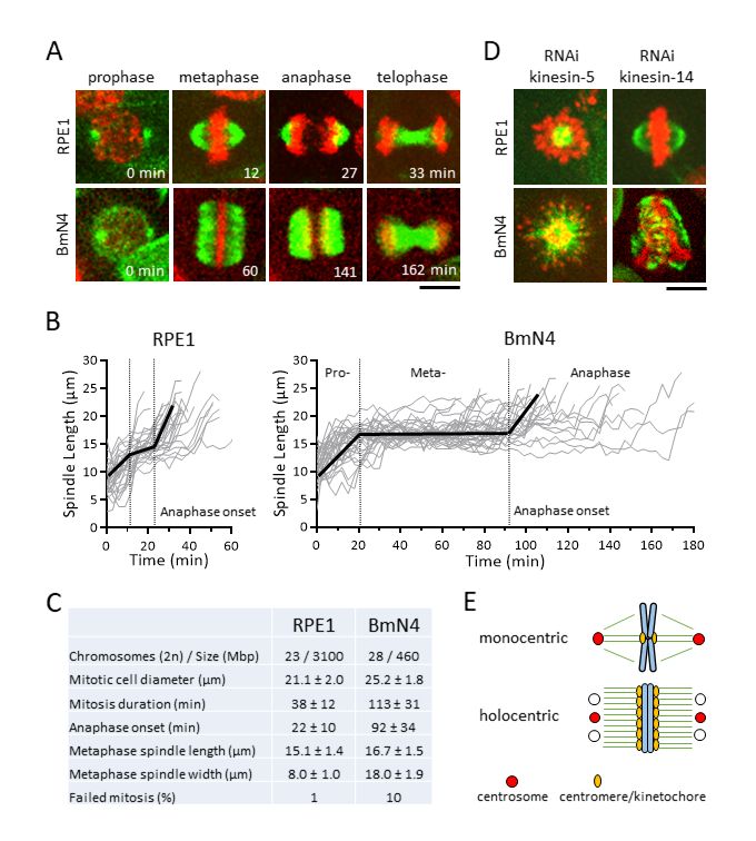
spindle architecture and dynamics, and highlighting differential kinesin function between mono- and holocentric cells.9/1/2021 - Open Access Figure 1. Holocentric BmN4 cells have “square” spindle architecture and long metaphase duration.: A. Time-lapse images of human hTERT-RPE1 and silkworm Bmn4-SID1 cells undergoing mitosis (green = microtubules; red = DNA). Scale bar, 10 μm. B. Spindle length versus time plot for RPE1 (n = 27 cells) and BmN4 (n = 31 cells) during mitosis (bold dark lines mark average spindle length; dashed lines mark transition of mitotic phases). C. Mitotic spindle parameters of wild-type RPE1 and BmN4 cells (mean ± standard deviation). D. Metaphase spindle phenotypes of RPE1 and BmN4 cells RNAi-mediated depletion of kinesin-5 and kinesin-14. Scale bar, 10 μm. E. Cartoon model of monocentric and holocentric spindle and chromosome architecture – square spindle may have multiple centrosome-like microtubule organizing centers. Description Human hTERT-RPE1 cells represent wild-type monocentric cells as they were immortalized, via telomerase expression, from normal somatic retinal pigment epithelium cells (Jiang et al. 1999). Silkworm BmN4-SID1 cells represent wild-type holocentric cells as they were spontaneously immortalized from somatic ovary cells, and modified with the SID1 gene from C. elegans for RNAi-mediated depletion (Mon et al. 2012). Interestingly, cancer such as leukemia and lymphoma often exhibits dicentric or tricentric chromosomes (Herry et al. 2007; M’Kacher et al. 2020), a condition reminiscent of holocentric cells. Further, monocentric cells induced to be dicentric showed transformed cancerous properties (Gascoigne and Cheeseman 2013). We seek to understand the differences in spindle assembly dynamics and chromosome segregation
9/1/2021 - Open Access
   in wild-type monocentric RPE1 and holocentric BmN4 cells, to potentially reveal mechanisms of how di- or tricentric
   cancer cells divide.
   We first established protocols for optimal live-cell imaging of RPE1 and BmN4 cells undergoing mitosis (see Methods).
   Using the vital dyes SPY-tubulin and SPY-DNA to label cells, and the spinning disk confocal microscope, we imaged
   RPE1 cells for 3 hr and BmN4 cells for 9 hr with no discernible photo damage. In comparing mitosis between RPE1 and
   BmN4 cells, striking differences in spindle dynamics and spindle architecture were observed. The RPE1 spindles
   exhibited the canonical mitotic phase transition (Fig. 1A). At prophase we observed the initial assembly of the bipolar
   spindle from two foci, likely the centrosome; at metaphase we observed the stable-length spindle with compacted
   metaphase plate-aligned chromosomes and the canonical “oval” shape spindle, with an aspect ratio of ~2 (15.1 μm
   length/8.0 μm width, Fig. 1C); at anaphase we observed chromosome separation concurrent with spindle elongation; at
   telophase we observed the compacted microtubule midzone, the future midbody (Fig. 1A). Total mitosis duration, from
   prophase to telophase, in RPE1 cells was ~38 min, with the onset of anaphase at ~22 min (Fig. 1B, 1C). In contrast,
   BmN4 spindles also exhibited clear mitotic phase transition, but with distinct differences. At prophase we mostly observed
   the assembly of the bipolar spindle from two foci, but in ~14% of cells spindle microtubules nucleated from more than
   two foci, with small microtubules present everywhere near the chromosomes, indicating potential centrosomal and
   acentrosomal MT nucleation (Mon et al. 2014); at metaphase we observed the stable-length spindle with compacted
   metaphase plate-aligned chromosomes and striking “square” shape spindle, with an aspect ratio of ~1 (16.7 μm
   length/18.0 μm width, Fig. 1C), and no clear foci indicative of spindle poles; at anaphase we observed chromosome
   separation concurrent with spindle elongation; at telophase we observed the compacted microtubule midzone (Fig. 1A).
   The square spindle of BmN4 cells is reminiscent of the square spindle of plant cells, which are also holocentric (de
   Keijzer et al. 2014; Zhang and Dawe 2011). Total mitosis duration in BmN4 cells was ~113 min, with the onset of
   anaphase at ~92 min (Fig. 1B, 1C). BmN4 cells took ~4X longer time to complete mitosis, with corresponding ~4X longer
   time spent in metaphase, compare to RPE1 cells (Fig. 1C). We also observed that mitotic failure, defined as spontaneous
   cell death or aneuploid cell division, occurred in ~1% (1 of 79 spindles) of RPE1 cells, compared to ~10% (22 of 207
   spindles) of BmN4 cells (Fig. 1C). Given that RPE1 and BmN4 cells have similar chromosome number and round mitotic
   cell size (Fig. 1C), we propose that holocentricity, where multiple kinetochores are present on one chromosome, requires
   longer time to make proper kinetochore-to-MT attachment at metaphase as compared to monocentricity. An exception
   maybe at embryogenesis or early development, where mitosis occurs relatively fast, compared to adult somatic cells such
   as RPE1 and BmN4. The extended metaphase duration and higher frequency of failed mitosis may be a consequence of
   the unusual “square” spindle architecture, or of the multiple kinetochores per chromosome.
   Spindle assembly dynamic is orchestrated by microtubule-associated proteins, many of which are kinesins (Mountain and
   Compton 2000). In human cells, kinesin-5 Kif11 is essential for bipolar spindle assembly and elongation, as its inhibition
   results in monopolar spindle and subsequent cell death (Mayer et al. 1999; Sawin et al. 1992); and Kinesin-14 HSET is
   required to focus the microtubule minus ends at the spindle poles, and its inhibition, while having little effect on the
   metaphase spindle in normal cells, causes catastrophic multipolar spindle defects in cancer cells with supernumerary
   centrosomes (Kleylein-Sohn et al. 2012; Kwon et al. 2008). To compare the function of kinesin-5 and -14 in RPE1 and
   BmN4 cells, we performed RNAi-mediated depletion. Consistent with previous reports, we observed monopolar spindles
   in kinesins-5 siRNA-treated RPE1 cells, and seemingly normal metaphase spindles in kinesin-14 siRNA-treated RPE1
   cells (Fig. 1D). In contrast, while we also observed monopolar spindles in kinesins-5 dsRNA-treated BmN4 cells, we
   observed mal-shaped metaphase spindles and unaligned chromosomes in kinesins-14 dsRNA-treated BmN4 cells (Fig.
   1D). The result indicates that BmN4 cells need kinesin-14 for proper spindle assembly and chromosome segregation,
   more so than RPE1 cells, and highlights the importance of differential motor function in the context of mono- and
   holocentricity.
   In summary, the holocentric silkworm cell BmN4 is an emergent model system to investigate novel pathways of spindle
   assembly and centromere-kinetochore assembly (Cortes-Silva et al. 2020; Li et al. 2019; Mon et al. 2014; Mon et al.
   2017; Senaratne et al. 2021). We define similarities and differences in spindle architecture and dynamics between BmN4
   and monocentric human RPE1 cells (Fig. 1E). Future work should probe the impact of the centrosome, the
   centromere/kinetochore, the spindle assembly checkpoint, on spindle architecture and dynamics and chromosome
   segregation. This work sets the stage for pinpointing and investigating specific genes/proteins affecting only BmN4 cells,
   but not RPE1 cells, in mono- and tricentric human cancer cells.

   Methods
   Request a detailed protocol
   Cells
   We used human hTERT-RPE1 (RRID:CVCL_4388) and silkworm BmN4-SID1 (RRID:CVCL_Z091) cells (Bodnar et al.
   1998; Mon et al. 2012). These immortalized cells are considered normal diploid cells, as they have proper number of
   chromosomes and do not exhibit transformed phenotypes. An added benefit is that they are easily amenable to RNAi-
   mediated depletion, enabling functional studies of protein-of-interest. Cell culture condition is well-established (Bodnar et
9/1/2021 - Open Access
   al. 1998; Mon et al. 2012). Briefly, hTERT-RPE1 cells were maintained in DMEM/F-12 medium (GIBCO Cat# 21041-
   025) supplemented with 10% fetal calf serum (Biowest Cat# S181T-500), 1% penicillin-streptomycin (GIBCO Cat#
   15140-22) in 5% CO2 incubator at 37°C; and BmN4-SID1 cells were maintained in Sf-900 II SFM medium (GIBCO Cat#
   10902-088) supplemented with 5% fetal calf serum (Biowest Cat# S181T-500), 1% penicillin-streptomycin (GIBCO Cat#
   15140-122) and 1% L-glutamine (GIBCO Cat# 25030-024) in a 27°C incubator.
   RNAi
   RNA-interference is well-established for RPE1 cells (Jackson et al. 2006). Briefly, we treated RPE1 cells with 20 nM of
   siRNA-mediated kinesin-5 or kinesin-14 depletion reagents (Dharmacon ON-TARGETplus siRNA SMARTPool) 48 hr
   prior to imaging. A subset of the treated-cells were used for RT-qPCR to determine the level of mRNA depletion. RNA-
   interference for BmN4 cells has been established (Mon et al. 2012), utilizing the ability of BmN4-SID1 cells to soak up
   dsRNA in the medium to perform RNA-interference. Briefly, ~300 bp of dsRNA targeting silkworm kinesin-5 and
   kinesin-14 were generated from DNA templates flanked by T7 promoter sequences on both sides using the MAXIscript
   T7 Transcription Kit (Thermo Fisher Scientific, Cat# AM1312) . We treated BmN4-SID1 cells with 20 nM of dsRNA 48
   hr prior to imaging. A subset of the treated-cells were used for RT-qPCR to determine the level of mRNA depletion. The
   cells were cultured in a 2 mL FluoroDish (World Precision Instruments, Cat# FD35-100).
   Live-cell imaging
   One hour prior to imaging, SPY-DNA-555nm (Spirochrome Cat# SC201) and SPY-tubulin-650nm (Spirochrome Cat#
   SC503) dyes were added to the medium at a 10,000X dilution. Cells were imaged using the spinning disk confocal
   microscope. Briefly, the Nikon Eclipse Ti-E perfect focus inverted microscope, with 40X/1.3 N.A. Plan Apo oil
   immersion objective lens and Mad City Piezo stepper stage, coupled to the Yokogawa CSU-X1 spinning disk confocal
   unit, the Photometrics Cascade EM-CCD camera, the Gataca Systems laser unit with 561 nm (100 mW) and 642 nm (100
   mW) lines, controlled by Molecular Devices software MetaMorph 7.8, enclosed within a thermal box to keep stable
   temperatures of 27°C (BmN4 cells) or 37°C (RPE1 cells). Movies were made with the following parameters: laser power
   5mW, EM-gain 300, Bin 1X, exposure time 100-200 ms, 13 optical z-sections, 2 µm spacing per 3D stack, 3 min time
   interval between stacks, and 3 hr movies (RPE1 cells) or 9 hr movies (BmN4 cells).

   Reagents
   Dharmacon ON-TARGETplus siRNA SMARTPool Cat# L-003317-00-0005 (human kinesin-5 KIF11)
   Dharmacon ON-TARGETplus siRNA SMARTPool Cat# L-004958-00-0005 (human kinesin-14 KIFC1)
   Bombyx mori primers to generate T7 template for kinesin-5 dsRNA generation:
   sense: GCTGGCTACAATTGCACTGT
   antisense: TCATGAGTGTCGAAGCCACT
   Bombyx mori primers to generate T7 template for kinesin-14 dsRNA generation:
   sense: TGCTGCACCTCGGATTAAGA
   antisense: CCTCTAGAAGCTTTGTCTTACGT
   Acknowledgments: We thank Sabine Bardin (Institut Curie) for technical advice on methods in cell cultures. We thank
   Vincent Fraisier and Lucie Sengmanivong for the maintenance of microscopes at the PICT-IBiSA Imaging facility (Institut
   Curie), a member of the France-BioImaging national research infrastructure. LV is Fellow of the Programme Médicine-
   Sciences ENS-PSL. IAD is a member of the Labex DEEP, PTT is a member of the Labex CelTisPhyBio, part of IDEX
   PSL.

   References
   Bodnar AG, Ouellette M, Frolkis M, Holt SE, Chiu CP, Morin GB, Harley CB, Shay JW, Lichtsteiner S, Wright WE.
   1998. Extension of life-span by introduction of telomerase into normal human cells. Science. 279: 349-352. PMID:
   9454332.
   Cortes-Silva N, Ulmer J, Kiuchi T, Hsieh E, Cornilleau G, Ladid I, Dingli F, Loew D, Katsuma S, Drinnenberg IA. 2020.
   CenH3-Independent Kinetochore Assembly in Lepidoptera Requires CCAN, Including CENP-T. Curr Biol. 30: 561-
   572.e10. PMID: 32032508.
   de Keijzer J, Mulder BM, Janson ME. 2014. Microtubule networks for plant cell division. Syst Synth Biol. 8: 187-94.
   PMID: 25136380.
   Gascoigne KE, Cheeseman IM. 2013. Induced dicentric chromosome formation promotes genomic rearrangements and
   tumorigenesis. Chromosome Res. 21: 407-418. PMID: 23793898.
9/1/2021 - Open Access
   Herry A, Douet-Guilbert N, Morel F, Le Bris MJ, Morice P, Abgrall JF, Berthou C, De Braekeleer M. 2007. Evaluation of
   chromosome 5 aberrations in complex karyotypes of patients with myeloid disorders reveals their contribution to dicentric
   and tricentric chromosomes, resulting in the loss of critical 5q regions. Cancer Genet Cytogenet. 175: 125-131. PMID:
   17556068.
   Jackson AL, Burchard J, Leake D, Reynolds A, Schelter J, Guo J, Johnson JM, Lim L, Karpilow J, Nichols K, Marshall
   W, Khvorova A, Linsley PS. 2006. Position-specific chemical modification of siRNAs reduces "off-target" transcript
   silencing. RNA. 12: 1197-1205. PMID: 16682562.
   Jiang XR., Jimenez G, Chang E, Frolkis M, Kusler B, Sage M, Beeche M, Bodnar AB, Wahl GM, Tlsty TD, Chiu CP.
   1999. Telomerase expression in human somatic cells does not induce changes associated with a transformed phenotype.
   Nat Genet. 21: 111-114. PMID: 9916802.
   Kleylein-Sohn J, Pollinger B, Ohmer M, Hofmann F, Nigg EA, Hemmings BA, Wartmann M. 2012. Acentrosomal spindle
   organization renders cancer cells dependent on the kinesin HSET. J Cell Sci. 125: 5391-5402. PMID: 22946058.
   Kwon M, Godinho SA, Chandhok NS, Ganem NJ, Azioune A, Thery M, Pellman D. 2008. Mechanisms to suppress
   multipolar divisions in cancer cells with extra centrosomes. Genes Dev. 22: 2189-2203. PMID: 18662975.
   Li B, Li Z, Lu C, Chang L, Zhao D, Shen G, Kusakabe T, Xia Q, Zhao P. 2019. Heat Shock Cognate 70 Functions as A
   Chaperone for the Stability of Kinetochore Protein CENP-N in Holocentric Insect Silkworms. Int J Mol Sci. 20: 5823.
   PMID: 31756960.
   M'Kacher R, Colicchio B, Borie C, Junker S, Marquet V, Heidingsfelder L, Soehnlen K, Najar W, Hempel WM, Oudrhiri
   N, Wilhelm-Murer N, Miguet M, Arnoux M, Ferrapie C, Kerbrat W, Plesch A, Dieterlen A, Girinsky T, Voisin P,
   Deschenes G, Tabet AC, Yardin C, Bennaceur-Griscelli A, Fenech M, Carde P, Jeandidier E. 2020. Telomere and
   Centromere Staining Followed by M-FISH Improves Diagnosis of Chromosomal Instability and Its Clinical Utility.
   Genes. 11: 475. PMID: 32349350.
   Mayer TU, Kapoor TM, Haggarty SJ, King RW, Schreiber SL, Mitchison TJ. 1999. Small molecule inhibitor of mitotic
   spindle bipolarity identified in a phenotype-based screen. Science. 286: 971-974. PMID: 10542155.
   Mon H, Kobayashi I, Ohkubo S, Tomita S, Lee J, Sezutsu H, Tamura T, Kusakabe T. 2012. Effective RNA interference in
   cultured silkworm cells mediated by overexpression of Caenorhabditis elegans SID-1. RNA Biol. 9: 40-46. PMID:
   22293577.
   Mon H, Lee JM, Mita K, Goldsmith MR, Kusakabe T. 2014. Chromatin-induced spindle assembly plays an important role
   in metaphase congression of silkworm holocentric chromosomes. Insect Biochem Mol Biol. 45: 40-50. PMID: 24291286.
   Mountain V, Compton DA. 2000. Dissecting the role of molecular motors in the mitotic spindle. Anat Rec. 261: 14-24.
   PMID: 10700732.
   Sawin KE, LeGuellec K, Philippe M, Mitchison TJ. 1992. Mitotic spindle organization by a plus-end-directed microtubule
   motor. Nature. 359: 540-543. PMID: 1406972.
   Senaratne AP, Muller H, K.A. Fryer, M. Kawamoto, S. Katsuma, Drinnenberg IA. 2021. Formation of the CenH3-
   Deficient Holocentromere in Lepidoptera Avoids Active Chromatin. Curr Biol. 31: 173-181e177. PMID: 33125865.
   Zhang H, Dawe RK. 2011. Mechanisms of plant spindle formation. Chromosome Res. 19: 335-44. PMID: 21424324.

   Funding: Fondation ARC pour la Recherche sur la Cancer (PTT); La Ligue Contre le Cancer, Ile-de-France (PTT);
   Institut National du Cancer (PTT); ATIP-AVENIR (IAD); ERC-Starting Grant (IAD)
   Author Contributions: Lucien Vanpoperinghe: Conceptualization, Methodology, Investigation, Formal analysis, Writing
   - original draft. Frederique Carlier-Grynkorn: Methodology, Investigation. Gaetan Cornilleau: Methodology, Investigation.
   Takahiro Kusakabe: Methodology. Ines A Drinnenberg: Conceptualization, Methodology, Funding acquisition,
   Supervision, Writing - review and editing. Phong T Tran: Conceptualization, Methodology, Funding acquisition,
   Supervision, Writing - review and editing.
   Reviewed By: Stefanie Redemann
   History: Received June 29, 2021 Revision received August 4, 2021 Accepted August 21, 2021 Published September
   1, 2021
   Copyright: © 2021 by the authors. This is an open-access article distributed under the terms of the Creative Commons
   Attribution 4.0 International (CC BY 4.0) License, which permits unrestricted use, distribution, and reproduction in any
   medium, provided the original author and source are credited.
9/1/2021 - Open Access
   Citation: Vanpoperinghe, L; Carlier-Grynkorn, F; Cornilleau, G; Kusakabe, T; Drinnenberg, IA; Tran, PT (2021). Live-
   cell imaging reveals square shape spindles and long mitosis duration in the silkworm holocentric cells. microPublication
   Biology. https://doi.org/10.17912/micropub.biology.000441
You can also read