Injury of the optic radiation in patients with mild TBI: A DTT study
←
→
Page content transcription
If your browser does not render page correctly, please read the page content below
Translational Neuroscience 2020; 11: 335–340
Research Article
Sung Ho Jang, Seong Ho Kim, You Sung Seo*
Injury of the optic radiation in patients with
mild TBI: A DTT study
https://doi.org/10.1515/tnsci-2020-0108
received April 02, 2020; accepted August 23, 2020
1 Introduction
Abstract Traumatic brain injury (TBI) can be classified as mild,
Objectives ‒ We investigated injuries of the optic moderate, or severe based on assessment of the injury,
radiations (ORs) in patients with mild traumatic brain and mild TBI comprises approximately 75% of all TBIs
injury (TBI) by using diffusion tensor tractography (DTT). [1,2]. The visual system is vulnerable to TBI because
Methods ‒ Fifty-two consecutive patients who com- several cranial nerves and about 30–40 cortical areas are
plained of visual problems showed abnormal visual involved in vision [1,2]. The prevalence of visual
evoked potential (VEP) latency but no abnormality on problems (e.g., oculomotor problems, visual field de-
conventional brain MRI after mild TBI, and fifty normal fects, visual information processing dysfunction, and
control subjects were recruited for this study. Subjects’ ORs visual attention deficits) in patients with TBI is high,
were reconstructed using DTT, and three DTT parameters approximately 50% of cases [3–6]. However, research on
(fractional anisotropy [FA], apparent diffusion coefficient
visual problems in mild TBI is rare; to the best of our
[ADC], and tract volume) were measured for each OR.
knowledge, only one study has reported on the
Results ‒ Mean FA value and tract volume of the OR
prevalence of visual problems in mild TBI. In that study,
were significantly lower in the patient group than in
about 15% of mile TBI patients had visual problems
the control group (p < 0.05). However, there was no
(light sensitivity, 7%; blurred vision, 6%; double vision,
significant difference in the ADC values of the OR
2%) [7]. Since the introduction of diffusion tensor
between the patient and control groups (p > 0.05). A
imaging (DTI), several studies have described neural
weak negative correlation was detected between VEP
tract injuries in patients with mild TBI [8–10]. The
latency and OR fiber number (r = 0.204, p < 0.05).
demonstration of injury of neural tracts in patients with
Conclusions ‒ DTT revealed that OR injuries were not
mild TBI is clinically important because such patients
detected on the conventional brain MRI scans of patients
usually show no abnormality on conventional brain MRI
who complained of visual problems and had abnormal
[8,10–13]. Injury of the neural tracts after mild TBI has
VEP latency after mild TBI. Our results suggest that DTT
been demonstrated in the corticospinal tract, as well as
would be a useful technique for detecting OR injury in
in the fornix and cingulum [8,10–13]. However, little has
patients with mild TBI.
been reported on mild TBI and the neural tracts involved
Keywords: diffusion tensor imaging, diffusion tensor in visual function, such as the optic radiation (OR) [11].
tractography, optic radiation, visual evoked potential, The OR is not easily distinguishable from adjacent
mild traumatic brain injury, head trauma neural structures. Therefore, a precise diagnosis of an OR
injury is difficult when using conventional MRI or
positron emission tomography [14–16]. However, diffu-
* Corresponding author: You Sung Seo, Department of Physical
sion tensor tractography (DTT), derived from DTI, allows
Medicine and Rehabilitation, College of Medicine, Yeungnam
University, 317-1, Daemyungdong, Namku, Taegu, 705-717, Republic three-dimensional reconstruction and evaluation of
of Korea, e-mail: yousung1008@hanmail.net neural tracts, including the OR [14,16]. Although many
Sung Ho Jang: Department of Physical Medicine and Rehabilitation, studies have used DTI or DTT to describe OR injuries in
College of Medicine, Yeungnam University, 317-1, Daemyungdong, patients with various brain pathologies including TBI
Namku, Taegu, 705-717, Republic of Korea,
[17–19], little has been reported about such injuries in
e-mail: strokerehab@hanmail.net
Seong Ho Kim: Department of Neurosurgery, College of Medicine
mild TBI [9,20].
Yeungnam University, 317-1, Daemyungdong, Namku, Taegu, 705- In this study, we used DTT to investigate OR injuries
717, Republic of Korea, e-mail: shkim@medical.yeungnam.ac.kr in patients with mild TBI.
Open Access. © 2020 Sung Ho Jang et al., published by De Gruyter. This work is licensed under the Creative Commons Attribution 4.0
International License.336 Sung Ho Jang et al.
2 Subjects and methods (3) more than 1 month after TBI onset; (4) age ranging
from 18 to 75 years; (5) delayed visual evoked potential
(VEP) latency; (6) complaints related to visual problems
2.1 Subjects
(e.g., visual defect, poor vision, or blurred vision);
and (7) no history of head trauma or neurologic or
Fifty-two patients (20 males, 32 females; mean age: psychiatric disease. No significant differences in age or
45.9 ± 15.2 years, range: 18–72 years) with TBI who sex compositions were detected between the patient and
complained of visual problems and visited the rehabili- normal control groups (p > 0.05).
tation department of a university hospital and 50 normal The VEP latent period was used to evaluate the
control subjects (23 males and 27 females; mean age: status of the visual pathway. The normal VEP latency
42.2 ± 15.6 years, range: 21–75 years) were recruited for period reference values, by age and gender, were as
this study. The patients were recruited according to the follows:Optic radiation injuries in patients with mTBI 337
Ethical approval: The research has been complied with Table 1: Visual problems of individual patients
all the relevant national regulations, institutional poli-
cies, and in accordance the tenets of the Helsinki No. Age Sex Visual defect Poor vision Blurred vision
Declaration. The data were collected retrospectively, 1 45 F ○ ○
and the study protocol was approved by the appropriate 2 23 F ○
institutional review board. 3 56 F ○
4 52 F ○ ○
5 13 F ○ ○
6 50 F ○ ○
7 60 F ○ ○
2.2 DTI 8 40 F ○
9 35 F ○ ○
DTI data were acquired at an average of 5.2 ± 3.4 months 10 22 F ○ ○
after onset using a six-channel head coil on a 1.5 T 11 56 F ○ ○
12 72 F ○
Philips Gyroscan Intera MRI scanner. Imaging parameters
13 65 F ○ ○
were as follows: acquisition matrix = 96 × 96; recon- 14 53 M ○ ○
structed to matrix = 192 × 192 matrix; field of view = 15 42 M ○ ○
240 mm × 240 mm; TR = 10,398 ms; TE = 72 ms; EPI factor = 16 18 M ○ ○ ○
5.9; b = 1,000 s/mm2; NEX = 1; slice gap = 0 mm; slice 17 62 F ○ ○ ○
18 30 M ○ ○
thickness = 2.5 mm.
19 46 M ○ ○
Eddy current-induced image distortions were re- 20 50 F ○ ○
moved by using affine multiscale two-dimensional 21 49 F ○ ○
registration as provided in the Oxford Centre for 22 61 F ○
Functional Magnetic Resonance Imaging of Brain 23 56 F ○ ○
Software Library (FSL; www.fmrib.ox.ac.uk/fsl) [23]. 24 58 F ○ ○
25 33 F ○
DTI-Studio software (CMRM, Johns Hopkins Medical
26 39 M ○ ○ ○
Institute, Baltimore, MD, USA) was used for OR evalua- 27 41 M ○ ○
tion [24]. Fiber tracking was based on the fiber assign- 28 56 M ○ ○
ment continuous tracking algorithm and the multiple 29 58 M ○ ○
regions of interest (ROI) approach. To delineate the OR, a 30 63 F ○ ○
31 35 F ○ ○
seed ROI was placed on the lateral geniculate body (LGB)
32 65 F ○ ○
on the color map, whereas the target ROI was placed on 33 38 F ○ ○
the color map at the OR bundle located in the middle 34 38 M ○
portion between the LGB and the occipital pole [14,15]. 35 19 M ○
Fiber tracking was performed based on a fractional 36 25 F ○
anisotropy (FA) threshold of >0.15 and a direction 37 35 F ○
38 59 F ○ ○
threshold of338 Sung Ho Jang et al.
visual cortex-to-vertex) with OS (both eyes), OU (the Table 2: Comparison of DTT parameters between the patient and
right eye), and OD (the left eye) stimulation. control groups
Patient group Control group p-Value
FA 0.47 (±0.11) 0.50 (±0.05) 0.042a
2.3 Statistical analysis ADC 0.64 (±0.18) 0.60 (±0.04) 0.602
Fiber number 544.55 (±347.44) 1253.25 (±306.20) 0.001*
DTT data were analyzed by performing group-based FA: fractional anisotropy, ADC: apparent diffusion coefficient,
analyses of the DTT parameters of the ORs in the patient values represent patients: mean ± standard deviation (controls:
and control groups. SPSS software (v. 15.0; SPSS, Inc., mean ± standard deviation).
a
IBM Company, Chicago, Illinois, USA) was used for data Significant difference between the patient and control groups,
p < 0.05.
analysis. The chi-squared test was used to examine the
difference in sex compositions of the patient and control
groups, and an independent t-test was used to assess
age differences between the patient and control groups. Table 3: Correlation between DTT parameters and VEP latency
Paired t-tests were used to assess the differences in DTT
FA ADC Fiber number
parameter values of the ORs of the patient and control
groups. Pearson correlation coefficients were calculated VEP latency 0.005 0.026 −0.214a
to quantify the correlation between DTT parameters of
FA: fractional anisotropy, ADC: apparent diffusion coefficient, VEP:
the OR and clinical data (i.e., VEP latency). Null visual evoked potential.
hypotheses of no difference were rejected if p-values a
Significant difference between DTT parameter and VEP latency,
were less than 0.05. A correlation coefficient of more p < 0.05.
than 0.60 indicated a strong correlation, a correlation
coefficient between 0.40 and 0.59 indicated a moderate
correlation, a correlation coefficient between 0.20 and the following: (1) FA and tract volume values of the ORs
0.39 indicated a weak correlation, and a correlation were significantly lower in the patient group than in the
coefficient less than 0.19 indicated a very weak relation- control group; (2) tract volume of the OR in the patient
ship [25]. group was weakly negatively correlated with the VEP
latency period.
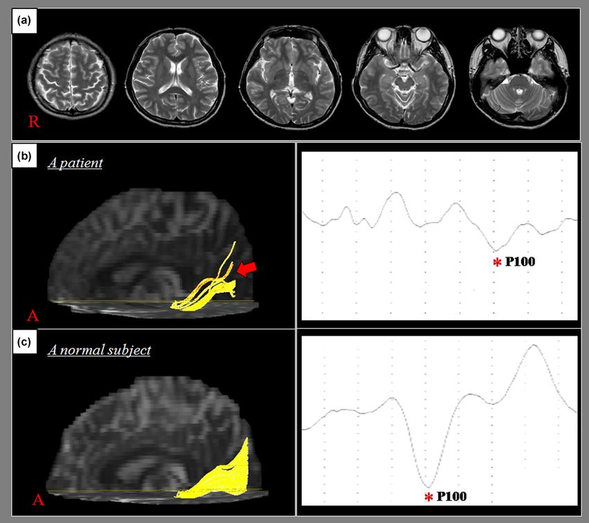
The FA value represents the degree of directionality
of microstructures, while the ADC value represents the
3 Results magnitude of water diffusion [26–28]. Tract volume
represents the number of voxels in a neural tract and
A summary of the comparisons of the DTT parameters of is considered to indicate the number of fibers in that
the patient and control groups is presented in Table 1. tract; therefore, a decrease in the fiber number indicates
The mean FA value and average tract volume of the ORs injury to a neural tract [29]. Decrements in FA and fiber
of the patient group were significantly lower than those number without a similar decrement in ADC in the
in the control group (p < 0.05). However, no significant patient group appears to indicate the presence of OR
difference was detected between the ADC values of the injuries in the patient group. Because the conventional
ORs of the patient and control groups (p > 0.05) (Table 2). brain MRI scans of the subjects in the patient group were
A weak negative correlation was observed between normal, we suggest that the injury of these neural tracts
the VEP latency values and fiber numbers of the ORs of was the result of traumatic axonal injury [30,31].
the patient group (r = 0.204, p < 0.05) (Table 3). Furthermore, the weak negative correlation between
VEP latency and OR fiber number suggests that a change
in VEP latency reflects a change in the severity of an OR
injury.
4 Discussion Since the introduction of DTI, many studies have
used that imaging approach to document OR injury in
In this study, we investigated injury to the ORs in patients with TBI [9,11,20,30–35]. The majority of these
patients with visual problems and abnormal VEP studies demonstrated the presence of OR injuries in
latencies after the onset of mild TBI and determined patients with moderate or severe TBI [11,30–35]. Only aOptic radiation injuries in patients with mTBI 339
few similar studies of mild TBI have been reported [2] Elisevich KV, Ford RM, Anderson DP, Stratford JG,
[9,20]. In 2015, Jang and Seo investigated two patients Richardson PM. Visual abnormalities with multiple trauma.
with visual field defects in whom OR injuries were Surg Neurol. 1984;22:565–75.
[3] Kelts EA. Traumatic brain injury and visual dysfunction: a
revealed on DTT [9]. During the same year, Vigneswaran
limited overview. NeuroRehabilitation. 2010;27:223–9.
et al. reported the decrements of FA values and [4] Ripley DL, Politzer T. Vision disturbance after TBI.
increments of ADC values on DTI of the ORs in 61 NeuroRehabilitation. 2010;27:215–6.
patients with mild TBI compared with 19 normal control [5] Schlageter K, Gray B, Hall K, Shaw R, Sammet R. Incidence
subjects [20]. To the best of our knowledge, this study is and treatment of visual dysfunction in traumatic brain injury.
Brain Inj. 1993;7:439–48.
the first DTT-based study on patients with visual
[6] Van Stavern GP, Biousse V, Lynn MJ, Simon DJ, Newman NJ.
problems and abnormal VEP latencies after the onset Neuro-ophthalmic manifestations of head trauma.
of mild TBI. However, some limitations of this study J Neuroophthalmol. 2001;21:112–7.
should be considered. First, the fiber tracking technique [7] Lannsjo M, Af Geijerstam JL, Johansson U, Bring J, Borg J.
is operator dependent. Second, DTT can produce false- Prevalence and structure of symptoms at 3 months after mild
traumatic brain injury in a national cohort. Brain Inj.
negative results throughout the white matter of the brain
2009;23:213–9.
due to fiber crossing or the partial volume effect [36,37].
[8] Arfanakis K, Haughton VM, Carew JD, Rogers BP, Dempsey RJ,
Third, we could not investigate the relationship between Meyerand ME. Diffusion tensor MR imaging in diffuse axonal
the severity of visual problems and the magnitude of the injury. AJNR Am J Neuroradiol. 2002;23:794–802.
DTT parameters because the clinical records presented [9] Jang SH, Seo JP. Damage to the optic radiation in patients with
presence or absence information, not severity-related mild traumatic brain injury. J Neuroophthalmol. 2015;35:270–3.
[10] Shenton ME, Hamoda HM, Schneiderman JS, Bouix S,
information. Therefore, further prospective studies in-
Pasternak O, Rathi Y, et al. A review of magnetic resonance
cluding detailed data related to visual problems should imaging and diffusion tensor imaging findings in mild
be encouraged. traumatic brain injury. Brain Imaging Behav. 2012;6:137–92.
In conclusion, by using DTT, we investigated OR [11] Huang MX, Theilmann RJ, Robb A, Angeles A, Nichols S,
injuries in patients who complained of visual problems Drake A, et al. Integrated imaging approach with meg and dti
and had VEP latency abnormalities after the onset of to detect mild traumatic brain injury in military and civilian
patients. J Neurotrauma. 2009;26:1213–26.
mild TBI. Our analysis of DTT parameters revealed the
[12] Jang SH, Kim SY. Injury of the corticospinal tract in patients
presence of OR injuries that were not detected on with mild traumatic brain injury: a diffusion tensor tracto-
conventional brain MRI scans. Our results suggest that graphy study. J Neurotrauma. 2016;33:1790–5.
DTT would be a useful technique for detecting OR [13] Yang DS, Kwon HG, Jang SH. Injury of the thalamocingulate
injuries in patients with mild TBI. tract in the papez circuit in patients with mild traumatic brain
injury. Am J Phys Med Rehabil. 2016;95:E34–E38.
[14] Glass HC, Berman JI, Norcia AM, Rogers EE, Henry RG, Hou C,
Author contributions: Sung Ho Jang: study concept and et al. Quantitative fiber tracking of the optic radiation is
design, manuscript development, and writing; Seong Ho correlated with visual-evoked potential amplitude in preterm
Kim: acquisition and analysis of data; You Sung Seo: infants. AJNR Am J Neuroradiol. 2010;31:1424–9.
study concept and design, acquisition and analysis of [15] Hofer S, Karaus A, Frahm J. Reconstruction and dissection of
data, and manuscript authorization, the entire human visual pathway using diffusion tensor MRI.
Front Neuroanat. 2010;4:15.
[16] Staempfli P, Rienmueller A, Reischauer C, Valavanis A,
Source of funding: This work was supported by the Boesiger P, Kollias S. Reconstruction of the human visual
National Research Foundation of Korea (NRF) grant system based on dti fiber tracking. J Magn Reson Imaging.
funded by the Korean Government(MSIP) (No. 2007;26:886–93.
2018R1A2B6000996)., [17] Seo JP, Choi BY, Chang CH, Jung YJ, Byun WM, Kim SH, et al.
Diffusion tensor imaging findings of optic radiation in patients
with putaminal hemorrhage. Eur Neurol. 2013;69:236–41.
Conflict of interest: The authors state no conflict of [18] Taoka T, Sakamoto M, Nakagawa H, Nakase H, Iwasaki S,
interest. Takayama K, et al. Diffusion tensor tractography of the meyer
loop in cases of temporal lobe resection for temporal lobe
epilepsy: correlation between postsurgical visual field defect
and anterior limit of meyer loop on tractography. AJNR Am J
References Neuroradio. 2008;29:1329–34.
[19] Yogarajah M, Focke NK, Bonelli S, Cercignani M, Acheson J,
[1] De Kruijk JR, Twijnstra A, Leffers P. Diagnostic criteria and Parker GJ, et al. Defining meyer’s loop-temporal lobe
differential diagnosis of mild traumatic brain injury. Brain Inj. resections, visual field deficits and diffusion tensor tracto-
2001;15:99–106. graphy. Brain. 2009;132:1656–68.340 Sung Ho Jang et al.
[20] Veeramuthu V, Narayanan V, Kuo TL, Delano-Wood L, [30] Johnson VE, Stewart W, Smith DH. Axonal pathology in
Chinna K, Bondi MW, et al. Diffusion tensor imaging traumatic brain injury. Exp Neurol. 2013;246:35–43.
parameters in mild traumatic brain injury and its correlation [31] Povlishock JT. Traumatically induced axonal injury: patho-
with early neuropsychological impairment: a longitudinal genesis and pathobiological implications. Brain Pathol.
study. J Neurotrauma. 2015;32:1497–509. 1992;2:1–12.
[21] Alexander MP. Mild traumatic brain injury: pathophysiology, [32] Alhilali LM, Yaeger K, Collins M, Fakhran S. Detection of
natural history, and clinical management. Neurology. central white matter injury underlying vestibulopathy
1995;45:1253–60. after mild traumatic brain injury. Radiology. 2014;
[22] Liveson JA, Ma DM. Laboratory reference for clinical neuro- 272:224–32.
physiology. Philadelphia: F.A. Davis Company; 1992. [33] Caeyenberghs K, Leemans A, Geurts M, Taymans T, Linden CV,
[23] Smith SM, Jenkinson M, Woolrich MW, Beckmann CF, Smits-Engelsman BCM, et al. Brain-behavior relationships
Behrens TE, Johansen-Berg H, et al. Advances in functional in young traumatic brain injury patients: fractional
and structural MR image analysis and implementation as FSL. anisotropy measures are highly correlated with dynamic
Neuroimage. 2004;23(Suppl 1):S208–19. visuomotor tracking performance. Neuropsychologia.
[24] Jiang H, van Zijl PC, Kim J, Pearlson GD, Mori S. Dtistudio: 2010;48:1472–82.
resource program for diffusion tensor computation and fiber [34] Palmer HS, Garzon B, Xu J, Berntsen EM, Skandsen T,
bundle tracking. Comput Methods Prog Biomed. 2006;81:106–16. Haberg AK. Reduced fractional anisotropy does not change the
[25] Evans JD. Straignt forward statistics for the behavioural shape of the hemodynamic response in survivors of
science. Pacific Grove, CA: Brooks, Cole Pub; 1996. severe traumatic brain injury. J Neurotrauma.
[26] Assaf Y, Pasternak O. Diffusion tensor imaging (DTI)-based 2010;27:853–62.
white matter mapping in brain research: a review. J Mol [35] Yeo SS, Kim SH, Kim OL, Kim MS, Jang SH. Optic radiation
Neurosci. 2008;34:51–61. injury in a patient with traumatic brain injury. Brain Inj.
[27] Mori S, Crain BJ, Chacko VP, van Zijl PC. Three-dimensional 2012;26:891–5.
tracking of axonal projections in the brain by magnetic [36] Lee SK, Kim DI, Kim J, Kim DJ, Kim HD, Kim DS, et al. Diffusion-
resonance imaging. Ann Neurol. 1999;45:265–9. tensor MR imaging and fiber tractography: a new method of
[28] Neil JJ. Diffusion imaging concepts for clinicians. J Magn Reson describing aberrant fiber connections in developmental CNS
Imaging. 2008;27:1–7. anomalies. Radiographics. 2005;25:53–65.
[29] Pagani E, Agosta F, Rocca MA, Caputo D, Filippi M. Voxel- [37] Parker GJM, Alexander DC. Probabilistic anatomical connec-
based analysis derived from fractional anisotropy images of tivity derived from the microscopic persistent angular
white matter volume changes with aging. Neuroimage. structure of cerebral tissue. Philos T R Soc B.
2008;41:657–67. 2005;360:893–902.You can also read