Case Report The Challenges of ST-Elevation Myocardial Infarction in COVID-19 Patients
←
→
Page content transcription
If your browser does not render page correctly, please read the page content below
Hindawi
Case Reports in Cardiology
Volume 2021, Article ID 9915650, 5 pages
https://doi.org/10.1155/2021/9915650

Case Report
The Challenges of ST-Elevation Myocardial Infarction in
COVID-19 Patients

          Ju Young Bae ,1 Khalil Ian Hussein ,1 Christopher John Howes ,1,2
          and John Francis Setaro 1,2
          1
              Department of Medicine, Greenwich Hospital, Yale-New Haven Health System, 5 Perryridge Road, Greenwich, CT 06830, USA
          2
              Section of Cardiovascular Medicine, Department of Internal Medicine, Yale University School of Medicine, 333 Cedar St,
              New Haven, CT 06510, USA

          Correspondence should be addressed to John Francis Setaro; john.setaro@yale.edu

          Received 4 April 2021; Accepted 2 August 2021; Published 21 August 2021

          Academic Editor: Vincenzo Russo

          Copyright © 2021 Ju Young Bae et al. This is an open access article distributed under the Creative Commons Attribution License,
          which permits unrestricted use, distribution, and reproduction in any medium, provided the original work is properly cited.

          By July 2021, the United States had over 34.4 million confirmed COVID-19 cases. Various cardiovascular manifestations of
          COVID-19 have been reported including ST-elevation myocardial infarction (STEMI), and there is concern that SARS-CoV-2
          may be associated with a higher thrombus burden. We performed a retrospective chart review of 535 adult patients with
          COVID-19 admitted at Yale-New Haven Health Greenwich Hospital from February 1, 2020, to May 13, 2020. All admitted
          patients had undergone testing for serum troponin I and various inflammatory markers, and we identified three patients who
          were diagnosed with acute STEMI. Data was collected via manual chart review and included patient demographics,
          comorbidities, laboratory tests, electrocardiogram (ECG) results, echocardiography results, diagnoses during hospitalization,
          inpatient therapies, and outcomes including length of hospital stay, revascularization results, and mortality. Three of our
          patients had obstructive coronary artery disease confirmed via angiography. One subject was noted to display vasospasm in
          addition to coronary atherosclerotic obstruction and refractory thrombus formation. Among our patients with COVID-19 and
          STEMI, presentations were variable in terms of timing of onset of ECG changes, age, gender, race, comorbidities,
          symptomology, and outcomes.

1. Introduction                                                        2. Case Presentation
As of July 2021, the United States has over 34.4 million               We performed a single-center retrospective chart review of
confirmed cases of COVID-19, which is an infectious                     535 adult patients with COVID-19 admitted to our suburban
disease caused by severe acute respiratory syndrome coro-              community hospital, Yale-New Haven Health Greenwich
navirus 2 (SARS-CoV-2) [1]. SARS-CoV-2 infection has                   Hospital, from February 1, 2020, to May 13, 2020, and we
variable manifestations in humans, and there are reported              identified three cases of acute STEMI who underwent coro-
cases of patients with COVID-19 presenting with a broad                nary angiography. These patients diagnosed with COVID-19
spectrum of cardiovascular manifestations including acute              had a positive result on polymerase chain reaction (PCR)
coronary syndrome, arrhythmia, myopericarditis mimick-                 testing of a nasopharyngeal sample. After being granted
ing ST-elevation myocardial infarction (STEMI), ischemic               Institutional Review Board approval, subjects were identified
and stress-induced cardiomyopathy, coronary vasospasm,                 by requesting access to a list of all hospitalized patients with
and pericardial effusion [2–4].                                         COVID-19 from our Infection Control department.
2 Case Reports in Cardiology
I aVR V1 V4
II aVL V2 V5
III aVF V3 V6
II
(a)
(b) (c)
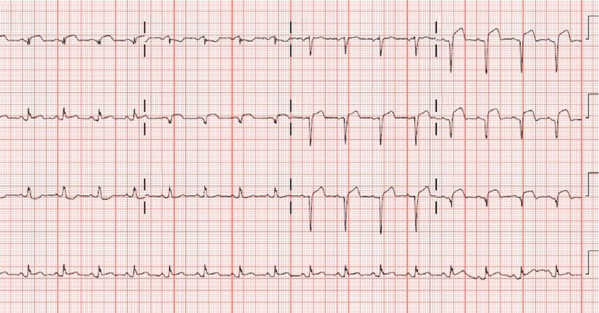
Figure 1: ECG on admission and LHC findings. (a) ECG showing ST elevations in anterior leads. (b) Initial coronary angiography.
(c) Coronary angiography after PCI. Arrows indicate the course of LAD.
3. Case 1 Course was further complicated by elevated D-dimer of
33.35 mg/L FEU for which he was started on heparin drip,
A 59-year-old Caucasian male was admitted for hypoxia in new atrial flutter, and septic shock in setting of prolonged
the setting of COVID-19. His past medical history was ventilator requirement. He received digoxin 62.5 mcg daily,
notable for type 2 diabetes mellitus, hyperlipidemia, and metoprolol tartrate 37.5 mg every 6 hours, and amiodarone
overweight BMI, and he took aspirin 81 mg daily for primary loading dose followed by maintenance at 200 mg daily for
prevention. On admission, blood pressure (BP) was atrial flutter. He eventually required the initiation of
148/79 mmHg, respiratory rate was 24, and oxygen satura- norepinephrine for blood pressure support, which also opti-
tion was 89% on room air. There were no notable findings mized the patient’s heart rate, so metoprolol was discontin-
on the physical exam. Chest X-ray (CXR) showed patchy ued. For management of COVID-19, the patient received a
bilateral pulmonary infiltrates. Hospitalization was compli- 5-day course of azithromycin, a 9-day course of hydroxy-
cated by acute chest pain on hospital day five with ECG chloroquine, one dose of tocilizumab, and a 5-day course of
showing anterior wall STEMI (Figure 1(a)). He was intubated Lopinavir-Ritonavir, as was recommended by the Yale-New
and left heart catheterization (LHC) revealed 40% stenosis of Haven Health system’s treatment guidelines at the time.
distal left main artery (LM), 100% stenosis of midleft anterior The patient expired on day 30 of hospitalization after the
descending artery (LAD), normal left circumflex (LCx), 80% family opted to transition to comfort care.
stenosis of proximal right coronary artery (RCA), and prox-
imal posterior descending artery (Figure 1(b)). The proce- 4. Case 2
dure was complicated by heavy thrombus burden in LAD
and “no-reflow” phenomenon despite the use of rescue A 47-year-old Hispanic male presented with a 2-week history
thrombectomy, multiple intracoronary vasodilators, and 3 of intermittent substernal chest pain with exertion and
drug-eluting stent (DES) placement; flow was not reestab- dyspnea. He had completed a 5-day course of azithromycin
lished to the apex (Figure 1(c)), which was concerning for as an outpatient. His past medical history was notable for
stent thrombosis. Troponin I peaked at 105.0 ng/mL (normal prediabetes, hypertension treated with amlodipine 5 mg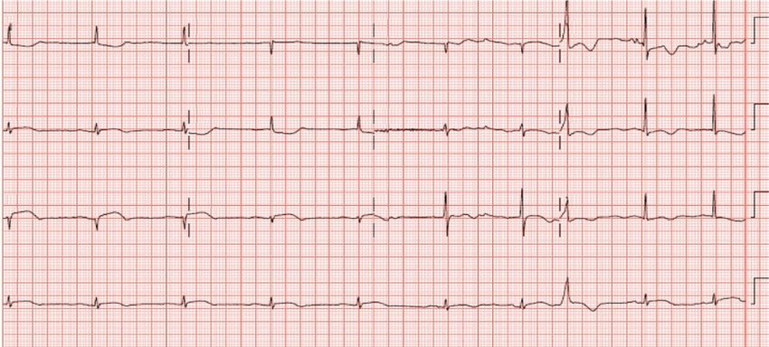
Case Reports in Cardiology                                                                                                            3

                           I                   aVR                  V1                   V4

                           II                  aVL                  V2                   V5

                           III                 aVF                  V3                   V6

                           II

                                                                    (a)

                                               (b)                                            (c)

Figure 2: ECG on admission and LHC findings. (a) ECG showing ST elevations in anterolateral leads. (b) Initial coronary angiography with
arrow showing occluded LAD. (c) Coronary angiography after PCI with arrow showing coronary vasospasm in distal LM.

of 15.3 ×1000/μL, Troponin I of 72.4 ng/mL, pro-BNP of                    5. Case 3
1,184 pg/mL, and total CK of 3,549 U/L. COVID-19 PCR
was positive. CXR showed mild heterogeneous bilateral                     A 75-year-old African American female presented with acute
ground-glass opacities. ECG demonstrated ST-elevation over                nausea, diaphoresis, and weakness while climbing stairs. Past
anterolateral wall distribution (Figure 2(a)). The patient was            medical history was notable for hypertension treated with
electively intubated, and LHC revealed late presenting ante-              amlodipine 10 mg and hydrochlorothiazide 25 mg daily,
rior wall MI with 30% stenosis of distal LM, 100% stenosis                overweight BMI, and illness four weeks prior to this admission
of LAD, normal LCx, and normal RCA. Left ventricular                      that was suspected to be COVID-19. CXR showed diffuse inter-
(LV) filling pressures were moderately elevated at 18 mmHg                 stitial changes. ECG performed by EMS showed atrial fibrilla-
(Figure 2(b)). Coronary vasospasm was noted and was                       tion with ST segment elevation. Vitals on presentation
treated using intracoronary vasodilators. Two DES were                    included HR 58 bpm, RR 22, BP 99/56mmHg, and SpO2 of
placed in the proximal and distal LAD, but no reflow was                   100% on room air. Physical exam was noncontributory. Labo-
observed with noteworthy thrombus burden likely from stent                ratory work showed WBC 12.7 ×1000/μL, D-dimer 0.6 mg/L,
thrombosis (Figure 2(c)). Troponin I peaked at 131.2 ng/mL.               Troponin I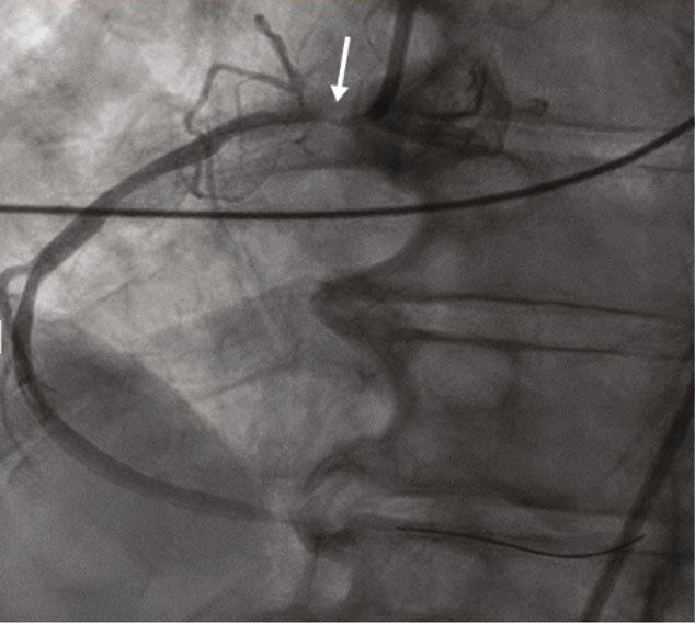
4                                                                                                             Case Reports in Cardiology

                          I                      aVR                V1                      V4

                          II                     aVL                V2                     V5

                          III                    aVF                V3                     V6

                         II

                                                                    (a)

                                           (b)                                              (c)

Figure 3: ECG on admission and LHC findings. (a) ECG showing ST elevations in inferolateral leads. (b) Initial coronary angiography with
arrow showing RCA ostium. (c) Coronary angiography after PCI.

    The patient’s hospital course was eventful for development            any oral anticoagulation therapy prior to hospitalization.
of bilateral neck swelling and angioedema without airway                  Ultimately, two patients were started on anticoagulation
compromise considered to be an atypical presentation of                   due to new atrial flutter or significantly elevated D-dimer
angioedema from ticagrelor use. These symptoms were treated               worrisome for venous thromboembolism. This highlights
with intravenous methylprednisone. The patient was dis-                   the need for clinicians to consider the role of oral anticoagu-
charged home after six-day hospitalization. She was comfort-              lation started before or during hospital admission for
able on room air throughout this hospitalization, and Yale-               COVID-19 patients. A recent retrospective study found that
New Haven Health system’s COVID-19 treatment protocols                    clinical outcomes were no better for COVID-19 patients who
did not recommend specific treatments for COVID-19 in                      were previously on oral anticoagulation prior to hospital
patients who did not require supplemental oxygen.                         admission compared to those not on any anticoagulation,
                                                                          even after balancing for confounders such as differences in
6. Discussion                                                             age and chronic disease burden [5]. While some of the most
                                                                          feared complications of COVID-19 are managed with antic-
Key characteristics of each case varied including the timing of           oagulation, anticoagulation does not offer proven benefit for
presentation, age, gender, race, and symptomology. How-                   other serious complications of COVID-19 such as acute
ever, all three patients had risk factors for CVD, including              respiratory distress syndrome, and anticoagulation therapy
hypertension, hyperlipidemia, diabetes mellitus, and/or over-             carries risks. Therefore, we do not recommend therapeutic
weight or obese BMI. One patient expired during their hospi-              anticoagulation for COVID-19 patients in the absence of
talization, and yet our two cases that presented with delayed             well-established indications.
symptom onset both survived. One subject displayed vaso-                       A case series by Bangalore et al. reported a total of 18 cases
spasm in addition to coronary atherosclerotic obstruction                 of STEMI in patients with COVID-19 [6]. Of the 18 subjects, 9
and refractory thrombus formation. In addition, we noted                  underwent invasive intervention with angiography, but only
signs of hypercoagulability such as flow not being reestab-                two-thirds of those procedures confirmed obstructive coro-
lished fully in two cases as well as all three cases having ele-          nary disease. That finding suggested that in addition to
vated D-dimer, which is nonspecific but may signify that                   obstructive disease, there were nonobstructive cardiovascular
our patients were experiencing undetected thrombotic events               diseases associated with COVID-19, which may represent
or tendencies. None had prior history of venous thromboem-                perimyocarditis, ischemic/stress cardiomyopathy, or coronary
bolism, atrial fibrillation, or atrial flutter, so they were not on         vasospasm. There are numerous hypotheses regarding the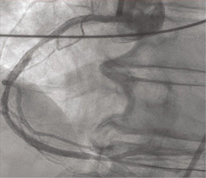
Case Reports in Cardiology                                                                                                               5

mechanism of myocardial injury caused by SARS-CoV-2 infec-         Acknowledgments
tion. These include roles for (i) SARS-CoV-2 binding of angio-
tensin enzyme 2, which is found on myocytes and type 2             We offer our very sincere thanks to Donna Belcinski, Green-
pneumocytes and which can directly cause toxicity to myocar-       wich Hospital medical librarian, for her assistance in finding
dial cells [7]; (ii) oxygen supply versus demand mismatch cre-     relative literature and acquiring articles.
ated secondary to hypoxemic respiratory failure arising from
COVID-19-induced lung injury, which then causes type 2 myo-
cardial infarction [8]; and (iii) COVID-19 induction of a pro-     References
thrombotic effect related to cytokine-mediated inflammation
                                                                    [1] C. Disease, (COVID-19) Weekly Epidemiological Situation
and endotheliopathy, which can then lead to plaque instability,
                                                                        Report, 2019, https://www.who.int/publications/m/item/
vasospasm, and rupture [9]. A recent case report describes the          weekly-epidemiological-update-on-covid-19—20-july-2021.
autopsy results of a patient with suspected STEMI and
                                                                    [2] E. Mahmud, H. L. Dauerman, W. FGP et al., “Management of
COVID-19, which found “extensive microvascular thrombosis
                                                                        Acute Myocardial Infarction During the COVID-19 Pan-
in the absence of epicardial coronary obstruction despite no            demic: A Position Statement From the Society for Cardiovas-
detectable virus in several sections of myocardial tissue [10]”.        cular Angiography and Interventions (SCAI), the American
    The unprecedented nature of the COVID-19 pandemic                   College of Cardiology (ACC), and the American College of
justifies a reevaluation of standard of care for treating                Emergency Physicians (ACEP),” Journal of the American Col-
patients with STEMI. Recent commentary written by experts               lege of Cardiology, vol. 76, no. 11, pp. 1375–1384, 2020.
reveals an openness to debate the use of fibrinolytic therapy        [3] V. Russo, R. Bottino, A. Carbone et al., “COVID-19 and heart:
versus PCI as the primary intervention [11]. In a recent mul-           from clinical features to pharmacological implications,” Jour-
ticenter case series by Hamadeh et al., 21% of subjects with            nal of Clinical Medicine, vol. 9, no. 6, 2020.
COVID-19 and STEMI treated with PCI experienced stent               [4] V. Russo, A. Rago, A. Carbone et al., “Atrial fibrillation in
thrombosis, and overall in-hospital mortality risk was 12%              COVID-19: from epidemiological association to pharmaco-
[12]. An observational study by Choudry et al., which com-              logical implications,” Journal of Cardiovascular Pharmacology,
pared outcomes between STEMI patients with COVID-19                     vol. 76, no. 2, pp. 138–145, 2020.
to those without the disease, also demonstrated increased           [5] V. Russo, R. Bottino, A. D’Andrea et al., “Chronic oral anticoa-
thrombus burden in those who were infected [13]. This phe-              gulation and clinical outcome in hospitalized COVID-19
nomenon correlated with what was observed in our group of               patients,” Cardiovascular Drugs and Therapy, pp. 1–8, 2021.
STEMI patients. Given the evidence that COVID-19 patients           [6] S. Bangalore, A. Sharma, A. Slotwiner et al., “ST-segment ele-
with STEMI are at increased risk of stent thrombosis and                vation in patients with Covid-19 - a case series,” New England
potentially are more difficult revascularize, interventional              Journal of Medicine, vol. 382, no. 25, pp. 2478–2480, 2020.
strategies for these patients should be revisited in light of       [7] M. Hoffmann, H. Kleine-Weber, S. Schroeder et al., “SARS-
the fact that fibrinolytic therapy can be an effective treatment          CoV-2 cell entry depends on ACE2 and TMPRSS2 and is
and exposes healthcare workers to lower theoretical risk of             blocked by a clinically proven protease inhibitor,” Cell,
COVID-19 transmission.                                                  vol. 181, no. 2, 2020.
    There are several domains of investigation that are             [8] F. Castagna, R. Cerrud-Rodriguez, M. A. Villela, and A. E.
needed, including (i) determining the relative risk of STEMI            Bortnick, “SARS-COV-2 infection presenting as ST-elevation
and other myocardial injury in patients with COVID-19                   myocardial infarction,” Catheterization and Cardiovascular
compared to those without active infection, (ii) etiologies to          Interventions, vol. 97, 2020.
explain any potential increase in relative risk so that focused     [9] F. A. Klok, M. J. H. A. Kruip, N. J. M. van der Meer et al., “Inci-
therapies can be developed, and (iii) relative effectiveness of          dence of thrombotic complications in critically ill ICU patients
fibrinolytic therapy versus PCI, given that complications                with COVID-19,” Thrombosis Research, vol. 191, pp. 145–147,
                                                                        2020.
from COVID-19 may hamper the effectiveness of PCI.
                                                                   [10] G. Guagliumi, A. Sonzogni, I. Pescetelli, D. Pellegrini, and
                                                                        A. V. Finn, “Microthrombi and ST-segment-elevation myo-
Data Availability                                                       cardial infarction in COVID-19,” Circulation, vol. 142, no. 8,
                                                                        pp. 804–809, 2020.
Figures used are available from the corresponding author
                                                                   [11] M. J. Daniels, M. G. Cohen, A. A. Bavry, and D. J. Kumbhani,
on request.
                                                                        “Reperfusion of ST-segment-elevation myocardial infarction
                                                                        in the COVID-19 era: business as usual?,” Circulation,
Consent                                                                 vol. 141, no. 24, pp. 1948–1950, 2020.
                                                                   [12] A. Hamadeh, A. Aldujeli, K. Briedis et al., “Characteristics and
No written consent has been obtained from the patients                  Outcomes in Patients Presenting With COVID-19 and ST-
as there is no patient identifiable data included in this                Segment Elevation Myocardial Infarction,” The American
case report.                                                            Journal of Cardiology, vol. 131, pp. 1–6, 2020.
                                                                   [13] F. A. Choudry, S. M. Hamshere, K. S. Rathod et al., “High
Conflicts of Interest                                                   Thrombus burden in patients with COVID-19 presenting with
                                                                        ST-segment elevation myocardial infarction,” Journal of the
The authors declare that there is no conflict of interest                American College of Cardiology, vol. 76, no. 10, pp. 1168–
regarding the publication of this article.                              1176, 2020.
You can also read