Metaphyseal Sleeve Failure in Revision Total Knee Arthroplasty
←
→
Page content transcription
If your browser does not render page correctly, please read the page content below
Open Access Original
                               Article                                                   DOI: 10.7759/cureus.18054

                                                Metaphyseal Sleeve Failure in Revision Total
                                                Knee Arthroplasty
Review began 09/03/2021
                                                Theodoros Bouras 1 , Peter Fennema 2 , Rhidian Morgan-Jones 1 , Sanjeev Agarwal 1
Review ended 09/10/2021
Published 09/17/2021                            1. Department of Trauma & Orthopaedics, Cardiff & Vale University Health Board, University Hospital Llandough,
© Copyright 2021                                Cardiff, GBR 2. Epidemiology and Public Health, AMR Advanced Medical Research, Männedorf, CHE
Bouras et al. This is an open access article
distributed under the terms of the Creative     Corresponding author: Theodoros Bouras, theo_bouras@hotmail.com
Commons Attribution License CC-BY 4.0.,
which permits unrestricted use, distribution,
and reproduction in any medium, provided
the original author and source are credited.
                                                Abstract
                                                Introduction
                                                A significant percentage of patients require re-revision surgery regardless of the demonstrated durable
                                                short- and mid-term clinical results using metaphyseal sleeves in revision total knee arthroplasty (TKA).
                                                The aim of this study was to identify the association between sleeve alignment and contact zones, with
                                                loosening in patients with revision TKA.

                                                Materials & Methods
                                                Of a series of 103 patients who underwent revision TKA, at a mean follow-up of eight years, six patients were
                                                re-revised for tibial loosening. These patients were compared with 19 unrevised control subjects in a 1:3
                                                ratio. We calculated and compared the cumulative number of contact zones between the porous-coated part
                                                of the sleeve and bone on immediate postoperative X-rays between re-revised and unrevised patients. The
                                                main hypothesis was that neutral positioning and absolute circumferential contact between trabecular
                                                metaphyseal bone and porous-coated part of the sleeve would lead to a better outcome.

                                                Results
                                                The use of a conservative (nonparametric) approach indeed revealed better circumferential contact between
                                                trabecular metaphyseal bone and porous-coated part of the sleeve among the survivors, i.e., survivors:
                                                median (interquartile range [IQR]): 3 (2-4); failures: 3 (1-3), p = 0.003 (Mann-Whitney [MW] test). The
                                                difference was borderline significant for coronal alignment, i.e., survivors: median (IQR): −1 (−4 to 2);
                                                failures: 0 (−1 to 3), p = 0.0569 (MW test).

                                                Conclusion
                                                A circumferential bony contact of the metaphyseal sleeve would lead to better survival of the revision
                                                implant, whereas the degree of varus fixation did not seem to influence the longevity of the implant.

                                                Categories: Orthopedics
                                                Keywords: contact zones, implant survival, porous coated, metaphyseal sleeves, revision total knee arthroplasty

                                                Introduction
                                                The need for revision total knee arthroplasty (RTKA) is constantly increasing over the years. The projected
                                                estimates for England and Wales suggest an increase in the volume of RTKA by 332% by the year 2030 [1]. In
                                                the challenging task of reconstruction and fixation in RTKA, especially in the presence of bone defects, the
                                                concept of zonal fixation provides the surgeon a working methodology toward the achievement of best
                                                results and lower re-revision rates [2,3]. The use of porous-coated metaphyseal sleeves is a modern
                                                technique addressing bone deficiency. It creates a stable platform for the femoral and/or tibial components,
                                                offering long-term biologic fixation to host bone. Reliable stability of the metaphyseal construct decreases
                                                torsional and shear stresses at the cement-bone interface [4]. Recent evidence suggests that the use of
                                                metaphyseal sleeves demonstrates excellent short-, mid-, and long-term clinical results and radiographic
                                                fixation [5-15]. Different modes of constraint, or sleeve and stem fixation, such as cemented or cementless,
                                                do not appear to impact the survivorship and the osseous integration of the porous-coated part of the
                                                metaphyseal sleeve. This has deprived the literature of studies looking specifically for failure reasons of the
                                                metaphyseal sleeve fixation. Proper assessment of indications and contraindications, as well as accurate
                                                surgical technique, is of utmost importance. The hypothesis of this study was that neutral positioning and
                                                complete circumferential contact between trabecular metaphyseal bone and porous-coated part of the
                                                sleeve would lead to a better outcome.

                                                Materials And Methods
                                                A retrospective review of a prospectively maintained longitudinal revision total knee arthroplasty (TKA)
                                                database was performed. There were 103 patients (104 knees) identified as having undergone revision TKA

                               How to cite this article
                               Bouras T, Fennema P, Morgan-Jones R, et al. (September 17, 2021) Metaphyseal Sleeve Failure in Revision Total Knee Arthroplasty. Cureus
                               13(9): e18054. DOI 10.7759/cureus.18054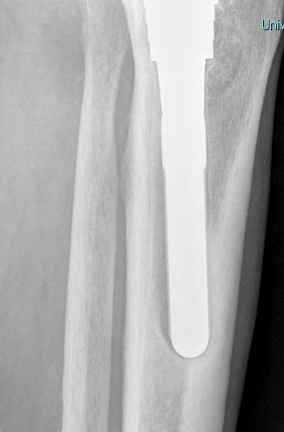
using porous-coated metaphyseal sleeves (DePuy, Warsaw, Indiana) [8]. At a mean follow-up of 95.7 months,
                                                 there were 23 (22.1%) re-revisions for any reason. All patients who had index revision and re-revision for
                                                 infection, stiffness, instability, peri-prosthetic fracture, or undiagnosed pain were excluded from the 23
                                                 cases, 6 patients (26%) were re-revised for tibial loosening, and 1 (4.3%) for femoral loosening. All six cases
                                                 of aseptic tibial loosening were included in the study. The patient with femoral loosening had
                                                 disengagement at the stem sleeve junction and was not included in the study as seen in Figure 1.

                                                    FIGURE 1: Case-control population study group.
                                                    TKR, total knee replacement.

                                                 Control patients were sampled in a 1:3 ratio among the patients with similar follow-up but without
                                                 loosening. Two adult reconstructive knee orthopedic surgeons (one consultant and one senior fellow)
                                                 independently assessed and compared the immediate postoperative X-rays, using a digital viewing platform,
                                                 at the time of index revision, between the six tibial re-revised patients and a group of 19 patients that had
                                                 maintained the sleeve in situ. Radiographs included standard weight-bearing anteroposterior (AP) and
                                                 lateral views of the knee and were undertaken based on a standard protocol by experienced radiographers.
                                                 The degree of coronal plane alignment and the porous-coated part of the sleeve was assessed. This
                                                 assessment was based on the presence of radiolucency at the metal bone interface of the metaphyseal sleeve.
                                                 On the tibial side, only the proximal third of the sleeve is porous-coated. This was the zone of interest for
                                                 metal-bone contact.

                                                 The two standard radiographic views provided four regions of interpretation: anterior and posterior on the
                                                 lateral view; and medial-lateral on the AP view (Figure 2).

                                                    FIGURE 2: The zones used for the scoring system.

2021 Bouras et al. Cureus 13(9): e18054. DOI 10.7759/cureus.18054                                                                                                  2 of 9
Absolute contact between trabecular metaphyseal bone and porous-coated part of the sleeve in each region
was graded as “1” and any gap in the bone-sleeve interface as “0.” A total score of “4” meant absolute press-
fit circumferential contact between trabecular metaphyseal bone and porous-coated part of the sleeve. This
score is similar to methods used to quantify fracture healing. The degree of varus or valgus fixation of the
sleeve was also measured in all patients. We did not routinely use CT scans for assessing osseointegration or
loosening in our patients. This was a retrospective study and no patient intervention or contact was
involved. Formal ethical approval was not required.
Statistical analysis
Statistical analyses were performed using Stata 15.1 software (StataCorp, College Station, Texas).
Continuous data are presented as mean ± standard deviation (range); the Mann-Whitney (MW) test was used
to compare between-group differences. Categorical variables are presented as frequencies, and Fisher's exact
test was used to determine between-group differences. Two-sided p-values of 0.05 were deemed to indicate
statistical significance.
Results
Baseline characteristics of cases and controls are summarized in Table 1.
Patients with tibial loosening (cases) Patients without tibial loosening (controls) p-value
Sample size 6 19
Age at inclusion (years) 72.0 ± 1.7 77.7 ± 1.7 0.056
Sex (female:male) 2:4 7:12 0.637
TABLE 1: Baseline characteristics of cases and controls.
The radiological scores of patients in the study group and control group are summarized in Table 2.
Score Study group (number of patients) Control group (number of patients)
0
1 1
2 2 3
3 3 2
4 14
TABLE 2: Radiological scores of study group and control group.
None of the six re-revised patients (study group) demonstrated absolute press-fit circumferential contact
between trabecular metaphyseal bone and porous-coated part of the sleeve. Three out of six (50%) scored
“3” with inadequate contact in the medial (AP) region of the sleeve while two patients (33.3%) scored “2.” In
one patient (16.7%), absolute contact was seen only in the lateral region of the sleeve (fixation score 1),
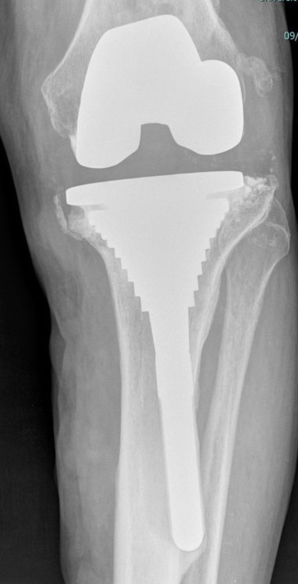
which finally collapsed in varus (Figures 3-6).
2021 Bouras et al. Cureus 13(9): e18054. DOI 10.7759/cureus.18054 3 of 9FIGURE 3: Early postoperative X-ray anteroposterior view showing lack
of metal-bone contact in the medial aspect of the metaphyseal sleeve.
2021 Bouras et al. Cureus 13(9): e18054. DOI 10.7759/cureus.18054 4 of 9FIGURE 4: Early postoperative X-ray lateral view showing lack of metal-
bone contact in the anterior and posterior aspect of the metaphyseal
sleeve.
2021 Bouras et al. Cureus 13(9): e18054. DOI 10.7759/cureus.18054 5 of 9FIGURE 5: A 72-month follow-up X-ray anteroposterior view. The tibial
component has loosened and migrated into varus.
2021 Bouras et al. Cureus 13(9): e18054. DOI 10.7759/cureus.18054 6 of 9FIGURE 6: A 72-month follow-up X-ray lateral stem tip view. The tibial
component has loosened and migrated into varus.
From the control group, there were 14 patients (73.7%) who had absolute contact in all four regions. A gap in
the anterior interface was seen in two patients (10.5%). The remaining three patients (15.8%) scored
“2.” Alignment between 3° varus and 1° valgus was found in the six re-revised cases. The alignment for the
unrevised patients was between 1° varus and 4° valgus. Taking a conservative (nonparametric) approach,
better circumferential contact between trabecular metaphyseal bone and porous-coated part of the sleeve
among the survivors was found, i.e., survivors: median (interquartile range [IQR]): 3 (2-4); failures: 3 (1-3), p
= 0.003 (MW test). For coronal alignment, the difference did not reach the significance threshold of 0.05,
i.e., survivors: median (IQR): −1 (−4 to 2); failures: 0 (−1 to 3), p = 0.0569 (MW test).
Discussion
Achieving secure long-lasting fixation of the implant in the bone is an important component of revision
2021 Bouras et al. Cureus 13(9): e18054. DOI 10.7759/cureus.18054 7 of 9knee replacement. Both cemented and cementless fixations principles exist in order to achieve this goal.
Maximizing contact with host bone is desirable in either fixation method. The most important finding of
this study was that a circumferential bony contact of the porous-coated part of the metaphyseal sleeve
would lead to better survival of the revision implant, whereas the degree of varus fixation did not seem to
influence the longevity of the implant. A press-fit insertion of the sleeve would require sequential broaching
with progressively increasing sizes until axial and rotational stability is achieved [4]. Depending on the bone
loss, impaction grafting or morselized allograft could be used to fill the remaining defects [16]. We did not
use allograft in any of our patients and are unable to comment on the impact of graft usage and sleeve
contact with host bone, or its influence on the long-term survival of the sleeve. Bone coverage of
uncemented sleeves, with a minimum of 75%, to achieve optimal osseointegration was reported by studies
included in a recent systematic review [17]. Another one reported a high osseointegration rate demonstrated
by the use of metaphyseal sleeves [18]. A recent case series reported proximal radiolucent lines in nine tibial
sleeves, one year following revision surgery compared to immediate postoperative X-rays. These lines had
not progressed in the five-year follow-up and the sleeves did not require revision. In the same study, 12
tibial sleeves had subsided >1 mm compared to the original postop X-ray, without further progression and
subsequent re-revision surgery [5]. Another recent study reported minor radiolucent lines detectable in a
few sleeves but no implant migration [9]. A retrospective cohort of osseointegrated sleeves reported no
radiolucencies in the porous coated part of the sleeves at the five-year follow-up. Despite the well-fixed
implants, radiolucencies were seen in the uncoated part of the sleeve. This was attributed to possible
pivoting especially in combination with long diaphyseal stems [6]. Ihekweazu et al. in 2019 published a
retrieval analysis study reporting bone ongrowth coverage of the entire porous surface of the tibial sleeve on
average of 14.7 ± 3.4%. The authors correlated the radiological findings with the bone ongrowth in the
retrieved specimens suggesting that plain radiology was highly unreliable to detect biologic fixation [19]. In
our study, the coronal alignment for the six revised cases was between 3° varus and 1° valgus, and for the
unrevised patients was between 1° varus and 4° valgus. This was borderline significant to the final outcome.
Martin-Hernandez et al. reported a similar outcome with satisfactory alignment between 3° varus and 3°
valgus [10]. With the relatively small number of patients, we are unable to comment about contact of specific
aspects-medial/lateral/anterior/posterior-of the sleeve, which would influence osseointegration and
stability. A malaligned sleeve, loaded more on one side, couples with lack of osseous contact on the same
side can lead to cantilever failure as shown in Figures 3-6. The current literature on metaphyseal sleeves is
mainly comprised of case series with limited information on failure mechanisms; hence, our study adds to
the body of evidence on the subject. However, this is limited by the small sample size, which decreases the
options such as a stratified or multiple logistic regression analysis to assess the presence of confounding.
Conclusions
Absolute circumferential bony contact of the porous-coated part of the metaphyseal sleeve at the time of
index revision TKA surgery can lead to better implant survival. Coronal alignment does not seem to play a
significant role in the final outcome.
Additional Information
Disclosures
Human subjects: Consent was obtained or waived by all participants in this study. issued approval NA. This
was a retrospective study and no patient intervention or contact was involved. Formal ethical approval was
not required. Animal subjects: All authors have confirmed that this study did not involve animal subjects or
tissue. Conflicts of interest: In compliance with the ICMJE uniform disclosure form, all authors declare the
following: Payment/services info: All authors have declared that no financial support was received from
any organization for the submitted work. Financial relationships: All authors have declared that they have
no financial relationships at present or within the previous three years with any organizations that might
have an interest in the submitted work. Other relationships: All authors have declared that there are no
other relationships or activities that could appear to have influenced the submitted work.
References
1. Patel A, Pavlou G, Mújica-Mota RE, Toms AD: The epidemiology of revision total knee and hip arthroplasty
in England and Wales: a comparative analysis with projections for the United States. A study using the
National Joint Registry dataset. Bone Joint J. 2015, 97-B:1076-81. 10.1302/0301-620X.97B8.35170
2. Morgan-Jones R, Oussedik SI, Graichen H, Haddad FS: Zonal fixation in revision total knee arthroplasty.
Bone Joint J. 2015, 97-B:147-9. 10.1302/0301-620X.97B2.34144
3. Mancuso F, Beltrame A, Colombo E, Miani E, Bassini F: Management of metaphyseal bone loss in revision
knee arthroplasty. Acta Biomed. 2017, 88:98-111. 10.23750/abm.v88i2-S.6520
4. Angerame MR, Jennings JM, Holst DC, Dennis DA: Management of bone defects in revision total knee
arthroplasty with use of a stepped, porous-coated metaphyseal sleeve. JBJS Essent Surg Tech. 2019, 9:e14.
10.2106/JBJS.ST.18.00038
5. Bloch BV, Shannak OA, Palan J, Phillips JR, James PJ: Metaphyseal sleeves in revision total knee
arthroplasty provide reliable fixation and excellent medium to long-term implant survivorship. J
Arthroplasty. 2020, 35:495-9. 10.1016/j.arth.2019.09.027
6. Wirries N, Winnecken HJ, Lewinski GV, Windhagen H, Skutek M: Osteointegrative sleeves for metaphyseal
defect augmentation in revision total knee arthroplasty: clinical and radiological 5-year follow-up. J
2021 Bouras et al. Cureus 13(9): e18054. DOI 10.7759/cureus.18054 8 of 9Arthroplasty. 2019, 34:2022-9. 10.1016/j.arth.2019.04.024
7. Fedorka CJ, Chen AF, Pagnotto MR, Crossett LS, Klatt BA: Revision total knee arthroplasty with porous-
coated metaphyseal sleeves provides radiographic ingrowth and stable fixation. Knee Surg Sports Traumatol
Arthrosc. 2018, 26:1500-5. 10.1007/s00167-017-4493-y
8. Agarwal S, Neogi DS, Morgan-Jones R: Metaphyseal sleeves in revision total knee arthroplasty: minimum
seven-year follow-up study. Knee. 2018, 25:1299-307. 10.1016/j.knee.2018.09.010
9. Thorsell M, Hedström M, Wick MC, Weiss RJ: Good clinical and radiographic outcome of cementless metal
metaphyseal sleeves in total knee arthroplasty. Acta Orthop. 2018, 89:84-8.
10.1080/17453674.2017.1398013
10. Martin-Hernandez C, Floria-Arnal LJ, Muniesa-Herrero MP, Espallargas-Doñate T, Blanco-Llorca JA,
Guillen-Soriano M, Ranera-Garcia M: Mid-term results for metaphyseal sleeves in revision knee surgery .
Knee Surg Sports Traumatol Arthrosc. 2017, 25:3779-85. 10.1007/s00167-016-4298-4
11. Watters TS, Martin JR, Levy DL, Yang CC, Kim RH, Dennis DA: Porous-coated metaphyseal sleeves for
severe femoral and tibial bone loss in revision TKA. J Arthroplasty. 2017, 32:3468-73.
10.1016/j.arth.2017.06.025
12. Chalmers BP, Desy NM, Pagnano MW, Trousdale RT, Taunton MJ: Survivorship of metaphyseal sleeves in
revision total knee arthroplasty. J Arthroplasty. 2017, 32:1565-70. 10.1016/j.arth.2016.12.004
13. Dalury DF, Barrett WP: The use of metaphyseal sleeves in revision total knee arthroplasty . Knee. 2016,
23:545-8. 10.1016/j.knee.2016.02.005
14. Graichen H, Strauch M, Scior W, Morgan-Jones R: Knee revision arthroplasty: cementless, metaphyseal
fixation with sleeves. (Article in German). Oper Orthop Traumatol. 2015, 27:24-34. 10.1007/s00064-014-
0333-0
15. Barnett SL, Mayer RR, Gondusky JS, Choi L, Patel JJ, Gorab RS: Use of stepped porous titanium metaphyseal
sleeves for tibial defects in revision total knee arthroplasty: short term results. J Arthroplasty. 2014,
29:1219-24. 10.1016/j.arth.2013.12.026
16. Sheth NP, Bonadio MB, Demange MK: Bone loss in revision total knee arthroplasty: evaluation and
management. J Am Acad Orthop Surg. 2017, 25:348-57. 10.5435/JAAOS-D-15-00660
17. Zanirato A, Cavagnaro L, Basso M, Divano S, Felli L, Formica M: Metaphyseal sleeves in total knee
arthroplasty revision: complications, clinical and radiological results. A systematic review of the literature.
Arch Orthop Trauma Surg. 2018, 138:993-1001. 10.1007/s00402-018-2967-0
18. Bonanzinga T, Akkawi I, Zahar A, Gehrke T, Haasper C, Marcacci M: Are metaphyseal sleeves a viable option
to treat bone defect during revision total knee arthroplasty? A systematic review. Joints. 2019, 7:19-24.
10.1055/s-0039-1697611
19. Ihekweazu UN, Weitzler L, Wright TM, Padgett DE: Distribution of bone ongrowth in metaphyseal sleeves
for revision total knee arthroplasty: a retrieval analysis. J Arthroplasty. 2019, 34:760-5.
10.1016/j.arth.2018.12.033
2021 Bouras et al. Cureus 13(9): e18054. DOI 10.7759/cureus.18054 9 of 9You can also read