Wearing High Heel and Planter Fasciitis: MRI Evaluation
←
→
Page content transcription
If your browser does not render page correctly, please read the page content below
International Journal of Medical Imaging
2020; 8(1): 1-5
http://www.sciencepublishinggroup.com/j/ijmi
doi: 10.11648/j.ijmi.20200801.11
ISSN: 2330-8303 (Print); ISSN: 2330-832X (Online)

Wearing High Heel and Planter Fasciitis: MRI Evaluation
Sameh Ahmad Khodair1, *, Rasha Loutfy Younes2
Radiology Department, Faculty of Medicine, Tanta University, Tanta, Egypt

Email address:
*
    Corresponding author

To cite this article:
Sameh Ahmad Khodair, Rasha Loutfy Younes. Wearing High Heel and Planter Fasciitis: MRI Evaluation. International Journal of Medical
Imaging. Vol. 8, No. 1, 2019, pp. 1-5. doi: 10.11648/j.ijmi.20200801.11

Received: December 13, 2019; Accepted: December 30, 2019; Published: January 7, 2019

Abstract: Purpose: To determine the type and frequency of characteristic MRI changes of the plantaris fascia in patients with
painful heel and wear high heel shoes. Materials and Methods: 40 patients with painful heel, wearing high heels, their age raged from
25-50 years, underwent MR imaging. A control group included 20 subjects with no history of painful heel & not using high heels
were included. Associations between the presence of palnter fasciitis, high heel, and body mass index, height of the heel, MRI
imaging, self-reported co-morbidities and current heel pain were then explored. MR images were obtained with a 1.5-T
superconducting MR imager with a 5-inch (13-cm) standard small flexible surface coil. Results: Thirty cases (75%) of the clinically
suspected of plantar fasciitis was established by MR imaging. The most common finding was a peritendinous edema at the calcaneal
insertion site which was found in all 30 patients. In 16 patients (53%), an intratendinous signal intensity increase of the plantar fascia
could be observed. Compared to the control group (mean thickness 3.3 mm) the plantar fascia showed significant thickening in the
thirty MR positive patients (mean thickness 7.7 mm). Conclusion: Planter fasciitis is common in high heel using women. Besides
thickening of the plantar fascia, intratendinous signal intensity increase and peritendinous edema close to the plantar fascia are
characteristic signs of plantar fasciitis on MRI. Both signs can reliably be seen on STIR sequences only.
Keywords: MRI, Plantaris Fascia, High Heel Shoes, Painful Heel

                                                                       newer studies have demonstrated structural changes more
1. Introduction                                                        consistent with a degenerative process or repetitive minor
   Women are interested to wear high-heeled shoes to                   trauma. As a result of this new observation, many in the
increase their attractiveness. High-heeled shoes might create          academic community have stated that the condition should be
harmful effects to the musculoskeletal system. [1]                     renamed plantar fasciosis. [5]
Furthermore earlier studies proved that the function of foot              Understanding of the normal anatomy of the plantar
and lower extremity will be changed due to wearing high                aponeurosis (PA) and familiarity with pathologic conditions are
heeled shoes. [2] Plantar fasciitis (also known as plantar             required for an accurate evaluation of the patient with heel pain.
fasciopathy or jogger's heel) is a common painful disorder             [6]
affecting the heel and underside of the foot. [3] It is a                 The output of this research is describing the effect of wearing
disorder of the insertion site of ligament on the calcaneous           high heel on plantar fascia. In this study, we evaluated the
bone and is characterized by scarring, inflammation, or                diagnostic capabilities of magnetic resonance (MR) imaging in
structural breakdown of the foot's plantar fascia. It is often         the assessment of the PA in correlation with the use of high heel.
caused by overuse injury of the plantar fascia. [4]
   Plantar fasciitis is the most common injury of the plantar          2. Material & Method
fascia and is the most common cause of heel pain. It is
commonly associated with excessive inward rolling of the                  MRI study was conducted on 40 patients with a mean age
foot as in high heel. Individuals with plantar fasciitis often         of 40±7.2 years. Twenty control volunteers, age-matched and
have difficulty with dorsiflexion of the foot. Though plantar          healthy also were underwent the same protocol of MRI study.
fasciitis was originally thought to be an inflammatory process,        All our 40 patients were using high heels as the following
                                                                       criteria: length of the heel not less that 2.5 inches, a duration
2 Sameh Ahmad Khodair and Rasha Loutfy Younes: Wearing High Heel and Planter Fasciitis: MRI Evaluation
mean of 4 hours /day, 5 days/ week, with average body mass other bony lesions as enthesopathy, spurs & tarsal tunnel
index 25-30%. The control group were not wearing high heel syndrome, also of the 20 control cases one case showed mild
& had no painful heel. edema denoting mild form of plantar fascitis.
Patients with the following criteria were excluded from our MR imaging criteria in 3 patients, one patient aged 30
study: over weight, abnormal shape of the foot as high arched years, the other two at age group (46-55) aging 47 & 50 years
foot, and sportive runners. All the examined patients were respectively, these patients were having partial tear of the PS,
laboratory investigated for blood glucose level, rheumatoid showed partial high T2-weighted signal intensity of the
factor, uric acid & ESR. Also plain x-ray foot was done in plantar fascia. In the three patients, abnormal thickening of
different direction for exclusion of other causes of painful heel. the PA at the site of partial disruption was evident. Tears
Four patients were suspected to have tarsal tunnel syndrome & involved the proximal PA was present in one case and no tear
nerve conduction study was done to exclude this cause. at the middle PA was observed. The three cases showed
Clinical orthopedic examination revealed heel pain with the edema in perifascial soft tissue with high signal intensity on
following criteria: the pain starts with initial weight bearing after T2 WI. Fascial thickening & scar tissue was depicted with
a period of time & increases upon rising in morning, on low signal intensity on T2 WI, and STIR images.
palpation, tenderness was at the inferior aspect of the heel, MR imaging studies in the 27 patients with plantar fasciitis,
MR images were obtained with a 1.5-T superconducting 10 patients at age group (25-35), 11 at age group (36-45) & 6
MRI (Signa; GE Medical Systems, Milwaukee, Wis) and at age group (46-55) revealed the signal intensity changes of
( Optima MR 360, Toshiba Medical Systems) at MRI units, perifascial edema either superficial to (n = 11) or both
with a 5-inch (13-cm) standard small flexible surface coil. superficial and deep to (n = 16) the PA. In 13 patients, the PA
Routine ankle MR imaging was performed in the axial, exhibited abnormal intrafascial high signal intensity on T2-
coronal, and sagittal planes. The foot is imaged in the oblique weighted images, STIR images, or both. The PA was
coronal plane, oblique axial plane, and oblique sagittal plane. abnormally thickened (5–7 mm) in 20 patients. (Table 1)
T1-weighted (repetition time msec/echo time msec = 600/20)
and T2-weighted (2,000/20,80) Fat suppression techniques, Table 1. MRI findings in patients and control group.
fat-suppressed proton-density–weighted imaging & short- MRI findings Pt. (no. 40 ) Control (no. 20)
inversion-time inversion recovery (STIR) sequences Rupture of PA 3
(3611/48; inversion time msec = 100–150). Partial disruption 3
Soft tissue edema 3
The patients were in supine position with the foot in about 20°
Facial thickening 3
of plantar flexion; this is specially helpful for the following Plantar faciaitis 27
reasons: it accentuates the fat plane between the tendons specially Intra fascial edema 13
the peroneal tendons & decreases the effect of the magic angle. Perifascial edema 27 1
The imaging data were reviewed by two radiologists (with Fascial thickening 20
30 1
more than five years of experience) blinded of the patients
clinical pictures; and then they nearly reached a consensus 20 healthy non complaining control group were examined,
opinion. The regional ethics committee approved the study one person showed superficial edema of the plantar fascia
and written informed consent were obtained from all denoting mild fasciitis, while the rest of the control group
participants. showed no abnormality.
Accordingly, out of the 40 patients; 10 cases (25%) were
3. Results clinically complaining but radiologically free. On the other
hand, one of the control non complaining group turned to
The MR imaging features of our 40 clinical cases are listed have mild plantar fasciitis that accounts for 1/20 = 0.05%.
in the Table 1. Ten cases were excluded due to presence of
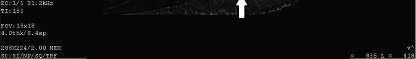
Figure 1. MRI right ankle joint, Sagittal STIR, shows abnormal peri-plantaris high signals of edema.International Journal of Medical Imaging 2020; 8(1): 1-5 3
Figure 2. MRI Right ankle, Sagittal STIR, shows abnormal high signals of peri-plantaris edema deep to it (arrow) with mildly thickened plantaris facia (arrow
heads).
Figure 3. MRI Left ankle, Sagittal STIR, shows abnormal high signals of peri-plantaris edema superficial and deep to it as well as abnormal high signals
disrupting it partially (arrow) with mildly thickened plantaris facia (arrow head).
1
Figure 4. MRI Right ankle, Sagittal STIR, shows abnormal high signals of peri-plantaris edema superficial and deep to it as well as abnormal high signals
disrupting it partially (arrow).
Heel pain is a very common complaint in adults especially
4. Discussion in females. It is estimated that more than one million patients
The plantar fascia is a thick, ligamentous connective tissue seek treatment annually for this condition. The plantar
that runs from the calcaneus to the ball of the foot. This tight fasciitis is thought to be the most common cause & is mainly
tissue helps to maintain the arch of the foot and transmits due to biomechanical overuse, thus creating micro-tears at
weight across the foot as person walks or runs. That's why the calcaneal enthesis. [8]
tremendous stress is placed on the plantar fascia. [7] Some experts have deemed this condition “plantar4 Sameh Ahmad Khodair and Rasha Loutfy Younes: Wearing High Heel and Planter Fasciitis: MRI Evaluation
fasciosis” implying that its etiology is a more chronic lots & we tried to exclude some causes both by clinical
degenerative process versus acute inflammation. [9] Far and examination & laboratory investigations, so that we come to
away the most common cause of plantar fascia pain is faulty a conclusion that a relation is present between the presence of
biomechanics of the foot or leg. Faulty biomechanics causes plantar fascial pathology & high heel in absence of other
the foot to sustain increased or prolonged stresses over and obvious causes of heel pain.
above those of routine ground contacts. [10]
High-heeled shoes can limit proprioception where the heel 5. Conclusion
hits the ground first followed by the toe. It has been shown
that high heeled stiff shoes restrict the inversion/eversion of In conclusion, MRI was helpful in diagnosis of pathology
the foot, which can lead to an increase in rearfoot prontion, of the plantar fascia in our patients with a clinical diagnosis
therefore showing that the stiffer the shoe, the more the of rupture of the PA, MR imaging allowed demonstration,
natural foot motion is restricted. [11] exact localization, and determination of severity of the lesion
Not only is the normal foot motion restricted by shoes, it with regard to the thickness of the affected PA. Depicting the
has also been shown that wearing high heeled shoes restricts relation between the complain, physical examination & the
other biomechanics aspects of the foot. [11] Forefoot to presence of high heel in absence of any other cause for the
rearfoot eversion/inversion and abduction/adduction were presence of either PA rupture or fasciitis/fasciosis, & drawing
restricted by shoes, as well as forefoot spreading, which is a line correlating the high heel & plantar fascial pathology.
important for comfort as well as natural gait, and forefoot We recognize that although the patients show the similar
pronation during push-off. [12] symptoms & similar imaging findings, yet the implication of
Plantar fasciosis can be confused with a condition called this examination over a larger population may help support
tarsal tunnel syndrome. In tarsal tunnel syndrome, the tibial our conclusion.
nerve is trapped and pinched as it passes through the tarsal
tunnel. This may cause symptoms similar to the pain of a
plantar fasciosis/fasciitis, in this study, the suspected cases to References
have tarsal tunnel syndrome were investigated with nerve
conduction study & were all free. [13] [1] A. Ahmady, E. Soodmand, I. Soodmand, T. L Milani: The
effect of various heights of high-heeled shoes on foot arch
In all our patients suspected of having plantar fasciitis, deformation: Finite element analysis. Journal of Foot and
high signal intensity consistent with edema in the perifascial Ankle Research. 2014; 7 (Suppl 1): A78.
soft tissue was appreciated on T2-weighted images & STIR
images as well. Our findings are in agreement with those of [2] Yu J.: Biomechanical simulation of high-heeled shoe donning and
walking. Journal of Biomechanics. 2013, 46 (12): 2067-2074.
previous studies, in which the perifascial edema was
considered the most common manifestation of plantar [3] Sullivan J, Pappas E, Burns J.: Role of mechanical factors in
fasciitis. [14] In our patients with plantar fasciitis, soft-tissue the clinical presentation of plantar heel pain: Implications for
edema superficial and deep to the PA was the dominant management. Foot (Edinb). 2019 Sep 3; 42.
abnormal imaging finding in 90% of cases, that frequency for [4] J. D. Goff and R. Crawford: Diagnosis and Treatment of
edema superficial to the PA was reported previously. Plantar Fasciitis. American Family Physician. 2012, 84 (6).
The second most common MR imaging finding of plantar
[5] Thomas JL, Christensen JC, and Kravitz SR.: The diagnosis
fasciitis, seen in 66% of our patient, was mild thickening of and treatment of heel pain: a clinical practice guideline-
the PA, corresponding to the presence of granulation tissue. revision. J Foot Ankle Surg. 2010; 49 (3 suppl): S1-S19.
This finding was more common in our study than was
previously reported. [14] [6] Eugene G. McNally, and Shilpa Shetty.: Plantar Fascia:
Imaging Diagnosis and Guided Treatment. Semin
Then, the following MR imaging finding of plantar Musculoskelet Radiol. 2010; 14: 334–343.
fasciitis, observed in 23% of our cases, was increased signal
intensity within the involved plantar fascia on T2-weighted [7] Hedrick MR.: The plantar aponeurosis. Foot Ankle. 1996; 17:
and STIR images; this finding was consistent with edema, 646–649.
this findings did not match with the other studies, we believe [8] Riddle DL, Schappert SM. Volume of ambulatory care visits
that the difference may be contributed to the different number and patterns of care for patients diagnosed with plantar
of patient & the plenty of exclusion criteria. [14] fasciitis: a national study of medical doctors. Foot Ankle Int.
In patients with a clinical diagnosis of partial rupture of the 2004; 25 (5): 303-310.
PA, MR imaging allowed diagnosis of the case, exact [9] Karr SD.: Subcalcaneal heel pain. Orthop Clin North Am.
localization, and determination of severity of the lesion with 1994; 25: 161–173.
regard to the thickness of the affected PA.
[10] Lemont H, Ammirati KM, and Usen N.: Plantar fasciitis: a
In the examined volunteers with age matching not wearing degenerative process (fasciosis) without inflammation. J Am
high heels, one case for mild edema & plantar fasciitis was Podiatr Med Assoc. 2003; 93 (3): 234–237.
seen only among the group, raising the consideration to the
effect of high heel on the PA. [11] Yu J, et al: Development of a finite element model of female
foot for high-heeled shoe design. Clinical Biomechanics. 2008,
As we preceded, the causes of plantaris fascia pain were 23: S31-S38.International Journal of Medical Imaging 2020; 8(1): 1-5 5
[12] Esenyel M, et al: Kinetics of high-heeled gait. Journal of the [14] Grasel R, Schweitzer M, Kovalovich A, et al.: MR imaging of
American Pediatric Medical Association. 2003, 93 (1): 27-32. plantar fasciitis: edema, tears, and occult marrow
abnormalities correlated with outcome. AJR. 1999; 173: 699–
[13] Bailie DS, Kelikian AS.: Tarsal tunnel syndrome: diagnosis, 701.
surgical technique, and functional outcome. Foot Ankle. 1998;
19: 65–72.You can also read