Initial Clinical Experience with an Iterative Denoising Algorithm Applied to Reduced-data 2D Turbo Spin Echo Acquisitions
←
→
Page content transcription
If your browser does not render page correctly, please read the page content below
Clinical · Head to Toe Imaging                                                                      MAGNETOM Flash (78) 2/2021

Initial Clinical Experience with an Iterative
Denoising Algorithm Applied to Reduced-data
2D Turbo Spin Echo Acquisitions
Johan Dehem, M.D.1; Stephan Kannengießer, Ph.D.2; Uvo Christoph Hoelscher, Ph.D.2

1
    Jan Yperman Ziekenhuis, Ieper, Belgium
2
    Siemens Healthineers, Erlangen, Germany

Introduction
Magnetic Resonance Imaging (MRI) with standard 2D                 ing the practical acceleration factor to two or three. Note
Turbo Spin Echo (TSE) sequences is a trusted technique to         that it is common practice to combine parallel imaging and
guarantee excellent soft tissue contrast in musculoskeletal       multiple averages, since motion effects can be minimized
(MSK), neurological, and abdominal imaging. The signal            by the shorter acquisition times of the former, while
and contrast behavior are well understood and appreciated         regaining SNR with the latter, which has advantages
by the reading radiologist and referring physicians alike.        over un-accelerated acquisitions of the same duration.
The acquisition time of these 2D TSE sequences is in the               At the same time elaborate denoising methods have
order of several minutes, hence has always been a point           been developed which have to balance noise removal and
of attention. With the introduction of parallel imaging [1],      preservation of details [2]. A recently introduced accelera-
acquisition times could be sped up, typically by an acceler-      tion technique with strong data under-sampling, allowing
ation factor (p) of two or three, by only acquiring a fraction    for significantly higher acceleration factors, for example
of the data lines in k-space and calculating the missing          five or higher, is Compressed Sensing (CS) [3]. CS works
data lines taking into account the coil-sensitivity profiles.     best in combination with random undersampling of multi-
The well-known standard 2D TSE soft tissue contrast               dimensional data, and the reconstruction algorithm can
behavior is not affected by this acceleration. However, as a      achieve both image restoration and denoising. Pushed
rule of thumb, higher acceleration factors do induce some         to the limit, the CS images may appear unnatural, so that
noise in the image by a factor of √p*G where G stands for         both radiologist and referring physician need to get used
geometry factor (G is close to 1 in a perfect system), limit-     to this new sequence, to gain experience with a new

1A                               1B                              1C                               1  Parallel imaging p2: halving
                                                                                                      acquisition time with
                                                                                                      acceleration factor 2 comes
                                                                                                      with a signal loss by √2*G,
                                                                                                      best noticeable in the T2
                                                                                                      stir images; acquisition time
                                                                                                      46 seconds T1 TSE (1A),
                                                                                                      47 seconds T2 STIR TSE (1B),
                                                                                                      and 68 seconds T2 TSE (1C).

2        siemens-healthineers.com/magnetom-world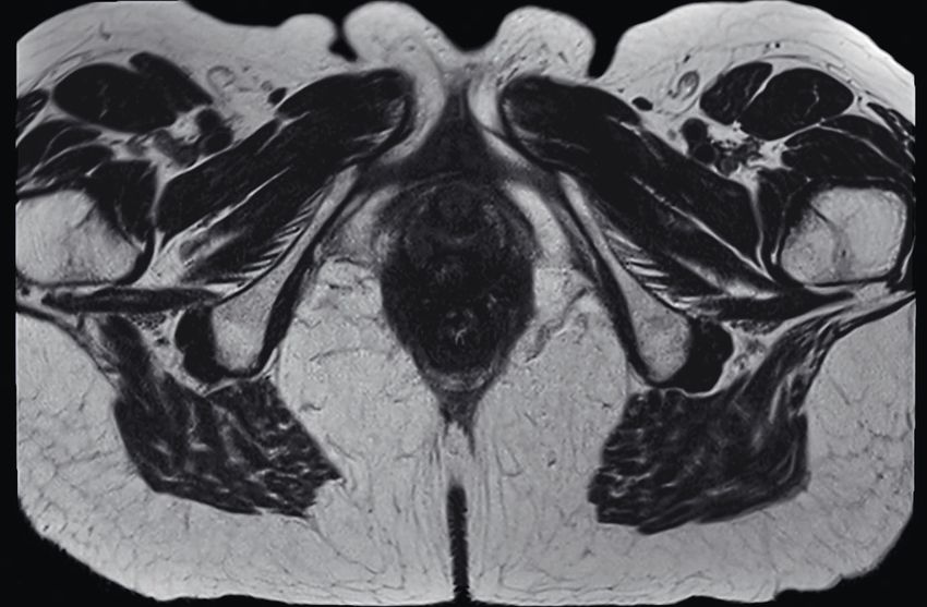
MAGNETOM Flash (78) 2/2021                                                                          Head to Toe Imaging · Clinical

signal and contrast behavior, a new look and feel of the        removal according to “Stein’s Unbiased Risk Estimator”
soft tissue contrast. Regular 2D TSE data does not seem         (SURE) [9]. This automated parameter selection allows the
to be optimal for CS reconstruction.                            algorithm to adapt to both the noise and signal characteris-
     Image reconstruction based on artificial intelligence      tics of each individual image and to generalize to multiple
(AI) / deep learning (DL) is the latest development capable     body regions and scan protocols without additional manual
of denoising [4]. These techniques, however, require large      tuning. Some edge enhancement was applied after the ID
amounts of training data, which was beyond the scope of         to compensate for perceived loss in sharpness.
this study.                                                          From each multi-average raw data set three different
     Iterative denoising (ID) is a technique which uses         versions of the images were reconstructed. The first
similar noise-suppressing operations as Compressed              version (called “original”) used all available averages and
Sensing, but which is specifically designed to be combined      corresponds to the regular reconstruction; it does not
with standard parallel imaging and other standard imaging       use the denoising algorithm. To simulate accelerated
techniques, allowing for shorter scan times and/or higher       acquisition, the second reconstruction version discarded
resolution while compensating for the resultant SNR             one signal average from the raw data sets, e.g. one out of
loss [5]. First applications focused on volumetric acquisi-     two; it also did not use the iterative denoising algorithm
tions [6]. This study presents initial experience with this     and is called “accelerated”. The third reconstruction version
technique applied to standard 2D TSE data in multiple           discarded the same signal average from the raw data sets,
body regions.                                                   and additionally applied the described iterative denoising
                                                                algorithm. These images are called “accelerated + ID”.
                                                                     The versions “accelerated” and “accelerated + ID”
Methods and materials                                           simulate accelerated acquisition by lowering the number of
The goal of this study was to investigate whether the           averages. With this simulation approach the three versions
new iterative denoising technique can compensate for            are based upon as similar data sets as possible so that they
the resultant SNR loss when using higher acceleration           can be directly compared. They derive from the same
in standard 2D TSE imaging. To make this comparison             acquisition so that all variations between repeated scans
between images with higher acceleration versus standard         can be avoided. However, the data is not identical because
acceleration as accurate as possible, instead of rescanning     version two and three only use a subset of the data. Hence
patient with higher acceleration factors, higher accelera-      effects like physiological motion still can have a different
tion was simulated by discarding one average from the raw       effect on the different versions – especially if the motion
data sets. By applying this simulated acceleration, it was      happens during the discarded part of the raw data.
possible to obtain datasets that are – except for the virtual        The described versions of the images were viewed
acceleration – completely identical.                            side-by-side on an open source DICOM viewer (HorosTM)
     Eleven clinical data sets from the perineum, uterus,       and ranked by an experienced radiologist according to
prostate, l-spine, and sacroiliac joint were acquired on a      image quality in terms of perceived signal to noise ratio
1.5T clinical MR scanner (MAGNETOM Sola, Siemens                as well as noticeable artifacts like blurring. Any non-
Healthcare, Erlangen, Germany) with the standard turbo          diagnostic image quality was marked. Given the obvious
spin echo (TSE) sequence. Raw data allowing retrospective       differences between the image versions, no effort at
image reconstruction with subsets of the originally             blinding was made.
acquired averages was collected from regular patient
examinations so that no additional or modified scans had
to be performed. Informed consent from patients was ob-
                                                                Results
tained to reprocess anonymized data for clinical research.      Table 1 lists the ranking results. In 10 out of 11 cases
All raw data sets featured Parallel Imaging under-sampling      (91%), the “original” version was ranked best. Of the
and comprised two or more signal averages.                      “accelerated” versions, study 2 (Figs. 2, 3) is no longer
     Data processing was performed offline and with the         diagnostic (marked as X). All “accelerated + ID” versions
help of a prototype implementation of the ID algorithm as       were ranked better than the “accelerated” versions,
described in [4], integrated into the image reconstruction      and all were diagnostic. In study 5 (Fig. 5) the image
pipeline. Quantitative noise measurements were drawn            quality in the “accelerated + ID” version equals the
from the system’s adjustment framework. Taking into             image quality of the “original” version. In study 11
account all noise-modifying operations, a noise map was         (Figs. 7, 8) the image quality of the “original” is less than
calculated which describes the spatial noise distribution       the image quality of both the “accelerated + ID” images
in coil-combined, complex-valued images after the parallel      and “accelerated” images. Example image features are
imaging reconstruction [7, 8]. Then the wavelet threshold-      described in the figure captions.
ing is automatically adjusted for MMSE-optimal noise

                                                                                siemens-healthineers.com/magnetom-world         3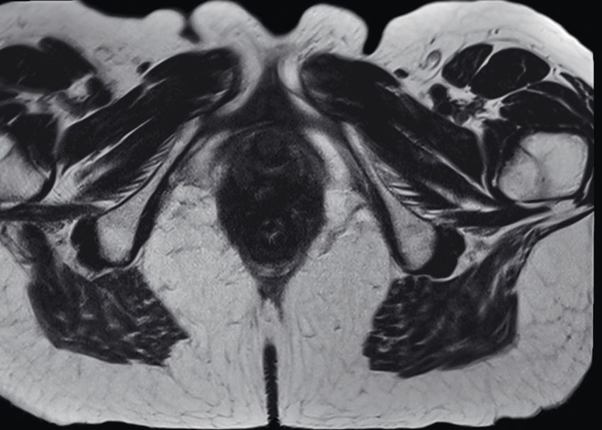
Clinical · Head to Toe Imaging                                                                                         MAGNETOM Flash (78) 2/2021

                                      # of averages,
study #           contrast                                          best image                     2nd best image             3rd best image
                                       [timesaving]
     1        STIR SI joint, cor           2 [50%]                    “original”                  “accelerated + ID”           “accelerated”

     2        STIR L-spine, sag            2 [50%]                    “original”                  “accelerated + ID”         “accelerated” (X)

     3         T1 L-spine, sag             2 [50%]                    “original”                  “accelerated + ID”           “accelerated”

     4         T1 L-spine, sag             2 [50%]                    “original”                  “accelerated + ID”           “accelerated”

     5         T2 uterus, cor              3 [33%]         “original” = “accelerated + ID”            “accelerated”                  –

     6         T2 prostate, ax             3 [33%]                    “original”                  “accelerated + ID”           “accelerated”

     7        STIR SI joint, cor           2 [50%]                    “original”                  “accelerated + ID”           “accelerated”

     8        STIR SI joint, cor           2 [50%]                    “original”                  “accelerated + ID”           “accelerated”

     9        STIR SI joint, cor           2 [50%]                    “original”                  “accelerated + ID”           “accelerated”

     10        T2 uterus, sag              2 [50%]                    “original”                  “accelerated + ID”           “accelerated”

     11       T2 perineum, ax              4 [25%]                “accelerated + ID”                  “accelerated”              “original”

Table 1: Results from the side-by-side reading of an experienced radiologist.

Study 2
2A                                    2B                                    2C

              original                         accelerated + ID                         accelerated

 2  The “original” version (2A) with two averages has the best image quality since two averages are
     effectively averaging out the ghosting artifact. On top of the ghosting, version “accelerated” (2C)
     has a very low SNR with a "grainy unsharpness" e.g. in the body of vertebra L1 (square box) or
     prevertebral space (red circle) and intervertebral space level L2–L3 making this image non-diagnostic. In
     version “accelerated + ID” (2B) the "grainy blurriness" is effectively removed (arrow points to substantial
     SNR gain in prevertebral space). This leads to a still challenging but more diagnostic image quality
     resembling the morphology and contrast of the “original” version, e.g. in the endplates L2–L3.
     Simulated acquisition time saving: 50%. Scanning parameters: TE 99 ms, TR 4570 ms, TI 140 ms,
     duration for original (two averages) 1:17 min.

4         siemens-healthineers.com/magnetom-world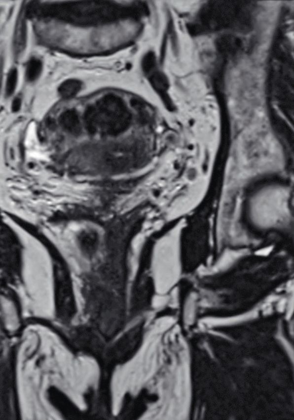
MAGNETOM Flash (78) 2/2021 Head to Toe Imaging · Clinical
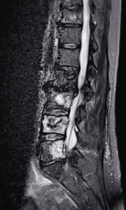
3A 3B 3C
original accelerated + ID accelerated
3 Same patient as Figure 1. Image zoomed in and slightly off midline. Version “accelerated” (3C) is
severely impaired by both low SNR and ghosting artifact. The exaggerated noise level makes it of
questionable diagnostic value (red circles indicate low SNR).Version “accelerated + ID” (3B) still suffers
from ghosting artifact, however the ID algorithm processing of the image effectively removes the grainy
pattern over the vertebral bodies and prevertebral fat plane. The resulting image quality improvement
makes it easy to delineate the intravertebral disc herniation (red arrows).
Study 3
4A 4B 4C
original accelerated + ID accelerated
4 The “original” version (4A) with two averages has crisp image quality in this “perfect patient”. Version
“accelerated” (4C) features an exaggerated noise level in comparison to the “original” (red squares). The
SNR of version “accelerated + ID” (4B) is still lower than in the “original” version; however, in comparison
to the “accelerated” version SNR is clearly higher. Simulated acquisition time saving: 50%. Scanning
parameters: TE 8 ms, TR 603 ms, duration for original 1:38 min.
siemens-healthineers.com/magnetom-world 5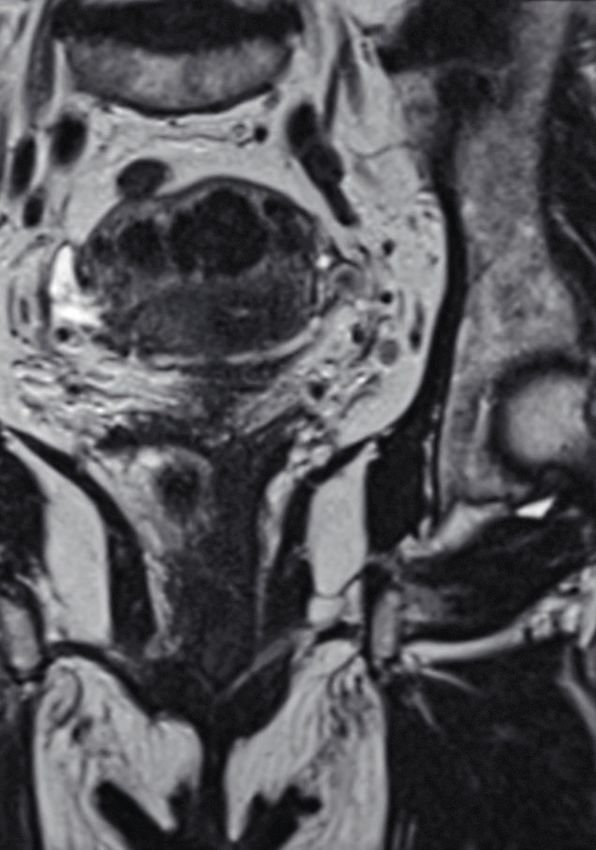
Clinical · Head to Toe Imaging                                                                                   MAGNETOM Flash (78) 2/2021

Study 5
5A                                    5B                                    5C

              original                          accelerated + ID                        accelerated

 5 All three images are of diagnostic quality with high signal. Some graininess indicating lower SNR is
   present in the “accelerated” image (5C) over the uterus (red oval) and vagina (red box). This grainy
   superposition is removed after ID in version “accelerated + ID” (5B). The resulting image in version
   “accelerated + ID” (center) matches very closely the image quality of the “original” version (5A). The
   version “accelerated + ID” (center) has the highest overall SNR without graininess. Taking a closer
   look at details e.g. the fatty streaks in the right ischiococcygeal muscle (red arrow): these small fatty
   streaks are depicted with the same confidence as on the original image, indicating that no image
   detail is lost during ID. Simulated time saving: 33%. Scanning parameters: TE 132 ms, TR 7780 ms,
   duration for original 1:25 min.

Study 7
6A                                    6B                                    6C

              original                          accelerated + ID                        accelerated

 6 The “original” version (6A) with two averages has the best image quality. Version “accelerated” (6C) has
   a low signal to noise with impressive "graininess" (square boxes) over the fifth lumbar and first sacral
   vertebra. In version “accelerated + ID” (6B) the overlying "graininess" is effectively removed resulting in
   an image with SNR resembling the “original” version. Simulated acquisition time saving: 50%. Scanning
   parameters: TE 87 ms, TR 3400 ms, TI 140 ms, duration for original 0:52 min.

6       siemens-healthineers.com/magnetom-world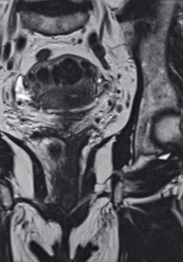
MAGNETOM Flash (78) 2/2021 Head to Toe Imaging · Clinical
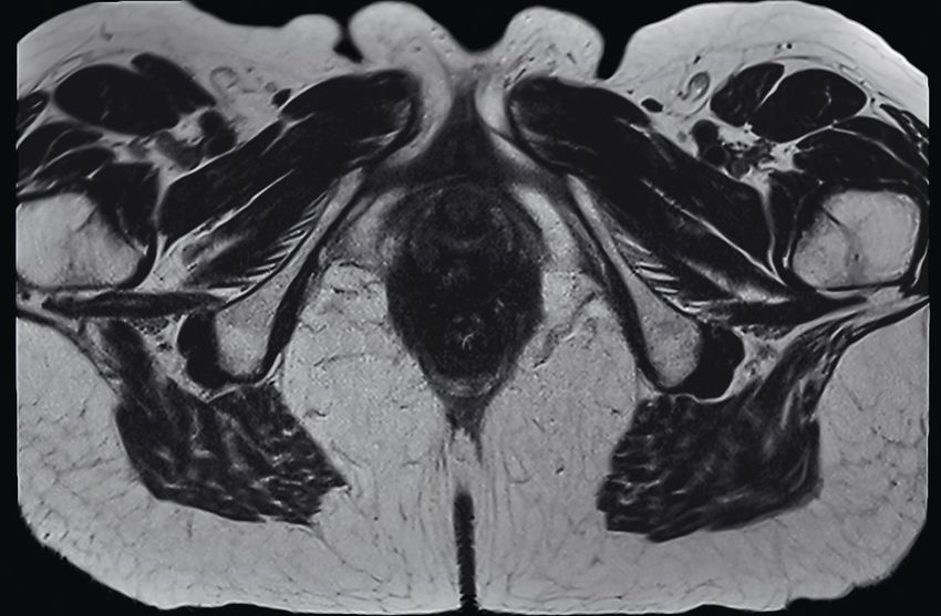
Study 11
7A 7B 7C
original accelerated + ID accelerated
7 Version “original” (7A) has abundant signal, however, some blurring is present. Version “accelerated”
(right) and “accelerated + ID” (7B) have less blurring since less measurement time leads to less patient
movement, but still have abundant signal. This abundance in signal results in an image quality where
for instance the veins in the ischiorectal fossa are better depicted in both versions “accelerated” (7C) and
“accelerated + ID” (center) than on the original version. Although high enough SNR is present in version
“accelerated” (right), ID further enhances the image quality in “accelerated + ID” (center) image: the
small venous bifurcation (red arrow, magnifying glass) in the left ischiorectal fossa is better depicted
after ID (center). Simulated time saving: 25%. Scanning parameters: TE 106 ms, TR 814 ms, duration for
original 4:12 min.
8A 8B 8C
original accelerated + ID accelerated
8 Same study as Figure 7: In version “original” (8A) the small fibrous strands in subcutaneous fat are hard
to depict even though they are clearly present on the ”accelerated” version (8C) and really stand out
on the “accelerated + ID” version (red arrows). The fatty streaks (red circle) in between the muscle fibers
of the external obturator cannot be seen on the “original” version, they are however visible on the
“accelerated” version and really stand out on the “accelerated + ID” version (8B). The ID algorithm does
not only increase SNR but also enhances image details, for example the fatty streaks in the iliac muscle
(red box). The internal structure with fat-containing hilum of the left inguinal lymph node (between the
red arrow and red box) is again best depicted on the “accelerated + ID” version. Simulated time saving:
25%. Scanning parameters: TE 106 ms, TR 814 ms, duration for original 4:12 min.
siemens-healthineers.com/magnetom-world 7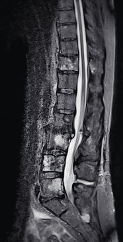
Clinical · Head to Toe Imaging                                                                          MAGNETOM Flash (78) 2/2021

Discussion                                                       over-filtering occurs: the small septae in the subcutaneous
                                                                 fat are better depicted on the accelerated images with
Acquiring a dataset that is substantially accelerated (by
                                                                 denoising when compared to the accelerated version as
reducing the number of averages) leads to a discernible
                                                                 well as to the original version. This can be seen in the fatty
drop in signal to noise ratio. This is rendering the resulting
                                                                 streaks in the external obturator muscle.
images grainy and harder or even sometimes impossible to
interpret, with an obvious drop in image quality in compar-
ison to the “original” images. The results of this small-scale   Conclusion
study provide evidence that the process of ID as described
                                                                 Image quality in standard 2D MRI sequences, accelerated
above can compensate for the drop in signal to noise ratio
                                                                 in simulation beyond the threshold of standard acceptable
in substantially accelerated 2D datasets.
                                                                 noise levels, can be substantially improved by applying
     In this study the perceived gain in image quality after
                                                                 an ID algorithm using supplementary information about
ID was obvious in images which are already by design
                                                                 the image noise level. It is expected that standard 2D MRI
inherently lower in signal to noise ratio like e.g. Short-TI
                                                                 can profit from ID when natively scanning with lower
Inversion Recovery (STIR) imaging. Studies 7 and 8 with
                                                                 numbers of averages and hence shorter acquisition times.
coronal STIR imaging of sacroiliac joints demonstrate
                                                                 More clinical studies with different clinical perspectives are
the benefit from the ID bringing image quality back to
                                                                 required to show if ID could become as indispensable a tool
the standard imaging quality. Apparently, ID is reducing
                                                                 in MRI as iterative reconstruction is in CT.
the noise level in the signal and image quality thereby
approaches the image quality of the “original” images.
     Less intuitive, even in images with abundant signal,        Acknowledgements
the image quality of “accelerated + ID” can be as good
                                                                 The authors are grateful to Dr. Boris Mailhe for developing
(study 5) or even better (study 11) than the original.
                                                                 the core of the iterative denoising technique and Dr. Carm-
A plausible explanation is that by removing an average
                                                                 el Hayes for her support in algorithm implementation and
also removes the blurring that can occur due to slight
                                                                 data processing.
patient movement between multiples averages.
     Iterative denoising, in contrast to conventional            References
noise-removing image filters, has the advantage of supple-
                                                                 1 Heidemann RM et al. A brief review of parallel magnetic resonance
mentary quantitative noise distribution information,
                                                                   imaging. European Radiology, Vol 13 2003: 2323–2337.
which would otherwise have to be estimated from the              2 Mohan J, Krishnavenib V,and Guo Y. A survey on the magnetic
images themselves. Consequently, over- or under-filtering          resonance image denoising methods. Biomed Signal Process
is inherently avoided. In combination with the SURE-opti-          Control, Vol 9 2014: 56-69.
mizing iteration, this is especially important for preserving    3 Geethanath S et al. Compressed sensing MRI: a review. Crit Rev
                                                                   Biomed Eng, Vol 41(1) 2013: 183-204.
small image details and sharpness, although some
                                                                 4 Lin DJ et al. Artificial Intelligence for MR Image Reconstruction:
additional edge enhancement appears to be beneficial.              An Overview for Clinicians. J Magn Reson Imaging 2020 Feb 12.
Preserved image details and sharp edges are striking in            Online ahead of print.
study 11 (Figs. 7) where the small venous bifurcation in         5 Kannengiesser SAR et al. Universal iterative denoising of
the left ischiorectal fossa is clearly sharper delineated in       complex-valued volumetric MR image data using supplementary
                                                                   information. Proc. Intl. Soc. Mag. Reson. Med., Vol 24 2016: 1779.
the “accelerated + ID” image than on the images without
                                                                 6 Kang HJ et al. Clinical Feasibility of Gadoxetic Acid-Enhanced
denoising. Figure 8 is another excellent example that no           Isotropic High-Resolution 3-Dimensional Magnetic Resonance
                                                                   Cholangiography Using an Iterative Denoising Algorithm for
                                                                   Evaluation of the Biliary Anatomy of Living Liver Donors.
                         Contact                                   Invest Radiol., Vol 54(2) 2019: 103-109.
                                                                 7 Breuer FA et al. General formulation for quantitative G-factor
                         Johan Dehem, M.D.                         calculation in GRAPPA reconstructions. Magn Reson Med.,
                         Jan Yperman Ziekenhuis                    Vol 62(3) 2009: 739-46.
                         Briekestraat 12
                                                                 8 Kellman P, and McVeigh ER. Image reconstruction in SNR units:
                         8900 Ypres
                         Belgium                                   a general method for SNR measurement. Magn Reson Med.,
                         Phone: +32 57 35 74 00                    Vol 54(6) 2005: 1439-47.
                         johan.dehem@yperman.net                 9 Blu F, and Lusier T. The SURE-LET Approach to Image Denoising.
                                                                   IEEE Trans Im Proc, Vol 16:11 2007: 2778 - 2786.

8       siemens-healthineers.com/magnetom-world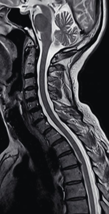
MAGNETOM Flash (78) 2/2021                       Head to Toe Imaging · Clinical

                             siemens-healthineers.com/magnetom-world         9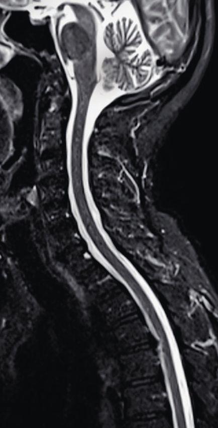
You can also read