Unique Immunological Profile In Patients With COVID- 19
←
→
Page content transcription
If your browser does not render page correctly, please read the page content below
Preprint: Please note that this article has not completed peer review.

Unique Immunological Profile In Patients With COVID-
19
CURRENT STATUS:                  UNDER REVIEW

Stefania Varchetta
Fondazione IRCCS Policlinico San Matteo

Dalila Mele
Fondazione IRCCS Policlinico San Matteo

Barbara Oliviero
Fondazione IRCCS Policlinico San Matteo

Stefania Mantovani
Fondazione IRCCS Policlinico San Matteo

Serena Ludovisi
Fondazione IRCCS Policlinico San Matteo

Antonella Cerino
Fondazione IRCCS Policlinico San Matteo

Marco Vecchia
Fondazione IRCCS Policlinico San Matteo

Silvia Roda
Fondazione IRCCS Policlinico San Matteo

Michele Sachs
Fondazione IRCCS Policlinico San Matteo

Raffaele Bruno
Fondazione IRCCS Policlinico San Matteo

Mario U. Mondelli
Fondazione IRCCS Policlinico san Matteo

 mario.mondelli@unipv.itCorresponding Author

                                                 1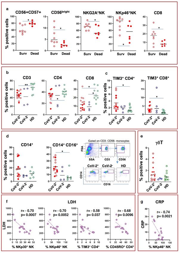
DOI:
  10.21203/rs.3.rs-23953/v1
SUBJECT AREAS
 Immunology      Infectious Diseases
KEYWORDS
 SARS-CoV-2, host immunity, altered immune phenotype in patients

                                       2
Abstract
The relationship between SARS-CoV-2 and host immunity is unknown. We show here that patients
with COVID-19 had an altered immune phenotype, with an expansion of adaptive FceRIgneg NK cells,
and inflammatory CD14+CD16+ monocytes. T cells were reduced and overexpressed the Tim-3
exhaustion molecule. Low frequencies of CD8 T cells and NKG2A+ NK cells, and expansion of mature
CD57+ NK cells were associated with poor prognosis. These findings unveil a unique immunological
profile in COVID-19 patients.
Introduction, Results And Discussion
Severe Acute Respiratory Syndrome Coronavirus-2 (SARS-CoV-2) is responsible for a pandemic thus
far responsible for nearly 1 million cases of Coronavirus Disease-19 (COVID-19) with a case/fatality
rate of 4.5% [1]. The infection usually causes mild symptoms, but may be responsible for severe
interstitial pneumonia, myocarditis, acute kidney injury, acute respiratory distress syndrome (ARDS),
multiorgan failure and death [2]. Laboratory tests indicate that patients with severe progression of
COVID-19 show signs of secondary haemophagocytic lymphohistiocytosis (sHLH), a
hyperinflammatory syndrome characterised by a potentially fatal cytokine storm with multiorgan
failure, which may be triggered by viral infections [3]. Akin to sHLH, COVID-19 is characterized by
lymphopenia, and increased serum ferritin, D-dimer, C-reactive protein (CRP), and lactic-
dehydrogenase (LDH), which are also considered predictors of poor outcome [4].
Moreover, several serum cytokine concentrations are increased during COVID-19, supporting the
hypothesis that virally driven hyperinflammation plays a key pathogenetic role [2].
Despite clear evidence of ongoing overexuberant inflammation, there are no systematic studies
addressing phenotypic and functional alterations of innate and adaptive immune cells, that are likely
exposed to a variety of stimuli in COVID-19 patients at presentation. The lack of a comprehensive
immunological analysis prompted us to assess the phenotypic and functional status of NK cells, γδT
cells, monocytes and CD4 and CD8 T cells in patients presenting with clinically moderate to severe
interstitial pneumonia emerging in the setting of COVID-19. Patient clinical details and laboratory
findings, as well as peripheral blood mononuclear cells (PBMC) flow cytometric analysis are reported
3in Supplementary Information, Patients and Methods.
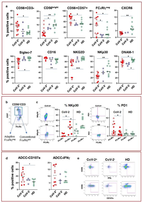
The frequency of NK cells was significantly higher in COVID-19 patients compared to healthy controls,
being significantly enriched in mature (CD56dimCD57+) NK cells (Fig.1a). Interestingly, there was a
relative expansion of CD57+/FcεRIγneg adaptive NK cells compared with non-COVID-19 disease
controls and healthy controls (Fig.1a) suggesting a SARS-CoV-2-related expansion of this population,
whereas the proportion of CD56bright NK cells was reduced. An increase in CD16+ NK cells was also
evident compared with healthy controls (Fig 1a). Notably, the frequency of CXCR6-expressing NK cells
was low in COVID-19 patients (Fig.1a), most likely since these cells home to the lungs where they
concentrate, their ligand CXCL16 being produced in large amounts by alveolar macrophages [5].
Additional changes in NK cells included significant reductions in the frequencies of Siglec-7, DNAM-1,
NKG2D, NKp30 (Fig.1a), the latter being particularly evident in the adaptive subset (Fig.1c).
Importantly, the frequency of PD-1 positive NK cells was significantly higher in the adaptive compared
with conventional NK cells in patients with COVID-19 (Fig. 1c). No changes were noted in bulk NK cell
expression of NKG2C, NKG2A, GITR, TRAIL, CD69, PD-1, TIGIT. The trend noted for TIM-3 was not
statistically significant (Suppl. Fig.1a). Of note, although no significant changes in degranulation
activity or IFNγ production were observed using K562 as target cells (Suppl. Fig.1b), there was an
increased ability of NK cells to exert antibody-dependent cell- mediated cytotoxicity (ADCC), a
function exquisitely performed by adaptive NK cells [6] (Fig. 1d). The proportions of CD56bright,
NKG2A and NKp46 positive NK cells were significantly lower and the proportion of mature CD57+ cells
significantly higher in patients who succumbed compared with those who survived (Fig.2a). The
relative frequencies of total CD3+, CD4+ and CD8+ T cells were significantly lower than healthy
controls, although no apparent differences were noted with disease controls (Fig.2b). Patients who
died showed a significantly lower frequency of CD8 T cells compared with those who survived (Fig.2a).
Moreover, both CD4 and CD8 T cells from COVID-19 patients overexpressed Tim-3 compared with
healthy controls, suggesting a pan T-cell exhaustion profile (Fig.2c). No differences were found in
CD45RO, HLA-DR, GITR expression or Treg population frequency (Suppl. Fig.2). Importantly, there was
4a clear relative expansion of CD14/CD16 double positive monocytes, a phenotype associated with an
inflammatory profile (Fig. 2d) [7]. There were no statistically significant changes in the frequency of
γδT cells (Fig. 2e). Negative correlations were found between laboratory indicators of severe or
progressive disease. Thus, NK cells expressing the activating receptors NKp30 and NKp46, as well as
CD45RO+ and Tim-3+ CD4 T cells, correlated negatively with LDH (Fig. 2f). A negative correlation was
also present between NKp46+ NK cells and CRP (Fig. 2g).
Information on PBMC phenotype and function are virtually lacking in patients with COVID-19. Here we
had the opportunity to evaluate patients admitted to hospital because of moderate to severe COVID-
19 interstitial pneumonia and compared them to a small group of SARS-CoV-2 negative pneumonia
and healthy We showed that patients with COVID-19 had a relative expansion of mature adaptive NK
cells endowed with ADCC function, which was increased in this setting in line with findings in other
viral infections, particularly cytomegalovirus [6]. Other phenotypic features were compatible with a
dysfunctional NK cell phenotype, namely the reduced frequencies of Siglec-7-, NKG2D- and NKp30-
expressing cells [8,9]. A recent study addressed the kinetics and breadth of immune responses
associated with clinical resolution of COVID-19 in a single patient with relatively mild disease [10].
Antibody-secreting cells appeared at the time of viral clearance together with follicular helper T cells
and activated CD4 and CD8 T cells. In contrast, in our patients with moderate to severe interstitial
pneumonia, some of whom sadly succumbed, Tim-3 positive exhausted CD4 and CD8 T cells largely
prevailed at presentation and lower frequencies of CD8+ T cells were linked to poor prognosis. A
recent study found lower frequencies of CD8 T cells and NK cells with a relative enrichment of NKG2A-
expressing cells which returned to normal after clinical recovery, suggesting rescue of impaired T and
NK cell function [11]. Interestingly, although no difference in the frequency of NKG2A- expressing NK
cells was found between patients and controls in the present study, NKG2A+ NK cells were lower in
patients who did not survive, suggesting that loss of this inhibitory receptor somehow unleashed NK
cells in patients with fatal outcome.
Our study provides important novel insights into the pathogenetic mechanisms of COVID- 19,
5characterized by a rapid expansion of phenotypically mature NK cells persisting at high frequency in
patient with poor prognosis. The simultaneously reduced frequency of CD4+ and CD8+ T cells
expressing the Tim-3 exhaustion marker unveils a multifaceted behavior of the two arms of immunity
in this clinical setting. The relative enrichment of inflammatory monocytes lends support to the
hypothesis that COVID-19 resembles in part to the macrophage-activation syndrome which is thought
to be closely related to hemophagocytic lymphohistiocytosis (HLH) [12], an uncommon life-
threatening disorder of severe hyperinflammation caused by uncontrolled proliferation of activated
lymphocytes and macrophages that secrete high levels of inflammatory cytokines. Of note patterns
similar to cytokine storm syndromes have been described for COVID-19 and SARS [2].
It is difficult at this early stage to precisely frame COVID-19 within an immunologically coherent
clinical entity. Indeed, several peculiarities have emerged that contribute to the uniqueness of its
immune profile. Understanding the dynamics and the quality of immune responses to SARS-CoV-2 will
provide invaluable translational information to design effective treatments for this potentially deadly
disease.
Methods
Provided as supplemental file.
Declarations
Author Contributions: SV and DL designed and performed experiments and critically contributed to
drafting the manuscript; BO, SM, AC performed experiments and critically read the manuscript; SL,
MV, SR, MS, RB recruited patients, prepared the database and critically read the manuscript, MUM
designed and discussed the experiments and wrote the manuscript.
Competing Interests: none.
Ethics: The study protocol conformed to the ethical guidelines of the 1975 Declaration of Helsinki and
was approved by the Institutional Review Board and Ethical Committee of Fondazione IRCCS
Policlinico San Matteo (Protocol number 20200033215). All patients provided written or, in case they
were unable to sign, verbally witnessed informed consent as per the above study protocol.
References
1. https://worldometers.info/coronavirus/
62. Huang, et al. Clinical Features of Patients Infected With 2019 Novel Coronavirus in
Wuhan, China. Lancet 395, 497-506 (2020).
3. Ramos-Casals, et al. Adult Haemophagocytic Syndrome. Lancet 383, 1503–16 (2014).
4. Ruan, et al. Clinical Predictors of Mortality Due to COVID-19 Based on an Analysis of
Data of 150 Patients From Wuhan, China. Intensive Care Med.
https://doi.org/10.1007/s00134-020-05991-x (2020).
5. Morgan, J. et al. Expression of CXCR6 and its ligand CXCL16 in the lung in health and
disease. Clin. Exp. Allergy 35, 1572–1580 (2005)
6. Schlums, H. et al. Cytomegalovirus Infection Drives Adaptive Epigenetic
Diversification of NK Cells with Altered Signaling and Effector Immunity 42, 443–456
(2015).
7. Ziegler-Heitbrock, The CD14+ CD16+ Blood Monocytes: Their Role in Infection and
Inflammation. J. Leukoc. Biol. 81, 584-92.
8. Varchetta, et al. Lack of Siglec-7 Expression Identifies a Dysfunctional Natural Killer
Cell Subset Associated With Liver Inflammation and Fibrosis in Chronic HCV Infection.
Gut 65, 1998-2006 (2016).
9. Vitale, et al. An Historical Overview: The Discovery of How NK Cells Can Kill Enemies,
Recruit Defense Troops, and More. Front. Immunol. 10, 1415 (2019).
10. Thevarajan, et al. Breadth of concomitant immune responses prior to patient
recovery: a case report of non-severe COVID-19. Nat. Med.
https://doi.org/10.1038/s41591-020-0819-2 (2020).
11. Zheng, M. et al. Functional exhaustion of antiviral lymphocytes in COVID-19 patients.
Mol. Immunol. https://doi.org/10.1038/s41423-020-0402-2 (2020).
12. Fisman, N. Hemophagocytic syndromes and infection. Emerging Infect. Dis. 6, 601–8
(2000).
7Covid19 Irccs San Matteo Pavia Task Force
ID Staff
Raffaele Bruno, Mario U Mondelli, Enrico Brunetti, Angela Di Matteo, Elena Seminari, Laura Maiocchi,
Valentina Zuccaro, Layla Pagnucco, Bianca Mariani, Serena Ludovisi, Raffaella Lissandrin, Aldo Parisi,
Paolo Sacchi, Savino FA Patruno, Giuseppe Michelone, Roberto Gulminetti, Domenico Zanaboni,
Stefano Novati, Renato Maserati, Paolo Orsolini, Marco Vecchia.
ID Residents
Marco Sciarra, Erika Asperges, Marta Colaneri, Alessandro Di Filippo, Margherita Sambo, Simona
Biscarini, Matteo Lupi, Silvia Roda, Teresa Chiara Pieri, Ilaria Gallazzi, Michele Sachs, Pietro Valsecchi.
Emergency Care Unit
ECU Staff: Stefano Perlini, Claudia Alfano, Marco Bonzano, Federica Briganti, Giuseppe Crescenzi,
Anna Giulia Falchi, Roberta Guarnone, Barbara Guglielmana, Elena Maggi, Ilaria Martino, Pietro
Pettenazza, Serena Pioli di Marco, Federica Quaglia, Anna Sabena, Francesco Salinaro, Francesco
Speciale, Ilaria Zunino.
ECU Residents: Marzia De Lorenzo, Gianmarco Secco, Lorenzo Dimitry, Giovanni Cappa, Igor Maisak,
Benedetta Chiodi, Massimiliano Sciarrini, Bruno Barcella, Flavia Resta, Luca Moroni, Giulia Vezzoni,
Lorenzo Scattaglia, Elisa Boscolo, Caterina Zattera, Tassi Michele Fidel, Capozza Vincenzo, Damiano
Vignaroli, Marco Bazzini.
Intensive Care Unit
Giorgio Iotti, Francesco Mojoli, Mirko Belliato, Luciano Perotti, Silvia Mongodi, Guido Tavazzi
Paediatric Unit
Gianluigi Marseglia, Amelia Licari, Ilaria Brambilla
Virology Staff
Barbarini Daniela, Bruno Antonella, Cambieri Patrizia, Campanini Giulia, Comolli Giuditta, Corbella
Marta, Daturi Rossana, Furione Milena, Mariani Bianca, Maserati Roberta, Monzillo Enza, Paolucci
Stefania, Parea Maurizio, Percivalle Elena, Piralla Antonio, Rovida Francesca, Sarasini Antonella,
Zavattoni Maurizio.
8Virology Technical staff
Adzasehoun Guy, Bellotti Laura, Cabano Ermanna, Casali Giuliana, Dossena Luca, Frisco Gabriella,
Garbagnoli Gabriella, Girello Alessia, Landini Viviana, Lucchelli Claudia, Maliardi Valentina, Pezzaia
Simona, Premoli Marta.
Virology Residents
Bonetti Alice, Caneva Giacomo, Cassaniti Irene, Corcione Alfonso, Di Martino Raffella, Di Napoli
Annapia, Ferrari Alessandro, Ferrari Guglielmo, Fiorina Loretta, Giardina Federica, Mercato Alessandra,
Novazzi Federica, Ratano Giacomo, Rossi Beatrice, Sciabica Irene Maria, Tallarita Monica, Vecchio
Nepita Edoardo.
Research Laboratories, Division of Infectious Diseases and Immunology
Antonella Cerino, Stefania Varchetta, Barbara Oliviero, Stefania Mantovani, Dalila Mele.
Pharmacy Unit
Monica Calvi, Michela Tizzoni
Hospital Management
Carlo Nicora, Antonio Triarico, Vincenzo Petronella, Carlo Marena, Alba Muzzi, Paolo Lago
Data Unit
Francesco Comandatore, Gherard Batisti Bissignandi, Stefano Gaiarsa, Marco Rettani, Claudio Bandi
Figures
910
Figure 1

NK cell characterization in SARS-CoV-2 infection. a) Frequency of NK cells and expansion of

mature CD57+ and adaptive (FcεRIγneg) NK cells in COVID-19 patients. Reduced frequency

of CXCR6, Siglec-7, NKG2D and NKp30, and increased proportion of CD16+ cells. b) Dot plot

showing gating on CD57+FcεRIγneg adaptive and CD57+FcεRIγpos conventional NK cells. c)

Representative dot plots and graphs showing NKp30 reduction and PD1 increase in adaptive

  compared with conventional NK cells in COVID-19 patients. Representative dot plots are

 gated on total CD57+ NK cells. Circles indicate adaptive NK; squares, conventional NK. d)

 Increased NK degranulation and IFNγ expression in COVID- 19 patients. e) Representative

 IFNγ and CD107a dot plots in patients and controls. Full red symbols indicate patients who

 subsequently died. Middle bars represent medians. The One Way Anova test was used to

                 compare three groups. *p
Figure 2 12
a) Expansion of mature CD57+ NK cells and reduction of CD56bright NK cells, NKG2A+ and

     NKp46+ NK cells and CD8 T cells in patients who survived and in those who succumbed. b)

       Frequencies of total CD3+, CD4+ and CD8+ T cells were reduced in COVID-19 patients

       compared to HD. c) Tim-3 expressing CD4 and CD8 T cells were increased in COVID-19

      patients. d) Expansion of CD14+CD16+ double positive monocytes in COVID-19 patients

    and representative dot plots. e) No differences were observed in the proportion of γδT cells.

       f & g) Correlations of NK and CD4 T receptor molecules with LDH and CRP. Middle bars

      represent median values. The Mann-Whitney test was used to compare survivors versus

      dead patients. The One Way Anova test was used to compare three groups. The Pearson

                     test was used to examine correlations. * p
You can also read