Expression of islet neogenesis-associated protein in islets of normal hamsters
←
→
Page content transcription
If your browser does not render page correctly, please read the page content below
243

Expression of islet neogenesis-associated protein in islets of
normal hamsters
L E Flores, M E García, M I Borelli, H Del Zotto, M E Alzugaray,
B Maiztegui and J J Gagliardino
CENEXA – Centre of Experimental and Applied Endocrinology (UNLP-CONICET, PAHO/WHO Collaborating Centre), National University of La Plata
  School of Medicine, La Plata, Argentina
(Requests for offprints should be addressed to J J Gagliardino, CENEXA (UNLP-CONICET), Facultad de Ciencias Médicas, UNLP, Calles 60 y 120,
   1900 La Plata, Argentina; Email: gagliardino@infovia.com.ar)

Abstract
The aim of the present study was to test the possible                             INGAP protein was identified by Western blot in
presence and expression of islet neogenesis-associated                         the cytosolic fraction of homogenates from fresh isolated
protein (INGAP) in islet cells of normal adult hamsters.                       islets, exocrine cells and whole fresh pancreas. INGAP-
   Pancreata from normal male Syrian hamsters were                             immunopositive cells were observed in duct, exocrine and
removed to perform the following studies. (i) Western blot                     islet cells in either fixed intact or digested pancreatic tissue.
analysis using the cytosolic fraction from homogenates of                      INGAP mRNA was identified in samples of total RNA
isolated islets, exocrine tissue and whole pancreas, and                       from fresh and cultured isolated islets and from exocrine
rabbit INGAP-specific antibody. (ii) Immunohisto-                              cells.
chemical identification of INGAP-positive cells in fixed                          Our data demonstrate that INGAP is present and
sections of intact pancreata, fresh and 72 h cultured islets                   expressed in islets and in exocrine pancreatic cells of
(isolated by collagenase digestion), and smears of exocrine                    normal hamsters. The ubiquitous localization of INGAP
pancreatic cells, using the same INGAP-specific antibody                       suggests its possible role in the physiological process of islet
and streptavidin-biotin complex. (iii) RT-PCR using total                      growth and its protective effect upon streptozotocin-
RNA extracted from isolated islets and from exocrine                           induced diabetes.
tissue as template, and a specific pair of primers. (iv)                       Journal of Endocrinology (2003) 177, 243–248
Control of the sequence of the PCR products.

Introduction                                                                   hamsters, and a significant increase of this peptide in
                                                                               hamsters with an insulin-resistant state induced by the
Several hormones, transcription and growth factors are                         administration of a sucrose-rich diet (Del Zotto et al.
involved in the control of islet development and growth                        2000). In all cases INGAP protein was detected in cells
(Edlund 2001). One of these factors, islet neogenesis-                         from the three pancreas subsectors, but mainly in islet
associated protein (INGAP), originally isolated from                           non--cells. Most islet INGAP-positive cells did not
pancreata of normal hamsters with previous cellophane                          co-stain with antibodies against any of the hormones
wrapping of the pancreas head (CW), has been identified                        expressed in the islets, while around 40% of them
as part of a protein complex called ilotropin (Pittenger et al.                co-stained with a glucagon antibody (Del Zotto et al.
1992). The immunocytochemical presence of INGAP has                            2000). However, Raffaelof et al. (1997) reported that
been shown not only in hamsters, but also in the pancreas                      INGAP was only expressed in the exocrine sector of the
of normal foetal mice (Rafaeloff-Phail et al. 1998a), adult                     CW pancreas.
rats (H Del Zotto & C Semakula, unpublished obser-                                On account of these apparent discrepancies, we
vations), and in human beings (Rafaeloff-Phail et al.                           have currently studied the presence of INGAP pro-
1998b, Semakula et al. 2002). It has further been reported                     tein (immunocytochemistry and Western blot analysis)
that INGAP administration to streptozotocin-treated ham-                       and its gene expression (RT-PCR) in isolated islets
sters decreased significantly the percentage of diabetic                       and in exocrine cells isolated from adult normal hamsters.
animals (Rafaeloff et al. 1997, Gold et al. 1998).                              Our results demonstrate that INGAP is not only
   Using quantitative immunocytochemical techniques                            present and expressed in exocrine cells, but also in islet
we have identified INGAP in pancreata from normal                              cells.

Journal of Endocrinology (2003) 177, 243–248                                                             Online version via http://www.endocrinology.org
0022–0795/03/0177–243  2003 Society for Endocrinology Printed in Great Britain
244 L E FLORES and others · INGAP expression in hamster islets
Materials and Methods to 100 V until the front was about 0·5 cm from
the bottom. The gels were then electroblotted onto a
Animals and pancreas digestion poly (vinylidene difluoride) microporous membrane
(Immobilon, Millipore) by means of a discontinuous buffer
Normal male Syrian hamsters from the Eppley colony (8
system. Before immunostaining, the Immobilon sheets
weeks of age, 80–100 g body weight; Omaha, NE, USA)
were treated with blocking solution and then incubated
were used. They were maintained in a room at 211 C
with appropriate dilutions of the primary specific antibody
and 505% humidity, with a 12 h light:12 h darkness
against INGAP (rabbit antibody IgG 1246, 1:1500;
cycle, and freely fed a commercial diet and tap water.
kindly provided by Dr Gerald Gold, Eli Lilly Company,
Animals were killed by cervical dislocation and the entire
Indianapolis, IN, USA) in PBS at room temperature for
pancreas was removed and digested (collagenase) to isolate
1 h. The specificity of this antibody has been previously
islets and exocrine tissue (Lacy & Kostianovsky 1967). For
tested (Rafaeloff-Phail et al. 1998a,b). Saturating antibody
that purpose, removed pancreata were rinsed in Krebs–
concentrations were used for all the immunostaining. After
Ringer bicarbonate (KRB) buffer, cut into small pieces,
rinsing with PBS, the streptavidin-peroxidase conjugate
and further incubated in the presence of collagenase; when
anti-rabbit IgG (Sigma Co., St Louis, MO, USA) was
islets were free of exocrine tissue (sequential observation
added for 1 h; this step was followed by rinsing. Colour
under a stereomicroscope), the reaction was stopped by
development was obtained using 3,3 -diamino-benzidine
adding cold KRB. Islets were picked up one-by-one with
(DAB) (Sigma).
capillary pipettes. Islet-free exocrine tissue was collected
when all islets were removed from the digested pancreas.
Immunohistochemical studies
Islet cultures Pancreata were fixed in Bouin’s liquid and embedded in
paraffin. Thin sections (5 µm) were mounted on silanized
Groups of 50 islets were cultured for 24 h at 37 C in (3-amino-propyltriethoxy-silane; Sigma) slides. Immuno-
RPMI 1640 (Gibco-BRL, Grand Island, NY, USA), pH cytochemical identification of INGAP-positive cells was
7·4, containing 10% (v/v) foetal calf serum and 4 mM performed in deparaffinized sections treated with normal
glucose in a humid atmosphere (5% CO2/95% O2). After porcine serum (30 min) to reduce non-specific staining,
24 h of culture the medium was removed and the islets and with methanol–hydrogen peroxide (30 min) to block
were cultured for another 48 h in the same medium. endogenous peroxidase. The slides were incubated for
24 h at 4 C in a humidified chamber, with appropriately
diluted rabbit INGAP-specific antibody (IgG 1246,
Western blotting
1:600), or normal rabbit serum. The final staining was
Isolated islets and exocrine tissue cells obtained by colla- performed using 30 min incubations with the streptavidin-
genase digestion (Lacy & Kostianovsky 1967) and whole biotin complex or alkaline phosphatase (Sigma, 1:40 and
pancreata were homogenized in 10 mM Tris–HCl, pH 1:20 respectively) and revealed with carbazole, DAB or
7·4, with protease inhibitors (4 µg aprotinin; 0·1 mM fast blue.
benzamidine, and 0·1 mM phenyl-methyl-sulphonyl
fluoride). The homogenates were then centrifuged at
5300 g for 15 min, and the supernatant was further Islet immunohistochemistry
centrifuged at 109 000 g for 90 min. Protein concentration Five hundred isolated islets were fixed in Bouin’s liquid
was measured in the supernatant by the method of and embedded in paraffin. Thin sections (5 µm) were
Bradford (BioRad, Hercules, CA, USA). Both 10 µg of processed as described above for the immunocytochemical
protein from isolated islets and 5 µg of protein from identification of INGAP-positive cells.
exocrine tissue cells or from whole pancreas homogenates
were placed into the wells of the stacking gel containing
4% acrylamide, 125 mM Tris–HCl, pH 6·8, 0·1% SDS, Smear immunocytochemistry
0·05% ammonium persulphate and 0·2% N,N,N ,N - Smears of pancreatic cells from the digested pancreas were
tetramethylethylenediamine (TEMED). The running gel fixed in 96% ethanol, and thereafter sequentially incubated
composition was 7·5% acrylamide, 375 mM Tris–HCl, with appropriate dilutions of INGAP antibody (1:600),
pH 8·8, 0·1% SDS, 0·2% ammonium persulphate, and and counterstained with haematoxylin.
0·2% TEMED. The reservoir buffer contained 0·025 M
Tris, 0·192 M glycine and 0·1% SDS. In all cases
Kaleidoscope polypeptide prestained standards (BioRad) Isolation of total RNA
were used as molecular mass markers. The gels were Total RNA was separately obtained from fresh or cultured
placed at room temperature and samples run at 60 V until isolated islets and exocrine pancreatic tissue using
they entered the gel; then, the voltage was increased up guanidinium isothiocynate (Chomczynski & Sacchi
Journal of Endocrinology (2003) 177, 243–248 www.endocrinology.orgINGAP expression in hamster islets · L E FLORES and others 245
1987). The integrity of RNA was checked by agarose-
formaldehyde gel electrophoresis (Sambrook et al. 1989),
while possible contamination with protein or phenol was
controlled by measuring the 260/280 nm absorbance ratio.
RT-PCR
RT-PCR was performed using the Access RT-PCR
single-tube, two-enzyme system (Promega Corporation,
Madison, WI, USA). The same amount of total RNA Figure 1 INGAP expression in the cytosolic fraction of islet cells,
(25 ng) from both endocrine and exocrine tissue was used pancreatic exocrine cells and whole pancreas. Western blotting
showed a single band of about 20 kDa when aliquots of
as template. A specific pair of primers based on the homogenates from isolated islets (lane 1), exocrine tissue (lane 2)
INGAP cDNA sequence in the hamster (sense primer: and whole fresh pancreas (lane 3) were incubated with INGAP
5 -CAAGACAGGTACCATGATGC-3 , and antisense antibody. The amount of protein used is shown below each lane.
primer: 5 -TGCTCTTCCTGAGTGAATCC-3 ) were In all cases, Kaleidoscope polypeptide prestained standards
used per reaction (Rafaeloff et al. 1997). Possible contami- (BioRad) were used as molecular mass markers.
nation with genomic DNA was checked in each isolation
by negative controls without the avian myeloblastosis
virus reverse transcriptase enzyme (Sambrook et al. 1989). we further identified a band corresponding to INGAP
A pair of primers for -actin (sense primer: 5 - cDNA in the islets and in exocrine cells (Fig. 3). The
CGTAAAGACCTCTATGCCAA-3 , and antisense quality and purity of each one of the samples used for this
primer: 5 -AGCCATGCCAAATGTCTCAT-3 ) were analysis was checked by its immunocytochemical staining
used for positive and negative controls (Mashima et al. (Fig. 2B and D). Identification of this band in previously
1996). cultured islets rules out the possible contamination of the
RT-PCR cycling conditions were: 48 C for 45 min; extracted material with exocrine cells attached to the
94 C for 2 min; 35 cycles of 94 C for 15 s, 60 C for isolated islets (Fig. 3). The sequencing study of this
30 s, 72 C for 1 min, and 72 C for 10 min. PCR band showed 100% homology (data not shown) with the
products were separated by gel electrophoresis on a 1·5% reported cDNA sequence of hamster INGAP (Gene Bank
(w/v) agarose gel. Sample identities were confirmed by accession No: U41738) (Rafaeloff et al. 1997).
sequencing on an ABI 373 A DNA sequencer system
(Applied Biosystems, Foster City, CA, USA) (Sambrook
et al. 1989). Discussion
Using different immune methods (Western blot and
Results immunocytochemistry) and different pancreas samples
(fresh whole pancreas, exocrine cells obtained by colla-
The Western blot analysis performed with homogenates of genase digestion and freshly isolated islets), we have
fresh isolated islets, exocrine cells, whole pancreas and currently shown the presence of INGAP protein in islets
INGAP antibody showed in all cases the presence of a and in exocrine cells. The immunocytochemical pattern
single band of about 20 kDa, compatible with INGAP’s observed was similar to that previously reported in fresh
molecular mass (Fig. 1). whole pancreas of hamsters (Del Zotto et al. 2000) and islet
Immunoreactive INGAP cells were observed in the neogenesis in human pancreas (Rafaeloff-Phail et al.
three subsectors of normal hamster pancreata – duct, 1998b, Semakula et al. 2002). These results and the
exocrine and islet cells – in either fixed intact or digested identification of INGAP mRNA demonstrate that, at least
pancreatic tissue (Fig. 2A–E). Fewer INGAP-positive cells in normal hamsters, INGAP is present and possibly
could be seen in digested islets than in the intact pancreas. synthesized in both endocrine and exocrine pancreatic
A similar decrease was observed in the number of cells. The previously reported failure to detect mRNA in
glucagon-positive cells in digested islets (data not shown). hamster islets may be ascribed to the lower sensitivity of
No stained cells were observed when the specific first the in situ hybridization technique used by the authors
antibody was replaced by normal rabbit serum (data not (Rafaeloff et al. 1997), as compared with the RT-PCR
shown). As previously reported, the majority of these currently used.
INGAP-positive cells did not react with antibody to The Western blot analysis of digested tissue showed a
insulin or with antibodies against the other islet hormones much more intense band in exocrine cells than in isolated
(Del Zotto et al. 2000). islets, even when half the amount of protein was used
Using the RT-PCR procedure with total RNA from in the former than in the latter (Fig. 1). Conversely,
either isolated islets or islet-free exocrine tissue as template, immunocytochemical studies of intact pancreas showed a
www.endocrinology.org Journal of Endocrinology (2003) 177, 243–248246 L E FLORES and others · INGAP expression in hamster islets
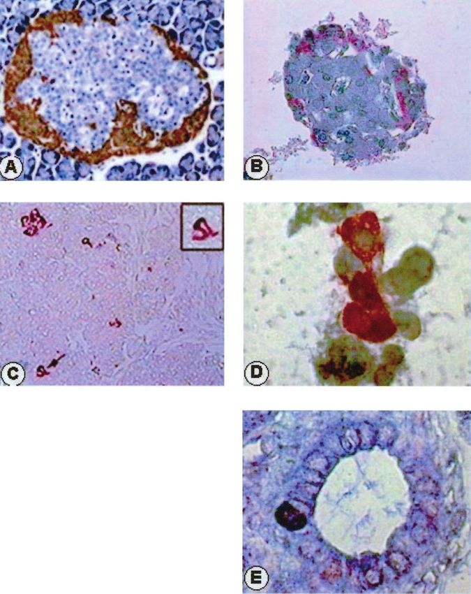
Figure 2 Immunocytochemical presence of INGAP in pancreatic cells. (A) Section of an entire fixed pancreas treated
with INGAP antibody and counterstained with haematoxylin. A continuous ring of INGAP-positive cells (stained in
brown (DAB reaction)) is observed at the islet periphery (non--cells) (200). (B) Section of an isolated fixed islet
treated with INGAP antibody and counterstained with haematoxylin. Scarce INGAP-positive cells (stained in red by
carbazole) can be seen at the islet periphery (non--cells) (200). (C) Section of an entire fixed pancreas treated with
INGAP antibody. Stained cells (DAB) have a pyramidal shape with the nucleus situated at the centre, characteristic of
acinar cells (100). Arrow shows two cells that are magnified in the inset. (D) Smears of acinar cells fixed with 96%
ethanol, treated with INGAP antibody (revealed with carbazole), and counterstained with haematoxylin. Stained cells
(in red) have the same characteristics as the acinar cells described in (C) (1000). (E) Section of an entire fixed
pancreas treated with INGAP antibody. An INGAP-positive cell (stained with alkaline phosphatase and fast blue) is
seen in the layer of cells of a pancreatic duct (1000).
Journal of Endocrinology (2003) 177, 243–248 www.endocrinology.orgINGAP expression in hamster islets · L E FLORES and others 247
Figure 3 RT-PCR performed using total RNA extracted from homogenates of islet and
exocrine tissue cells isolated by collagenase digestion (see Materials and Methods). Left
panel: expression of INGAP and -actin in freshly isolated islets (lanes 2 and 4
respectively), and in exocrine tissue (lanes 3 and 5 respectively). Negative controls for
-actin without reverse transcriptase were included (lanes 6 for islets and 7 for exocrine
tissue). Right panel: expression of -actin and INGAP in cultured islets (72 h, lanes 2 and 3
respectively). A negative control for -actin without reverse transcriptase was included
(lane 4). In both panels, lane 1 corresponds to the Low DNA Mass Ladder (100, 200, 400,
800, 1200 and 2000 bp; Gibco-BRL).
larger number of INGAP-positive cells in the islets rather human and rat genomes, but also in the hamster genome,
than in exocrine cells. The apparent discrepancies in these and that INGAP would be the hamster Reg III- itself
results may be due to the loss of cells from the islet (Abe et al. 2000).
periphery during collagenase digestion, exactly where In our immunocytochemical studies we used an
INGAP-positive cells are located. This assumption is INGAP antibody directed against a 20 amino acid
supported by the fewer number of INGAP- and glucagon- region (amino acids 20–39), having a 75% identity (and
positive cells observed in digested islets compared with 90% positives) with Reg III- (amino acids 21–40).
those seen in the intact pancreas (Fig. 2A and B). Such loss Thus, it could be argued that our antibody reacted not
of INGAP-positive cells could contribute to the increased only with INGAP but also with the Reg III- peptide.
apoptosis and reduced biological effectiveness of trans- There are, however, some facts arguing against such an
planted islets (Berney et al. 2001). Thus, in order to assumption, namely: (i) the Reg III- peptide is
preserve INGAP-positive cells, a gentler collagenase expressed predominantly in the exocrine pancreas of the
digestion should be used when islets are isolated for mouse, but not in normal islets (Terazono et al. 1990);
transplantation. Alternatively, even though individual islet (ii) other members of the Reg gene family are expressed
cells contain more INGAP than acinar cells, the greater in regenerating islets, but they are exclusively seen in
number of the latter could explain why the band from islet -cells (Terazono et al. 1990); and (iii) we have
exocrine tissue in Western blots is stronger than that consistently found that INGAP is present mainly in islet
corresponding to the islets. non--cells, and most of these INGAP-positive cells did
INGAP is a 175 amino acid protein with a molecular not stain with any of the other islet hormones (Del
mass of 19 940 Da and an isoelectric point of 7·86 Zotto et al. 2000). Moreover, preliminary data obtained
(Rafaeloff et al. 1997). Its amino acid sequence shows high using antibodies raised against other portions of the
homology with several proteins encoded by members of INGAP molecule showed that they reacted with - and
the Reg gene family: 58% with rat pancreatitis associated acinar cells exclusively (H Del Zotto, unpublished
protein (PAP)-I, 45% with PAP-II, 50% with PAP-III and observations). Thus, further evidence is needed to
54% with hepatocarcinoma–intestine–pancreas protein demonstrate whether INGAP and Reg III- are
(Rafaeloff et al. 1997). It also has high identity with the different proteins, or the same protein expressed in
mouse Reg III- protein (72% of identities and 82% of different cells in different animals.
positives). It has further been suggested that the counter- In conclusion, our data demonstrate that INGAP is
part of the mouse Reg III- gene exists not only in the expressed in both endocrine and exocrine pancreatic
www.endocrinology.org Journal of Endocrinology (2003) 177, 243–248248 L E FLORES and others · INGAP expression in hamster islets
cells of normal hamsters, a pattern which has also been Edlund H 2001 Developmental biology of the pancreas. Diabetes 50
demonstrated in islet neogenesis in humans (Rafaeloff- (Suppl 1) S5–S9.
Gold G, Broderick C, Carfagna M, Pittenger G, Rafaeloff R,
Phail et al. 1998b, Semakula et al. 2002). The ubiquitous Reifel-Miller A, Borts T, Hale J & Churgay L 1998 INGAP
localization of INGAP would lend support to the hypoth- treatment improves glycemic control in SZ diabetic hamsters.
esis of its possible role in the physiological process of islet Diabetes 47 (Suppl 1) A253.
growth, facilitating an understanding of its protective Lacy PE & Kostianovsky M 1967 Method for the isolation of intact
effect on streptozotocin-induced diabetes (Rafaeloff et al. islets of Langerhans from the rat pancreas. Diabetes 16 35–39.
Mashima H, Shibata H, Mine T & Kojima I 1996 Formation of
1997). insulin-producing cells from pancreatic acinar AR42J cells by
hepatocyte growth factor. Endocrinology 137 3969–3976.
Pittenger GL, Vinik AI & Rosenberg L 1992 The partial isolation and
Acknowledgements characterization of ilotropin, a novel islet-specific growth factor.
Advances in Experimental and Medical Biology 321 123–130.
This study was supported in part by grants from Rafaeloff R, Pittenger GL, Barlow SW, Qin XF, Yan B, Rosenberg
L, Duguid WP & Vinik AI 1997 Cloning and sequencing of the
FONCYT, CONICET and CICPBA. The authors are pancreatic islet neogenesis associated protein (INGAP) gene and its
most grateful to Adrián Díaz and César Bianchi for expression in islet neogenesis in hamsters. Journal of Clinical
technical assistance, and to Adriana Di Maggio for careful Investigation 99 2100–2109.
secretarial support. Rafaeloff-Phail R, Schmitt E, Edlund H, Gold G & Vinik AI 1998a
Expression of INGAP during ontogeny of the pancreas. Diabetes 47
(Suppl 1) A259.
Rafaeloff-Phail R, Schmitt E, Sandusky G, Rosenberg L, Gold G,
References Borts T, Duguid W & Vinik AI 1998b INGAP is a human
cytokine expressed only in the presence of islet neogenesis Diabetes
Abe M, Nata K, Akiyama T, Shervani NJ, Kobayashi S, 47 (Suppl 1) A259.
Tomioka-Kumagai T, Iso S, Takasawa S & Okamoto H 2000 Sambrook J, Fritsch EF & Maniatis TE 1989 Molecular Cloning: a
Identification of a novel Reg family gene, Reg III delta, and Laboratory Manual, edn 2. Cold Spring Harbor, NY: Cold Spring
mapping of all three types of Reg family gene in a 75 kilobase Harbor Laboratory Press.
mouse genomic region. Gene 246 111–122. Semakula C, Pambuccian S, Gruessner R, Kendall D, Pittenger G,
Berney T, Molano RD, Cattan P, Pileggi A, Vizzardelli C, Oliver R, Vinik A & Seaquist ER 2002 Clinical case seminar: hypoglycemia
Ricordi C & Inverardi L 2001 Endotoxin-mediated delayed islet after pancreas transplantation: association with allograft
graft function is associated with increased intra-islet cytokine nesidiodysplasia and expression of islet neogenesis-associated peptide.
production and islet cell apoptosis. Transplantation 71 125–132. Journal of Clinical Endocrinology and Metabolism 87 3548–3554.
Chomczynski P & Sacchi N 1987 Single-step method of RNA Terazono K, Uchiyama Y, Ide M, Watanabe T, Yonekura H,
isolation by acid guanidinium thiocyanate–phenol–chloroform Yamamoto H & Okamoto H 1990 Expression of Reg protein in rat
extraction. Analytical Biochemistry 162 156–159. regenerating islets and its co-localization with insulin in the beta
Del Zotto H, Massa L, Rafaeloff R, Pittenger GL, Vinik A, Gold G, cell secretory granules. Diabetologia 33 250–252.
Reifel-Miller A & Gagliardino JJ 2000 Possible relationship
between changes in islet neogenesis and islet neogenesis-associated
protein-positive cell mass induced by sucrose administration to Received 22 November 2002
normal hamsters. Journal of Endocrinology 165 725–733. Accepted 3 February 2003
Journal of Endocrinology (2003) 177, 243–248 www.endocrinology.orgYou can also read