THYROID TRANSCRIPTION FACTOR-1 (TTF-1): PROTEIN EXPRESSION IS NOT EXCLUSIVE TO LUNG AND THYROID TISSUE - SCORER PW, PINKNEY M, MCINTOSH GG. LEICA ...
←
→
Page content transcription
If your browser does not render page correctly, please read the page content below
Thyroid Transcription Factor-1 (TTF‑1): protein
expression is not exclusive to lung and thyroid tissue.
Scorer PW, Pinkney M, McIntosh GG.
Leica Biosystems Newcastle Ltd, Balliol Business Park West, Benton Lane, Newcastle upon Tyne, UK.

Living up to Life
Thyroid Transcription Factor-1 (TTF‑1): protein
expression is not exclusive to lung and thyroid tissue.
Scorer PW, Pinkney M, McIntosh GG.
Leica Biosystems Newcastle Ltd, Balliol Business Park West, Benton Lane, Newcastle upon Tyne, UK.

Abstract                                                                   Introduction
Thyroid Transcription Factor-1 (TTF‑1) is a member of the                  Carcinomas arising from lung and thyroid show frequent TTF‑1
homeodomain transcription factor family, is a tissue specific              expression.1 As lung is one of the most common sites of metastasis,
transcription factor, and plays a role in regulating proteins              TTF‑1 is considered as a reliable marker to distinguish between
expressed within the thyroid, lung and brain. These include                primary lung carcinoma and metastases within the lung, especially
thyroglobulin, thyroid peroxidase, Clara cell secretory protein and        when dealing with an adenocarcinoma or a large-cell carcinoma.1 It
surfactant proteins. Human TTF‑1 (38 kD) is a single polypeptide of        is also considered as a reliable marker in the differential diagnosis
371 amino acids sharing 98 percent homology with the equivalent            between pleural localization of a peripheral lung carcinoma and
rat and mouse proteins. TTF‑1 functions by binding to specific             malignant mesothelioma.1
recognition sites in a manner that may be regulated by both the            In normal tissue, TTF‑1 is reported to be expressed in epithelial cells
redox and phosphorylation status of the protein. In addition to its        of thyroid and type II pneumocytes and Clara cells in lung.1
role as a tissue-specific transcriptional activator in adult organs,
TTF‑1 may also function in organogenesis. Gene targeting studies           In cancerous tissue, TTF‑1 has been detected in pulmonary
have shown TTF‑1 to be essential for the proper development of the         adenocarcinoma2, large-cell carcinoma2, small cell carcinoma of
thyroid and lungs and abnormal expression may underline a number           lung3; medullary thyroid carcinoma4,5 and hepatocellular carcinoma.6
of congenital abnormalities.
Novocastra™ clone SPT24 was developed to a 123 amino acid                  Aim
sequence of the N-terminal region of the TTF‑1 molecule. The antibody      The introduction of sensitive antibodies and polymer detection
is effective in both manual and automated immunohistochemistry on          systems has lowered the threshold of immunohistochemical
formalin fixed, paraffin embedded tissue sections.                         detection of some proteins. This has resulted in the realization of
Novocastra clone SPT24 was evaluated on 428 normal and tumor               more widespread expression patterns by a number of proteins. The
tissues. Expression was noted particularly in the follicular epithelial    aim of this characterization was to review the expression of TTF‑1
cells of the thyroid, type II pneumocytes and Clara cells of the lung,     protein using Novocastra clone SPT24 and a sensitive polymer
thyroid and lung tumors.                                                   detection system in a broad range of tissues. RT-PCR was also
                                                                           used to identify the presence of TTF‑1 mRNA transcripts in order
Novocastra clone SPT24 also demonstrated TTF-1 expression
                                                                           to provide supporting evidence for protein expression in tissues in
in colon adenocarcinomas and thymomas. Whilst not widely
                                                                           which TTF‑1 has not been previously described
documented, RT-PCR analysis confirmed that cases detected
with the Novocastra clone SPT24 positively expressed fragments
of TTF-1 RNA. Novocastra clone SPT24 has a genuinely strong                Materials and Methods
affinity for TTF-1 protein, proven by its ability to identify genuine
TTF-1 expression in previously unrecorded tumors. Other clones,            Analysis of TTF‑1 RNA Transcripts
such as clone 8G7G3/1, when used at dilutions of 1:50 or lower also        RNA extraction
demonstrated staining in the same areas of tumor that were positive
with Novocastra clone SPT24.                                               RNA was extracted from 2 x 20 μm thick FFPE tissue sections using
                                                                           the PureLink FFPE total RNA isolation kit (Invitrogen, California)
The responsible validation of any antibody clone, including TTF-1, is      according to the manufacturer’s protocol. This procedure included
vital in assessing its potential diagnostic use. Specifically, awareness   a xylene-free protocol for deparaffinization followed by proteinase K
of the full range of normal tissue and tumor expression expected           digestion at 60 oC for up to 3 hours. The tissue lysate was further
for an antibody is pivotal to diagnostic application. Interpretation       processed by selectively binding the RNA to a silica-based
needs to take into account expression of a panel of appropriate            membrane spin cartridge, followed by several washing steps and an
immunohistochemical antibodies in addition to clinicopathological          elution step in 75 μl RNase free water.
features and most importantly tumor morphology before reaching
a diagnosis

                                                                                                                                                2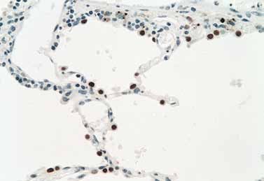
PCR Amplification Slides were washed in water and then sections were counterstained
with Hematoxylin. Finally sections were dehydrated, cleared and
Before conducting RT-PCR the total RNA extracted from each
mounted in DPX.
FFPE tissue sample was quantified by means of Qubit assay
using the Quant-iT RNA assay kit (Invitrogen). The quality of Automated Immunohistochemistry
the RNA extracted from each FFPE specimen was assessed by
conducting a control RT-PCR test on the RNA extracted using Automated immunohistochemical validation was performed on
primers specific for a 69 bp (base pair) fragment of the house a range of normal and tumor tissues, in the form of whole tissue
keeping gene GAPD (forward: 5’-CTCTCTGCTCCTCCTGTTCGAC3’; sections and tissue micro-arrays (TMA), using the automated Leica
reverse: 5’TGAGCGATGTGGCTCGGCT-3’). TTF‑1 RNA was BOND™ system, Leica BOND Dewax Solution (AR9222), Leica BOND
reverse transcribed into cDNA using a specific reverse primer R5 Epitope Retrieval Solution 1 (AR9961) and Leica BOND Polymer
(5’-GCTCGCCGGGCCCATGAAGC-3’) and the Reverse Transcription Refine Detection (DS9800). Epitope retrieval was performed for 20
System Kit (Promega, USA) in accordance with the manufacturer’s minutes followed by *IHC Protocol F using TTF‑1 primary antibody,
protocol. Amplification of a 93 bp fragment encoding for a region Novocastra clone SPT24 (Leica BOND Ready-to-Use PA0364).
bridging the exon boundary between exon 1 and 2 of TTF‑1 (Genbank
accession No NM_001079668.1) was carried out by PCR using a
second TTF‑1 specific primer (5’-ACCATGAGGAACAGCGCCTCTG-3’)
and half of the reverse transcription mix as a template in a 50 μl
PCR reaction mix (2 mM MgCl2, 0.2 mM dNTPs, 0.2 μM primer, 2.5 U
Taq-DNA polymerase and 1x RT transcription buffer reaction mix
(Promega, USA)). A total of 30 cycles of PCR (94°C/30 seconds,
55°C/30 seconds and 72°C/45 seconds) was performed.
Cloning and Sequencing
The PCR products were verified by agarose gel electrophoresis,
gel purified and then cloned into the pGEM-Teasy vector (Promega,
USA). Clones were identified by digestion with the restriction enzyme
EcoR1 and their identity confirmed by DNA sequencing (Beckman
Coulter Genomics, Takeley, Essex, UK).
Manual Immunohistochemistry
Manual immunohistochemical validation was performed on a range
of normal and tumor tissues, in the form of whole tissue sections
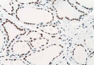
Figure 1. Immunohistochemical staining for TTF‑1 using Leica Novocastra clone
and tissue micro-arrays (TMA), using the mouse monoclonal SPT24 on normal lung (x400 original magnification). Leica BOND automated system
TTF‑1 antibody, Novocastra clone SPT24, in conjunction with the staining of a paraffin section. Insert shows nuclear staining localization in type II
Novolink™ Polymer Detection System RE7140-K (250 tests). Briefly, pneumocytes.
whole tissue paraffin sections, 4 μm thick, were cut onto Leica
Microsystems charged coated slides (S21.2113.A). To facilitate
adhesion, these slides were then dried overnight at 37°C and
finally baked for 1 hour at 56°C. TMA’s were prepared as directed
by the individual suppliers. Sections were deparaffinized in xylene
and rehydrated through graded alcohols. Heat induced epitope
retrieval was performed using Epitope Retrieval Solution pH 6.0
(RE7113) in a Prestige stainless steel pressure cooker for 8 minutes
at full pressure. Endogenous peroxidase activity was blocked by
incubation for 5 minutes in Peroxide Block solution. Slides were
washed in 50 mM Tris buffered saline (TBS, pH 7.6) for 5 minutes,
incubated with Protein Block for 5 minutes and washed in TBS for
a further 5 minutes. Sections were then incubated for 30 minutes at
25°C with TTF‑1 primary antibodies, Novocastra clone SPT24 and
Dako clone 8G7G3/1 both diluted 1:50 in Antibody Diluent (RE7133).
Following two sequential 5 minute wash steps in TBS, sections were
incubated in Post Primary for 30 minutes at 25°C. Two sequential
5 minute wash steps in TBS were again performed and sections
incubated for 30 minutes at 25°C with Novolink Polymer. Two further Figure 2. Immunohistochemical staining for TTF‑1 using Leica Novocastra clone
sequential 5 minute wash steps in TBS were performed and bound SPT24 on normal thyroid (x400 original magnification). Leica BOND automated system
staining of a paraffin section. Insert shows nuclear staining localization in thyroid
peroxidase visualized using DAB chromogen. The DAB working follicular epithelium.
solution was prepared by adding 50 μL of DAB chromogen per mL of
DAB Substrate Buffer.
3Tissue IHC Staining Results Abnormal tissue IHC positivity
Adrenal Negative Lung - adenocarcinoma 26/32
Bone Marrow Negative Lung - small cell carcinoma 4/7
Breast Negative
Lung - bronchioalveolar carcinoma 2/3
Bronchus Negative
Thyroid - papillary carcinoma 6/6
Cecum Negative
Cerebellum Negative Thyroid - follicular carcinoma 3/3
Cerebrum Weak nuclear staining in glial cells Thyroid - medullary carcinoma 4/4
Cervix Negative Thyroid - follicular adenoma 1/1
Colon Negative
Thyroid - Hashimoto’s thyroiditis 1/1
Endometrium Negative
Thymus - thymoma 19/59
Esophagus Negative
Thymus - atypical carcinoid 1/5
Eye Negative
Fallopian Tube Negative Colon - adenocarcinoma 4/49
Heart Negative Desmoplastic small round cell tumor 1/1
Ileum Negative Table 2. Immunostaining for TTF‑1 in abnormal tissues (whole sections and
Kidney Negative TMA’s)
Liver Negative
Lung Strong nuclear staining in type II
pneumocytes and Clara cells
The tissue involved in this study consisted of 31 whole tissue
Lymph Node Negative
cases and 7 TMA’s comprising 392 cases in the form of 1mm and
Mesothelium Negative
1.5mm cores. All scoring was carried out by senior scientist(s) at
Muscle, Skeletal Negative Leica Biosystems Newcastle Ltd experienced in the assessment
Ovary Negative of immunohistochemical staining and tissue histology. In normal
Parathyroid Negative tissues, nuclear staining of tissue elements was noted and the
staining intensity recorded as negative, weak, moderate or strong.
Pancreas Negative
In tumor tissues, any nuclear staining of tumor elements was
Peripheral Nerve Negative
classified as “positive” regardless of the staining intensity.
Pituitary Negative
Placenta Negative Results
Prostate Negative
Rectum Negative Immunohistochemistry
Salivary Gland Negative Normal Tissues
Spleen Negative
Table 1 shows the staining for TTF‑1 in normal tissues (TMA).
Skin Negative Novocastra clone SPT24 detected the TTF‑1 protein in the nucleus
Stomach Negative of type II pneumocytes and Clara cells of the lung (Figure 1), in the
Spinal Cord Negative nucleus of follicular epithelial cells of the thyroid (Figure 2) and in
Testis Negative glial cells of cerebrum. (Total cases = 140).
Thymus Negative Abnormal Tissues
Thyroid Strong nuclear staining in follicular
Novocastra clone SPT24 detected the TTF‑1 protein in 72/171
epithelial cells
abnormal tissues (whole sections and TMA’s). Staining was
Tongue Negative
particularly noted in the nucleus of tumors of the lung, thyroid, thymus
Tonsil Negative and colon (Table 2). No staining was observed in an additional 112
Umbilical Cord Negative abnormal tissues (Table 3).
Ureter Negative
Uterus (myometrium) Negative
Table 1. In positive tissues, only the tissue elements stated were positive
with Leica Novocastra clone SPT24, all other tissue elements were negative.
(Results from TMA tissue).
4Tumor tissue No of cases evaluated Tumor tissue No of cases evaluated
Lung – squamous cell carcinoma 18 Squamous cell carcinoma - esophagus 2
Lung – large cell carcinoma 5 Squamous cell carcinoma - larynx 1
Malignant Mesothelioma 5 Squamous cell carcinoma - tongue 2
Breast – carcinoma 6 Squamous cell carcinoma - cervix 1
Ovary – thecoma 1 Skin – basal cell carcinoma 1
Ovary – juvenile granulosa cell tumor 2 Malignant Melanoma 3
Ovary – serous carcinoma 3 Large B-cell lymphoma 1
Ovary – mucinous carcinoma 2 Thymic tumor – metastatic 1
Ovary – germ cell tumor 1 GIST 1
Ovary – clear cell carcinoma 1 Synovial sarcoma 1
Endometrium – adenocarcinoma 1 Leiomyosarcoma 1
Endometrium – clear cell carcinoma 1 Ewing’s sarcoma 1
Endometrium – stromal sarcoma 1 Spindle cell rhabdomyosarcoma 1
Cervix – adenocarcinoma 2 Omental fibrous tumor 1
Testis – seminoma 3 Ganglioneuroma 1
Testis – mixed germ cell tumor 1 Paraganglioma 1
Testis – embryonal carcinoma 1 Table 3. Abnormal tissues showing negative immunostaining for TTF‑1 (whole
sections and TMA’s)
Prostate – adenocarcinoma 1
Prostate – benign prostatic hyperplasia 1
Bladder – transitional cell carcinoma 2
Bladder – small cell carcinoma 1
Kidney – renal cell carcinoma 5
Kidney – transitional cell carcinoma 1
Adrenal – adenoma 1
Adrenal – pheochromocytoma 1
Liver – hepatocellular carcinoma 3
Liver – cholangiocarcinoma 2
Liver – adenoma 1
Pancreas – adenocarcinoma 3
Pancreas – endocrine tumor 2
Stomach – adenocarcinoma 4
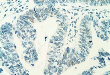
Figure 3. Immunohistochemical staining for TTF‑1 using clone SPT24 on colonic
Small bowel – adenocarcinoma 1 adenocarcinoma (x400 original magnification). Leica BOND automated system
staining of a paraffin section. Insert shows weak nuclear staining localization in
Small bowel - carcinoid 4
tumor cells (TMA).
Brain – astrocytoma 1
Brain – choroid plexus papilloma 1
Squamous cell carcinoma - skin 2
Squamous cell carcinoma - penis 2
Table 3. Continues
5The demonstration of TTF‑1 protein expression in the nuclei of
tumors in colon and thymus with Novocastra clone SPT24 is not
widely documented.
Novocastra clone SPT24 demonstrated TTF‑1 protein in the nuclei
of 3/49 colon adenocarcinomas (TMA) (6%) (Figure 3). TTF‑1 staining
was weakly positive and present in only a proportion of the tumor
nuclei. TTF‑1 positive staining in primary colon adenocarcinomas
(5%) has recently been identified and published.1 Genuine expression
A of TTF‑1 using Novocastra clone SPT24 has been cited in a small
proportion (10%) of colonic adenocarcinoma metastases found in
lung. The identification of this expression has been attributed to the
sensitivity of Novocastra clone SPT24.1
Novocastra clone SPT24 also demonstrated TTF‑1 protein in the
nucleus of 19/59 thymomas, in whole sections and a TMA (Table 4).
In 24% of thymomas (14/59), TTF‑1 staining was weakly positive in
a proportion of tumor nuclei (Figure 4a). In 8% of thymomas (5/59),
moderate to strong staining was noted in the majority of tumor nuclei
B (Figure 4b). Reported expression of TTF‑1 in thymoma and thymic
carcinoma has previously been documented as being negative.7,8,9
Figure 4. Immunohistochemical staining for TTF‑1 using Leica Novocastra clone
SPT24 on thymoma (x400 original magnification). Leica BOND automated system The TTF‑1 expression in cases of thymoma which have previously
staining of a paraffin section. (a) shows a case of thymoma with weak positive staining been reported as being TTF‑1 negative is possibly a reflection of
in a proportion of the tumor nuclei (b) shows a case of thymoma with moderate to
strong positive staining in majority of the tumor nuclei (whole sections and TMA). increased sensitivity in IHC detection.
From the literature published since 2002 it becomes apparent that
expression of TTF‑1 has now been identified as extending beyond
Abnormal tissue IHC positivity
the expected lung and thyroid tumors. This could possibly be due
Thymus - thymoma Type A 1/5 to a combination of the increased use of TTF‑1 in a wider range of
tumors and an increase in the sensitivity of IHC polymer detection
Thymus - thymoma Type AB 1/4
systems. Expression of TTF‑1 is now being identified in tissues and
Thymus - thymoma Type B1 6/22 tumors previously thought to be negative. Our experience with
Thymus - thymoma Type B2 4/10 Novocastra clone SPT24 in thymoma and colon adenocarcinomas
being a case in point.
Thymus - thymoma Type B3 (atypical thymoma) 1/4
Recently Novocastra clone SPT24 has been identified in primary
Thymus - thymoma Type C (thymic carcinoma) 3/11 colon adenocarcinoma and colon adenocarcinoma metastases in
Thymus - thymoma (unclassified) 3/3 lung1, uterine tumors10 – especially in malignant mixed Müllerian
tumor10 (82%), ovarian tumors10 and in hepatocellular carcinoma.6
Table 4. Immunostaining for TTF‑1 in thymoma (TMA)
Novocastra clone SPT24 was identified as being the most sensitive
TTF‑1 antibody in combination with Leica BOND Polymer Refine
Detection (DS9800) and the automated Leica BOND system.10
Discussion Novocastra clone SPT24 was also identified as possessing
TTF‑1 Novocastra clone SPT24, was shown to be effective at a consistent nuclear positivity and an absence of erratic cytoplasmic
dilution of 1:50 using Heat Induced Epitope Retrieval solution pH6.0 staining in comparison with other clones.11
(RE7113, Leica Microsystems) and the Novolink Polymer Detection Other clones, such as clone 8G7G3/1 and clone BGX-397A (available
System (RE7140-K, Leica Microsystems). Staining was unaffected from of a variety of suppliers), were found to be slightly less sensitive
by the position of the peroxide block step in the protocol or the use in the same studies.1,10,11 However clone 8G7G3/1 when used at a
of TBS or PBS-based diluents and wash buffers. TTF‑1, Novocastra dilution of 1:50 or lower demonstrated weak staining in the same
clone SPT24 (Leica BOND Ready-to-Use PA0364) was also effective areas of tumor that were positive with clone SPT24.1
on the automated Leica BOND system using Leica BOND Epitope
Retrieval Solution 1 and Leica BOND Polymer Refine Detection. The obvious question arising from this current Novocastra clone
SPT24 characterization work and the recently published new TTF‑1
Novocastra clone SPT24 demonstrated TTF‑1 protein expression expression data is whether the TTF‑1-positivity in primary colon
in the nuclei of tumors in lung, thyroid, thymus and colon (whole cancers and thymomas was specific and a result of true aberrant
sections and TMA’s). Expression of TTF‑1 in medullary, papillary expression of TTF‑1. To provide supporting evidence to answer this
and follicular carcinomas of thyroid5, and in non squamous cell question, analysis of the mRNA present in TTF‑1 positive cases of
carcinomas of lung2,3 was expected and has been well documented. primary colonic cancer and thymoma was performed to identify the
presence of TTF‑1 RNA transcripts.
6Six cases of primary colon adenocarcinoma (whole sections) which References
were positive with Novocastra clone SPT24 (two cases of which 1. Comperat E, Zhang F, Perrotin C et al. Variable sensitivity and specificity of TTF‑1
were also weakly positive with clone 8G7G3/1) were analyzed for the antibodies in lung metastatic adenocarcinoma of colorectal origin. Modern
Pathology. 2005;18,1371–1376.
presence of TTF‑1 RNA transcripts.11 RT-PCR analysis showed that 2. Nakamura N, Miyagi E, Murata S et al. Expression of thyroid transcription factor-1
all six Novocastra clone SPT24 positive cases expressed fragments in normal and neoplastic lung tissues. Modern Pathology. 2002;15(10):1058–1067.
of TTF‑1 RNA transcripts indicative of TTF‑1 gene expression. 3. Ordonez NG. Value of thyroid transcription factor-1 immunostaining in
Moreover, three smaller RNA fragments when sequenced provided distinguishing small cell lung carcinomas from other small cell carcinomas.
American Journal of Surgical Pathology. 2000;(9):1217-23.
evidence of the existence of a novel splice variant previously not 4. Katoh R, Miyagi E, Nakamura N et al. Expression of thyroid transcription factor-1
described in the literature.11 (TTF‑1) in human C cells and medullary thyroid carcinomas. Human Pathology.
2000;31(3): 386-393.
A case of thymoma (whole section), which was positive with
5. Miller R. Focus on immunohistochemistry: thyroid transcription factor-1 (TTF‑1).
Novocastra clone SPT24 and also weakly positive with clone ProPath Laboratory, Immunohistochemistry Divison. April 2001. Available from
8G7G3/1, was analyzed for presence of TTF‑1 RNA transcripts. RT- http://www.ihcworld.com/_newsletter/2001/focus_apr_2001.pdf.
PCR analysis showed that this Novocastra clone SPT24 positive 6. Pan CC, Chen PC, Tsay SH et al. Cytoplasmic immunoreactivity for
thyroid transcription factor-1 in hepatocellular carcinoma: a comparative
case expressed fragments of TTF‑1 RNA transcripts indicative of immunohistochemical analysis of four commercial antibodies using a tissue
TTF‑1 gene expression. array technique. American Journal of Clinical Pathology. 2004;121:343–349.
7. Pan CC, Chen PC, Chou TY et al. Expression of calretinin and other mesothelioma-
Overall, the SPT24 clone appears to have a genuinely strong related markers in thymic carcinoma and thymoma. Human Pathology. 2003
affinity for TTF‑1 protein, proven by its ability to identify genuine Nov;34(11):1155-62.
TTF‑1 expression in tumors previously considered TTF-1 negative. 8. Pomplun S, Wotherspoon AC, Shah G et al; Immunohistochemical markers in
Tumors now reported as TTF-1 positive, other than lung and thyroid the differentiation of thymic and pulmonary neoplasms. Histopathology. 2002
Feb;40(2):152-8.
tumors, include sub-sets of primary colorectal adenocarcinomas, 9. Saad RS, Landreneau RJ, Liu Y et al; Utility of immunohistochemistry in
thymomas, uterine tumors, ovarian tumors and hepatocellular separating thymic neoplasms from germ cell tumors and metastatic lung cancer
carcinoma as well as its established expression in medullary, involving the anterior mediastinum. Applied Immunohistochemistry & Molecular
Morphology. June 2003;11(2):107-112.
papillary and follicular carcinomas of thyroid, and in non squamous
10. Zhang PJ, Gao HG, Pasha TL et al TTF‑1 expression in ovarian and uterine
cell carcinomas of lung. epithelial neoplasia and its potential significance, an immunohistochemical
The responsible validation of any antibody clone, including TTF1, is assessment with multiple monoclonal antibodies and different secondary
detection systems. International Journal of Gynecological Pathology. 2009
vital in assessing its potential diagnostic use. Specifically, awareness Jan;28(1):10-8.
of the full range of normal tissue and tumor expression expected 11. Camilleri-Broet S, Smertenko T, McIntosh G et al; Detection of TTF‑1 RNA
for an antibody is pivotal to diagnostic application. Interpretation transcripts in primary colorectal adenocarcinoma. Submitted for publication
2010.
needs to take into account expression of a panel of appropriate
immunohistochemical antibodies in addition to clinicopathological
features and most importantly tumor morphology before reaching
a diagnosis.
Disclaimer
Unless specifically stated all references quoted refer to data on
TTF‑1 derived from established scientific publications involving
various antibodies and techniques and do not refer to IHC staining
expression directly attributable to Novocastra NCL-L-TTF‑1 or Leica
BOND Ready‑to‑Use PA0364.
Acknowledgement
Thank you to Dr Maurice Loughrey, Royal Group of Hospitals Trust,
Belfast, UK for his advice and support on this study.
www.Leica-microsystems.com
95.10112 • 05/2011 • Copyright © by Leica Biosystems Melbourne Pty Ltd, Melbourne, Australia, 2011
Leica and the Leica Logo are registered trademarks of Leica Microsystems IR GmbH
7You can also read