SKIN CANCER CRASH COURSE: RECOGNITION AND MANAGEMENT OF NONMELANOMA SKIN CANCER VICTOR NEEL, MD, PHD DIRECTOR, DERMATOLOGIC SURGERY, MGH
←
→
Page content transcription
If your browser does not render page correctly, please read the page content below
Skin Cancer Crash Course:
    Recognition and Management of
      Nonmelanoma Skin Cancer

           Victor Neel, MD, PhD
   Director, Dermatologic Surgery, MGH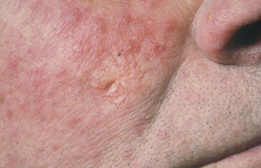
Disclosures

 Neither I nor my spouse/partner has a relevant
financial relationship with a commercial interest
                    to disclose.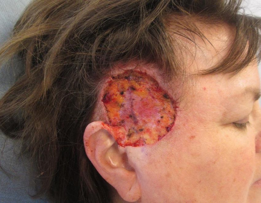
Blood Vessels
  sebaceous                     Pyogenic granuloma                           glomus tumor                     neural
sebaceous carcinoma
                                Kaposi’s sarcoma                             Angiosacoma                 Neurofibroma
sebaceous adenoma
                                Epitheliod hemangioendothelioma              Masson tumor                Neurothekeoma
sebaceoma
                                Targetoid hemosiderotic hemangioma           Angiokeratoma               Schwannoma
    immune cell                 Glomangioma                                  AV hemangioma               Palisaded encapsulated neuroma
    Lymphoma                                                                                             Merkel cell carcinoma
    Mast cell disease
    histiocytosis                                                                                   epithelial
                                                                                                   actinic keratosis
                                                                                                   squamous cell carcinoma
      eccrine                                                                                      keratoacanthoma
                                                                                                   seborrheic keratosis
   Microcystic adnexal ca
                                                                                                   porokeratosis
   Mucinous eccrine carcinoma
   Eccrine spriradenoma                                                                                  fibroblasts
   Cylindroma                                 follicular                                         Atypical fibroxanthoma
                                      Basal cell carcinoma                                       Malignant fibrous histiocytoma
   Poroma
                                      Trichoepithelioma
   Porocarcinoma
   Nodular hidradenoma
                                      Pilar sheath acanthoma           fat                   smooth muscle
                                      Tricholemmoma                    Lipoma
   Syringoma                                                                                 Leiomyoma
                                      Trichofolliculoma                angiolipoma
   Chondroid syringoma                                                                       Leiomyosarcoma
                                      Pilomatricoma                    Spindle cell lipoma
   Digital papillary adenocarcinoma                                                          angioleiomyoma
                                      Trichoblastoma                   liposarcoma
   Extramammarary Paget’s
                                      Fibrofolliculoma                 pleomorphic lipoma
                                                                                                                    metastatic
                                      Desmoplastic trichoepithelioma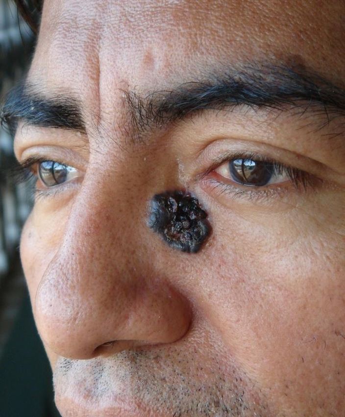
Goals of This Talk

• Discuss the most common NMSC tumors you
  will see and possibly diagnose & treat
• Convince you to consider performing skin
  biopsies in your practice
Primary Care & Dermatology

• Too many patients, too many tumors
• Delayed diagnosis, delayed treatment
• Many skin cancers can be diagnosed and
  treated in primary care setting
• PCPs must definitively diagnosis and have
  treatment algorithms in place
US Skin Cancer Incidence

• >5 million new cases of NMSC each year

• BCC about 80%, SCC about 20%

• About 15,000 deaths per year from SCC, more
  than twice as many than melanoma
Causes of Nonmelanoma Skin Cancer

 • Chronic UV exposure –> genetic mutations
 • Immunosuppression
    – Organ transplant patients and CLL patients
    – 80% of transplant patients develop skin cancers
    – 200-fold increased risk of SCC

 • Human papillomavirus - HPV 6,16 (vaccine may affect)

 • Inherited diseases - XP, BCNS, albinism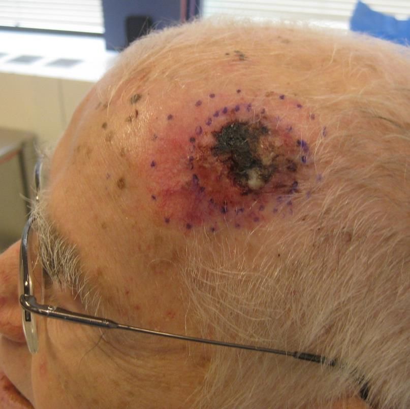
Basal Cell Carcinoma
                      Stats

• most common cancer in humans
• 3 million new cases a year, increasing 5%
  per year
• 1/3 of all Caucasians will develop at least
  one lesion
• billions of healthcare $$ spent
Basal Cell Carcinoma
Biology
• very indolent growth – perhaps decades until
clinically apparent
• rarely metastatic (75% most sporadic tumors have defects in
Sonic hedgehog signaling pathway (oral drug,
vismodegib, topicals in develpoment)Which Is BCC?
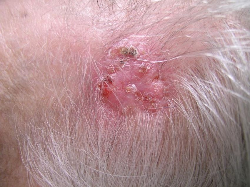
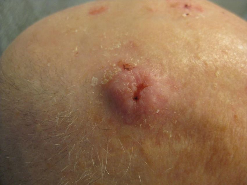
Basal Cell Carcinoma

• Subtypes
  – Nodulo-ulcerative (most common)
  – Morpheaform (sclerosing, infiltrative)
  – Micronodular
  – Metatypical (basosquamous)
  – Superficial (“multicentric”)
Basal Cell Carcinoma
• Subtypes
  – Nodulo-ulcerative (most common)
Basal Cell Carcinoma
• Pigmented BCC
• Can mimic MM
Basal Cell Carcinoma
• Subtypes
  – Morpheaform BCC
  – Can look like scar
Basal Cell Carcinoma
• Subtypes
  – Superficial “multicentric”
  – Can be misdiagnosed as psoriasis, tinea or
    eczema
  – Most common type on trunk and extremities
BCC or Tinea?
Tinea               BCC

 itchy & scaly   crusts/bleeds
often multiple   usually single
  antifungals    sun-exposed
BCCs?
Basal Cell Carcinoma
• Course
  – Slow, progressive growth
  – Bleeding, ulceration, superinfection
  – Enlarges over months to years
  – Is capable of extensive tissue destruction (invading
    into muscle, cartilage, and bone)
Suspected lesion Differential ?biopsy Treatment
diagnosis ?refer options
Actinic keratosis SK, wart, porokeratosis, NO 5-FU, imiquimod,
trichilemmoma NO cryotherapy, PDT-ALA
Squamous cell AK, discoid lupus, tinea YES 5-FU, imiquimod, PDT-ALA,
carcinoma, in situ psoriasis, SK YES cryotherapy, curettage
surgery
Squamous cell SK, AK, BCC, pyoderma YES surgery, Mohs surgery,
carcinoma, invasive gangrenosum YES radiation (rarely)
BCC, superficial tinea, SCC in situ, discoid lupus, ? 5-FU, imiquimod,
(body) porokeratosis, SCC in situ ? cryotherapy, curettage,
surgery
BCC, nodular nevus (melanocytic), molluscum YES cryo, curettage, surgery
(body) YES
BCC, infiltrative or scar YES
recurrent (body) YES
BCC, any type nevus, rosacea, angiofibroma, YES Mohs surgery
(head & neck) syringoma, sebaceous YES
hyperplasia, tinea, discoid lupus,
SCC, trichoepithelioma,
telangiectasia, scarSquamous Cell Carcinoma • Second most common skin cancer in the general population • Most common skin cancer in transplant recipients • Appears on sun-exposed skin • Red, scaly, firm, may ulcerate • 1-15% metastasize (lip & ear)
Squamous Cell Carcinoma
• Arises primarily on sun-damaged skin
  – Precursor is actinic keratosis (AK) on sun-exposed sites
  – 90% of AKs spontaneously resolve

• May occur anywhere on skin
      •   Face
      •   Lips (usually lower)
      •   Ears
      •   Dorsal hands
      •   Chest
Diffuse AKs? 5-FU!!
Two Weeks of Topical 5-FU
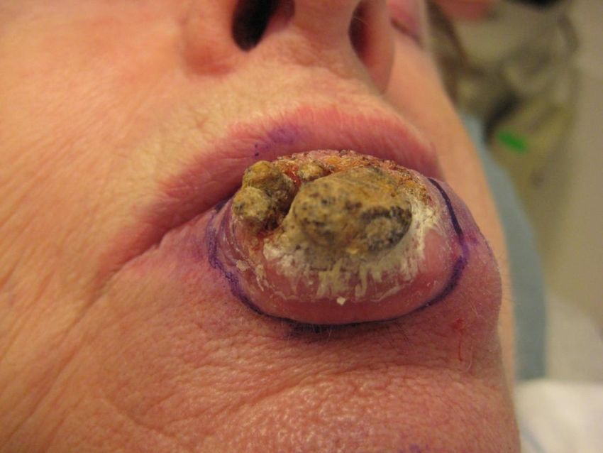
Squamous Cell Carcinoma
• Metastasis more likely in:
  –   Recurrent tumors
  –   Those with diameter > 2 cm
  –   Those with depth > 6 mm
  –   Mucosal sites, periauricular skin (lip & ear)
  –   SCC arising from chronic wounds (Marjolin’s ulcer)
  –   Perineural invasion of larger nerve fibers
  –   Immunocompromised patients
Squamous Cell Carcinoma
• Subtypes
  – Keratoacanthoma
    • Rapid initial growth
    • May be painful (unlike most NMSCs)
    • Exophytic nodule with central keratin-filled crater
    • Remains stable for a few months
    • May spontaneously resolve – new research!!
    • Dermpath reports as well-differentiated SCC
Time to get the derm surgeon
on the phoneSquamous Cell Carcinoma
• Subtypes
– Bowen’s Disease
• Squamous cell carcinoma in situ
• Thin, erythematous, scaling plaques
• Can progress into, and/or coincide with invasive SCC
• Can be misdiagnosed as psoriasis, tinea, eczema or BCCIncidence Ratios of Skin Cancer
in Transplant Recipients
• Squamous cell 100-fold increase
carcinoma
• Basal cell carcinoma 10-fold increase
• Melanoma 3.4-fold increaseMortality from Metastatic Skin Cancer
in Transplant Patients
Country Organ Cancer Mortality Rate
type
Australia Kidney SCC 5% of all patients with SCC
New Zealand
Australia Heart All 27% total deaths occurring
after the 4th yr post
transplant
USA All SCC 3 yr cause specific survival
54%, n = 71
USA All Melanoma 30% (compared to 15% in
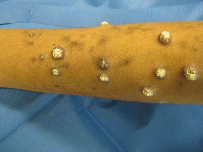
general population)A Lethal Tumor in a Transplant Patient
Please have your transplant
patients see a dermatologist for baseline evaluationSurgical Emergencies
in Dermatology
• SCC in immunosuppressed population
– Iatrogenic (organ transplant, anti-inflammatory states)
– CLL or other leukemias/marrow failures
AML – 80% blast, 0%PMNsSuspected lesion Differential ?biopsy Treatment
diagnosis ?refer options
Actinic keratosis SK, wart, porokeratosis, NO 5-FU, imiquimod,
trichilemmoma NO cryotherapy, PDT-ALA
Squamous cell AK, discoid lupus, tinea YES 5-FU, imiquimod, PDT-
carcinoma, in situ psoriasis, SK YES ALA, cryotherapy,
curettage
surgery
Squamous cell SK, AK, BCC, pyoderma YES surgery, Mohs surgery,
carcinoma, invasive gangrenosum YES radiation (rarely)
BCC, superficial tinea, SCC in situ, discoid lupus, ? 5-FU, cryotherapy.
(body) porokeratosis, ? curettage, surgery
BCC, nodular nevus (melanocytic), YES cryo, curettage, surgery
(body) molluscum YES
BCC, infiltrative or scar YES
recurrent (body) YES
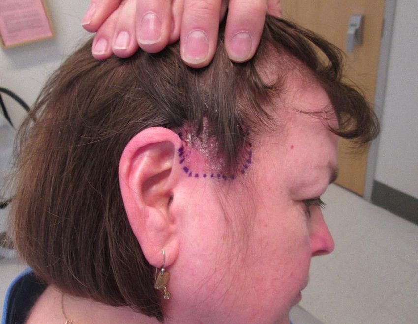
BCC, any type nevus, rosacea, angiofibroma, YES Mohs surgery
(head & neck) syringoma, sebaceous YES
hyperplasia, tinea, discoid
lupus, SCC, trichoepithelioma,
telangiectasia, scarLess Common Tumors
DFSP
Dermatofibroma
Extramammary Paget’s
Extramammary Paget’s
A Challenge to Primary Care:

 DO YOUR OWN BIOPSIES!
Primary Care & Dermatology
  Delay in Diagnosis & Treatment
• Community dermatology shortage: 2-6
  months
• Community surgical dermatology shortage
  (Mohs surgery): 1-3 months

Typical delay from Primary care to definitive
  treatment: 3-9 months!!!
Essentials for Serious PCPs

Do a “real” skin exam

Document lesions and take a pre-biopsy
photo & measurement

Do not be afraid to biopsy early – low-risk of
complications

If the biopsy is inadequate or doesn’t fit the
clinical picture, re-biopsy!
Essentials for Serious PCPs
• Don’t worry if your biopsies come back
  with benign diagnoses – steep learning
  curve
• If you treat a lesion, see the patient
  back to confirm improvement. If not
  improving biopsy or refer, DON’T KEEP
  TREATING!!
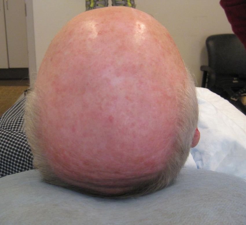
This Is Not an Actinic Keratosis!
In-Office Biopsy
      Cost: $1.50
      Time: 5-10 minutes. CLEAN not STERILE prep
      Reimbursement: $60-100 (CPT 11102)

sterile #15 blade
clean gauze & Q-tips
3cc lido/epi
 bottle Drysol in room
 vaseline & plaster

obtain signed consent
Biopsy Video
Please refer biopsy-proven skin cancers
 to dermatologic surgery, not plastics
Nicotinamide for Prevention
• Nicotinamide (vitamin B3) 500mg BID
• ~25% reduction of SCC/BCC in high risk skin
  cancer patients at 1 yr
• low side effect profile
• (NOT NIACIN)
The dermatologist will see you!

          vneel@partners.org
You can also read