Thymoma-Associated Paraneoplastic Myositis, Presenting with Rapidly Progressive Muscle Contractures
←
→
Page content transcription
If your browser does not render page correctly, please read the page content below
JCN Open Access LETTER TO THE EDITOR
pISSN 1738-6586 / eISSN 2005-5013 / J Clin Neurol 2021;17(3):496-498 / https://doi.org/10.3988/jcn.2021.17.3.496
Thymoma-Associated Paraneoplastic Myositis, Presenting
with Rapidly Progressive Muscle Contractures
Jin Hee Kima Dear Editor,
Hyemin Janga Thymic malignancy is associated with paraneoplastic neurological syndromes (PNSs). My-
Hee Jung Kwonb asthenia gravis (MG) is the most common PNS, and some thymomatous patients develop
Yeon-Lim Suhb MG (25–40%) or paraneoplastic myositis (0.5–9%), which usually coexists with MG.1,2 MG
Ju-Hong Mina and myositis mostly occur simultaneously, or myositis develops later in myasthenic patients.3
a
Department of Neurology, Here we report an atypical case of thymoma-associated paraneoplastic myositis presenting
Neuroscience Center, with rapidly progressive distal muscle contracture without apparent clinical weakness or
Samsung Medical Center,
Sungkyunkwan University
symptoms of MG.
School of Medicine, Seoul, Korea A 45-year-old female had developed painful swelling in her forearms without preceding
b
Department of Pathology, trauma or infection 1 month previously. Two days later she could not extend the right third
Samsung Medical Center,
Sungkyunkwan University finger, followed by all fingers and both elbow joints after 1 week. She denied other symp-
School of Medicine, Seoul, Korea toms such as myalgia or fever. At presentation her range of motion (ROM) was restricted in
all metacarpophalangeal, both proximal interphalangeal, elbow, and knee joints, and had
plantar flexed feet (Fig. 1A). A neurological examination revealed no remarkable weakness
in unaffected muscles and even in affected muscles within acceptable ROM. There was also
no evidence of atrophy or neuromyotonia. Serological results were normal except for mildly
elevated muscle enzymes [creatinine kinase (CK) at 273 IU/L]. She was seropositive for an-
ti-acethylcholine receptor (AChR) antibody (8.167 nmol/L) and titin (MG titin-30) antibody,
but negative for anti-leucine-rich glioma inactivated-1 and anti-contactin-associated protein-2
antibodies.4 Line immunoassays for myositis-specific antibodies and myositis-associated an-
tibodies only revealed positivity for anti-polymyositis scleroderma antigen (PM/Scl) 75. Nee-
dle electromyographic studies revealed myopathic changes in paraspinal, proximal, and distal
limb muscles, although a repetitive nerve stimulation test did not reveal any response decre-
ments. MRI of the forearm and lower extremities demonstrated inflammation and fibrosis
in all compartment muscles, suggestive of noninfectious inflammatory myositis involving
both arms and legs, but no abnormalities were observed in joint capsules or tendons. A mus-
cle biopsy demonstrated primary myopathy, with findings that were similar to but not diag-
nostic of polymyositis (Fig. 1B). Chest CT revealed a mediastinal mass, and video-assisted
Received February 22, 2021
Revised April 20, 2021
thoracoscopic surgery revealed thymoma (WHO type B2). She received intravenous methyl-
Accepted April 20, 2021 prednisolone (500 mg) for 5 days, and 3 months later her ROM had improved considerably
Correspondence and the CK level had normalized to 52 IU/L. She subsequently received intravenous immu-
Ju-Hong Min, MD, PhD noglobulin intermittently as maintenance therapy.
Department of Neurology, This patient represents an atypical case of thymoma-associated paraneoplastic myositis
Samsung Medical Center,
Sungkyunkwan University with double seropositivity for anti-AChR and titin antibodies who presented with rapidly
School of Medicine and progressive distal muscle contracture without apparent clinical manifestations of myopathy
Department of Health Sciences and
Technology, SAIHST,
or MG. The association of myositis with MG has been reported in patients with double se-
Sungkyunkwan University, ropositivity for anti-AChR and striational antibodies, and the presence of the striational an-
81 Irwon-ro, Gangnam-gu, tibodies may indicate concurrent MG and myositis.2,3 However, the pathogenetic role of these
Seoul 06351, Korea
Tel +82-2-3410-3590 cc This is an Open Access article distributed under the terms of the Creative Commons Attribution Non-Com-
Fax +82-2-3410-0052 mercial License (https://creativecommons.org/licenses/by-nc/4.0) which permits unrestricted non-commercial
E-mail juhongm@skku.edu use, distribution, and reproduction in any medium, provided the original work is properly cited.
496 Copyright © 2021 Korean Neurological Association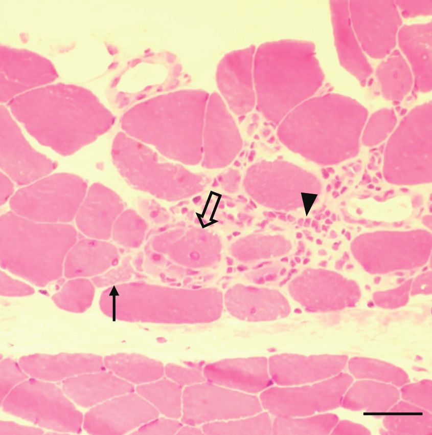
Kim JH et al. JCN
A
B
a b c
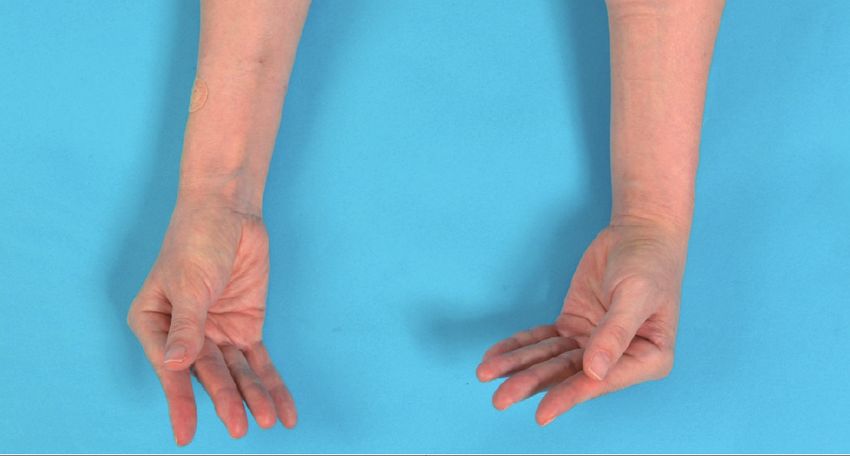
Fig. 1. Clinical and pathologic findings of the patient. A: Photograph of the patient’s hands. She had restricted ROMs in all metacarpophalangeal and
both proximal interphalangeal joints. B: Pathological findings of the vastus lateralis muscle biopsy. (a) Muscle fiber necrosis (solid arrow) and regenera-
tion (open arrow), and interstitial inflammatory cell infiltration (arrowhead) are evident (HE ×400, frozen section). (b) Necrotic muscle fibers (arrow)
with lymphocytes (HE, ×400). (c) Immunohistochemical staining of the same area in panel B (b) reveals predominantly CD8-positive T cells (arrow)
(×400). (Scale bar=100 micrometers). ROM: range of motion.
antibodies has not been demonstrated.3 A recent review of 13 MG, since symptoms of MG might not be obvious and could
cases with MG and myositis found that all patients had mild- be missed in myositis.2 The management of PNS in thymoma-
to-moderate MG symptoms, while thymoma was found in 10 tous patients includes treatment of the thymoma, immuno-
patients (8, 1, and 1 with WHO types B2, AB, and B3, respec- suppressive drugs, and symptom-specific management1.
tively), all of whom were positive for anti-RyR1 (ryanodine Our patient received complete thymectomy for thymoma,
receptor) and titin antibodies.3 The extent of muscle involve- immunotherapy, and rehabilitation. This case suggests that
ment varied, including isolated focal, distal predominance, or unexplained muscle contractures can be atypical symptoms
proximal predominance. MG and myositis occurred simulta- of paraneoplastic myositis, warranting further evaluation and
neously in 10 patients, and myositis subsequently developed in proper management.
2 myasthenic patients. However, MG developed 4 years after This article does not require IRB/IACUC approval because
the occurrence of myositis in only one patient, who had proxi- there are no human and animal participants.
mal weakness with high CK (2500 IU/l). This suggests that
close observation is needed to recognize newly developing
www.thejcn.com 497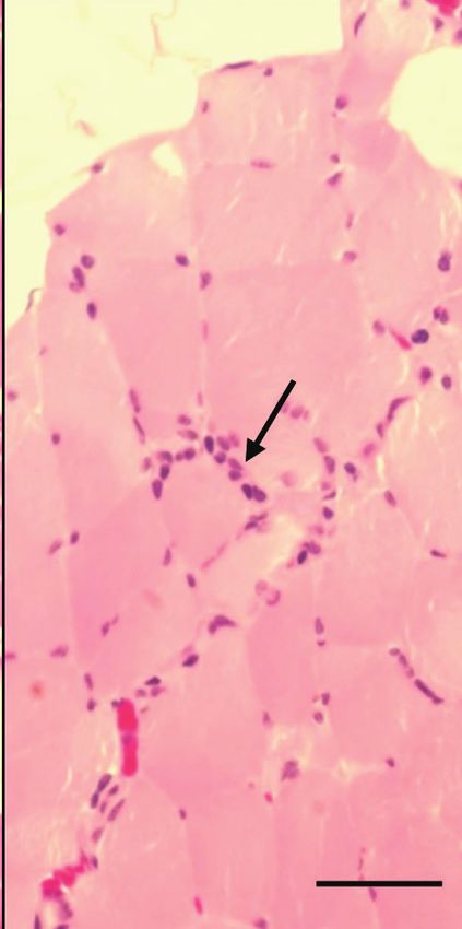
JCN                   Thymoma-Associated Paraneoplastic Myositis

Author Contributions                                                         al. A systematic review of paraneoplastic syndromes associated with
Conceptualization: Jin Hee Kim, Hyemin Jang, Ju-Hong Min. Visualiza-         thymoma: treatment modalities, recurrence, and outcomes in resect-
tion: Hee Jung Kwon, Yeon-Lim Suh. Writing—original draft: Jin Hee Kim.      ed cases. J Thorac Cardiovasc Surg 2020;160:306-314.e14.
Writing—review & editing: Ju-Hong Min.                                    2. Huang K, Shojania K, Chapman K, Amiri N, Dehghan N, Mezei M.
                                                                             Concurrent inflammatory myopathy and myasthenia gravis with or
ORCID iDs                                                                    without thymic pathology: a case series and literature review. Semin Ar-
                                                                             thritis Rheum 2019;48:745-751.
Jin Hee Kim                   https://orcid.org/0000-0002-9708-5420
                                                                          3. Garibaldi M, Fionda L, Vanoli F, Leonardi L, Loreti S, Bucci E, et al.
Hyemin Jang                   https://orcid.org/0000-0003-3152-1274
                                                                             Muscle involvement in myasthenia gravis: expanding the clinical spec-
Hee Jung Kwon                 https://orcid.org/0000-0002-8800-7690
                                                                             trum of myasthenia-myositis association from a large cohort of patients.
Yeon-Lim Suh                  https://orcid.org/0000-0001-5809-2401
                                                                             Autoimmun Rev 2020;19:102498.
Ju-Hong Min                   https://orcid.org/0000-0002-7338-9067
                                                                          4. Irani SR, Alexander S, Waters P, Kleopa KA, Pettingill P, Zuliani L, et
                                                                             al. Antibodies to Kv1 potassium channel-complex proteins leucine-
Conflicts of Interest
                                                                             rich, glioma inactivated 1 protein and contactin-associated protein-2
The authors have no potential conflicts of interest to disclose.             in limbic encephalitis, Morvan’s syndrome and acquired neuromyoto-
                                                                             nia. Brain 2010;133:2734-2748.
Acknowledgements
None

                            REFERENCES
 1. Zhao J, Bhatnagar V, Ding L, Atay SM, David EA, McFadden PM, et

498    J Clin Neurol 2021;17(3):496-498
You can also read