TOOLS AND METHODS OF TELEMEDICINE FOR EARLY DETECTION OF DENTAL ANOMALIES
←
→
Page content transcription
If your browser does not render page correctly, please read the page content below

De n t i s t r y                                                           | archiv euromedica | 2020 | vol. 10 | num . 3 |
                                                                                                                                                  127
http://dx.doi.org/10.35630/2199-885X/2020/10/3.31

TOOLS AND METHODS OF TELEMEDICINE
FOR EARLY DETECTION                                                                                  R eceived 23 July 2020;

OF DENTAL ANOMALIES                                                                                   Received in revised form 27 August 2020;
                                                                                                       Accepted 31 August 2020

Rakhman Nasrullaev1 , Tagir Magomedov1 ,                                                                                      Aim of study:
Gleb Mareev2 , Anna Petrova1 , Olga Popkova1 ,                            to investigate potential use of telemedicine technolo-
Irina Firsova1 , Valery Konnov3                                           gies and to develop software for early detection of
                                                                          dental anomalies under conditions of orthodontist
1
  Department of Pediatric Dentistry and Orthodontics, Saratov State       shortage.
Medical University, Saratov
2
  Department of Otorhinolaryngology, Saratov State Medical University,    MATERIALS AND MET H ODS
Saratov                                                                        We had carried out an analysis of research articles
3
  Department of Orthopedic Dentistry, Saratov State Medical University,   and academic issues, after which software to analyze
Saratov, Russia                                                           control and diagnostic models of the jaws were creat-
                                                                          ed. To do this, we developed a software program where
     nasrullaevrk@yandex.ru                                               the points on the images were placed following the
                                                                          instructions. After that, it analyzes the models based
                                                                          on common methods. Later on we compared the
A b s t r a c t — This work offers a view at the practical
application of telemedicine, especially in rural areas.
                                                                          calculation accuracy through matching the outcomes
There is a description and the principle of independent                   obtained by an analog method against those obtained
development. The item also contains comparative data on                   by using our software program. After that the analysis
the effectiveness of jaw model analysis performed in the                  was performed.
conventional way as matched against doing that with new
software employed to detect dental anomalies, with no
previously acquired skills. The software can be used by any
                                                                          RESULTS AND DIS C USSION
doctor as it features detailed and clear instructions. This                     One of the tasks within this work implied devel-
program will be useful in cases where orthodontists are not               oping software to detect early signs of dental anoma-
available.                                                                lies by analyzing control and diagnostic cast models in
                                                                          remote areas where residents cannot visit an orthodon-
K e y w o r d s — telemedicine, diagnostic software,
teledentistry, dentoalveolar issues detection.
                                                                          tist for various reasons.
                                                                                The major features for the developed software
                                                                          were to be the following ones:
                                                                             1) low system requirements;
Introduction                                                                 2) user-friendly interface and easy using (the soft-
      Availability of narrowly specialized medical                              ware is to be used by doctors of any profile);
care is an important feature attributing to its quality                      3) minimum time spent by the doctor when using
[7–11]. While in urban areas a specialist's consultation                        the software;
is available to most of the population, rural residents                      4) open source;
face certain deficits in this respect [1, 2]. Not only                       5) free distribution model.
rural areas have a worse access to medical centers,
but also a shortage of specialists and the insufficient                         The software allows performing analysis by using
technological infrastructure present a challenge there                    two photos of jaw cast models with minimal prelimi-
[3, 4]. The advance in communication technologies in                      nary preparation and no serious technical support. It
remote areas is still slow compared to what it could be                   takes placing the required dots on the image, which is
nowadays.                                                                 to be done following the detailed description appear-
      Telemedicine is one of the healthcare arrange-                      ing on the screen, after which the software will make
ments that may serve to improve medical services                          its own calculations.
offered in remote areas that face issues with access                            The software employs the main methods of analy-
to specialized care and appropriate equipment                             sis of control & diagnostic models, which require data
[5, 6].                                                                   on the teeth size, on the distance between them and
                                                                          the length of the dental rows. The software requires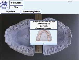
128   | archiv euromedica | 2020 | vol. 10 | num . 3 |                                                                       De n t i s t r y

        measuring 1 centimeter on a ruler, and then, in the im-
        age, marking the most prominent points on the teeth
        mesial and distal surfaces of each jaw (Fig. 1).
             After that, points are marked to identify the
        dentition width (Fig. 2). Further on, points are placed
        to identify the length of the apical basis, and then the
        software will require opening the models image in a
        frontal projection and place points to identify the api-
        cal basis width (Fig. 3).
             When all the data is specified, the software dis-
        plays the calculation outcomes based on the methods
        proposed by Pont, Linder-Harth, Gerlach, Bolton,
        Snagina, Korkhaus. The data is displayed in terms that
        are accessible to non-dental specialists (Fig. 4). Further
        options imply sending the report and save it on the PC.
             The major differences between our development             Fig.1. E xample of a popup window with a step-by-step instruction
        and those available on the market include:
          1) free distribution;
          2) easy to use; each stage of diagnostics offers
             graphic instructions in Russian;
          3) low system requirements; requires 6 megabytes of
             free hard disk space;
          4) can be used by doctors of any specialty.

              The second stage in the development was to
        check the analysis accuracy compared to the conven-
        tional traditional method of jaw models anthropom-
        etry with a caliper.
              The test involved ten undergraduate students
        majoring in Dentistry, who performed measurements
        on models — first in the traditional way, and then us-
        ing the respective software. The results showed that the
        differences between the measurements fell within the
        range of 2–4%, which is an acceptable tolerance.               Fig. 2. D iagnostic models with points placed on the mesial and distal
              The next step implied comparing the time spent           teeth surfaces, connected with reference lines
        on analog analysis and that using the software, which
        was done by untrained participants. We compared the
        time the students spent on analyzing 5 models in the
        traditional way and then — using our software. On
        average, the conventionally performed analysis took
        35–42 minutes, whereas it took 3–4 minutes only to
        do that with the software, which, actually, means 8–10
        times less than usual.

                          Putting the developed software to practice
          1) Using this, as well as other similar developments,
             allows early detection of dental issues, which
             offers an obvious advantage since the successful
             outcome of treatment depends largely on the
             starting time of orthodontic treatment.
          2) Many parents do not even suspect their children
             may be suffering from dental anomalies. Given
             that, another problem can be solved — the pa-             Fig. 3. O pening an image in a frontal projection to measure the apical
             tient’s awareness.                                        basis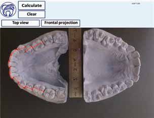
De n t i s t r y                                                             | archiv euromedica | 2020 | vol. 10 | num . 3 |
                                                                                                                                             129

                                                                                 of dental health in children and adolescents in Saratov
                                                                                 and the Saratov region // Saratov Journal of Medical
                                                                                 Scientific Research. 2013, T. 9; 3: 484–486
                                                                             2. Estai, M., Kanagasingam, Y., Mehdizadeh,
                                                                                M.et al. T eledentistry as a novel pathway to improve
                                                                                dental health in school children: a research protocol
                                                                                for a randomised controlled trial. BMC Oral Health
                                                                                20; 11 (2020). https://doi.org/10.1186/s12903-019-
                                                                                0992-1
                                                                             3. Suetenkov D.E., Kharitonova T.L., Danilov
                                                                                A.N., Popkova O.V., Kobets A.V. Dental morbid-
                                                                                ity in the school-age child population in the Saratov
                                                                                region. – Clinical dentistry. – 2019; 1: 96–99.
                                                                             4. Nichols, K. (2019). Teledentistry Overview: United
                                                                                States of America. Journal of the International Society
                                                                                for Telemedicine and EHealth, 7, e9 (1–6). https://
Fig. 4. Software compares measurements with table indices to make              doi.org/10.29086/JISf TeH.7.e9
further conclusions                                                          5. Popkova O.V., Suetenkov D.E. Egorova A.V.,
                                                                                Nasrullaev R.K. T      elemedicine technologies for
                                                                                the clinic of dentistry (literature review) – Clinical
Table 1. Comparison of the measuring results for the four upper incisors       dentistry. – 2018; 2: 93–96.
sum by each student using the software and the conventional analysis         6. Park, Jae. ( 2020). A licensed orthodontist versus do-
                                                                                it-yourself orthodontics. American Journal of Ortho-
                                                                                dontics and Dentofacial Orthopedics. 157. 591–592.
 Student 1 2                    3     4 5       6    7 8 9            10        DOI – 10.1016/j.ajodo.2020.02.003.
         28.5 28                28    29.5 28   28   28.5 28.5 28     29
Convention- Software

                                                                             7. Harutyunyan Yu. U        ndifferentiated connective
            (mm)

                                                                                tissue dysplasia as a key factor in pathogenesis of maxil-
                                                                                lofacial disorders in children and adolescents // Archiv
                       28   28.5 28   28.5 28.5 27   28   29.5 27     28.5      EuroMedica. 2020. Vol. 10; 2: 83–94. https://dx.doi.
                                                                                org/10.35630/2199-885X/2020/10/2.24
al method
(mm)

                                                                             8. Kondratyeva T. M    ethodological approaches to
                                                                                dental arch morphology studying // Archiv Euro-
                                                                                Medica. 2020. Vol. 10; 2: 95–100. https://dx.doi.
                                                                                org/10.35630/2199-885X/2020/10/2.25
                                                                             9. Shkarin V.V., Ivanov S.Yu., Lepilin A.V. Mor-
    3) Easy to use. Analyzing models using our software                         phological specifics of craniofacial complex in people
       takes having 2 photos of models with a ruler for                         with varioustypes of facial skeleton growth in case of
       zooming.                                                                 transversal occlusion anomalie // Archiv EuroMedica.
                                                                                2019. Vol. 9; 2: 5–16. https://doi.org/10.35630/2199-
    4) Maximum simplification of the diagnostics pro-                           885X/2019/9/2/5
       cedure and minimization of the time spent on it.
    5) Lower cost. Telemedicine technologies in ortho-                       10. Porfyriadis M.P. S canning electron microscopy
                                                                                 and X-ray spectral microanalysis in dental tissue resist-
       dontics may allow saving not only the doctor’s                            ance // Archiv EuroMedica. 2019. Vol. 9; 1: 177–185.
       time, yet also the patients’ money.                                       https://doi.org/10.35630/2199-885X/2019/9/1/177
                                                                             11. Shkarin V.V., Grinin V.M., Khalfin R.A. S pecific
C ON C LUSION                                                                    features of transversal and vertical parameters in lower
     The proposed development allows analyzing                                   molars crowns at various dental types of arches //
models by a dentist who has no special skills. In view of                        Archiv EuroMedica. 2019. Vol. 9; 2: 174–181. https://
a shortage of orthodontists to be observed in remote                             doi.org/10.35630/2199-885X/2019/9/2/174
areas with a low population density, this software, if
integrated into telemedicine systems, may facilitate de-
tection of dental anomalies, at the same time possibly
reducing the number of diagnostic errors.

RE F EREN C ES
1. Firsova I.V., Suetenkov D.E., Egorova A.V.,
   Magomedov T.B., Kharitonova T.L. I ndicators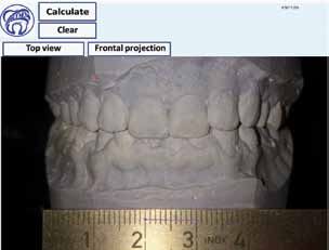
You can also read