Detection of Leukemia Using Machine Learning Algorithms
←
→
Page content transcription
If your browser does not render page correctly, please read the page content below

Journal of Physics: Conference Series

PAPER • OPEN ACCESS

Detection of Leukemia Using Machine Learning Algorithms
To cite this article: K P Jayavikash et al 2021 J. Phys.: Conf. Ser. 1916 012220

View the article online for updates and enhancements.

                                This content was downloaded from IP address 46.4.80.155 on 10/09/2021 at 22:38
ICCCEBS 2021                                                                                                    IOP Publishing
Journal of Physics: Conference Series                         1916 (2021) 012220          doi:10.1088/1742-6596/1916/1/012220

Detection of Leukemia Using Machine Learning Algorithms

                    K P Jayavikash1, P Gopinath 1, U Nandheeswaran 1, R Sivabalan 1, T Divya Bharathi1
                    1
                    Department of Biomedical Engineering, KPR Institute of Engineering and Technology,


                    Abstract. Detection of disorders of Blood usually include visual inspection through a microscope.
                    This may help to classify various conditions related to human blood. The paper details about
                    evolving a system that could help in diagnosis and detection of Leukemia at an early stage.
                    Only then controlling and prevention can be done. The proposed system focuses on White Blood
                    Cells as they are the affected ones in case of Leukemia. The affected cells differ in shape and
                    color features, which are used as classifier inputs.

1. Introduction
Medical Imaging involves technique and process that are used to generate images of different parts of our
body for diagnostic and treatment purposes. Medical Imaging has become an integral part of Medicine
overthe past decade or so. Advanced equipment has beendeveloped for detecting, analyzing and storing of
medical images. The main aim of analyzing through images is to formulate as much detail as possible
regarding identification, diagnosis, control, treatment, monitoring and assessment of diseases.
   Cancer is among the deadliest diseases. Leukemia is a cancer affecting the blood forming cells. It is a
blood cancer that happens when there is a lot of abnormal WBC’s are formed by the Bone marrow. Basic
components of Blood are - Plasma, RBC, WBC and also platelets. Blood is made of 45% RBC and 55%
plasma. WBC and platelets are present less than 1%. In case of Leukemia WBC’s will be produced more
in number and this would lead to disruption in the Blood system. The presence of unusual and extra white
blood cells can be exposed when the blood sample is collected and inspectedbya hematologist.
   Then the microscopic images would be visually checked by the hematologist. [1-3]The activity wouldbe
hectic and could take a lot of time. And moreover any work that involves human work is vulnerable to
errors because of emotional disturbance and the person’s physical capability limit. Also it is really tough
to get steady results from the perceptible inspection.
Automated system in image processing can really overcome the errors and problems in visual inspection.
The system developed will be mainly on microscopic images to differentiate normal cells from Leukemia
cells. Early detection helps providing fast and appropriate treatment for the condition.
With an automated system many images can be processed, scanning time can be reduced, accuracy can be
improved. [4-7] Examining and classifying leukemia is based on size, structure and color features of White

              Content from this work may be used under the terms of the Creative Commons Attribution 3.0 licence. Any further distribution
              of this work must maintain attribution to the author(s) and the title of the work, journal citation and DOI.
Published under licence by IOP Publishing Ltd                          1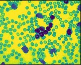
ICCCEBS 2021                                                                                 IOP Publishing
Journal of Physics: Conference Series           1916 (2021) 012220     doi:10.1088/1742-6596/1916/1/012220

Blood cells (WBC).

2. Literature review

A lot of algorithms are available and were used by various people to segment and classify medical Images.
By using the thresholding process it segments the image accurately from the white and black pixel it can
be clearly segmented. There are various algorithms like K means clustering and Watershed methods. Also
GFCVR (Gaussian feature convolutional visual recognition) was used [8-11]. Commonly used methods
were KNN classifier and SVM classifier.
We have used K-mean clustering algorithm for segmentation of color images of microscopic blood images
and VGG
Architecture was used to extract shape and color features of the image and then finally classify if the input
image is Normal or Leukemia. Figure 1 shows the flowchart.

3. Methodology

                                            Figure 1. Flowchart
i. Image Acquisition:

Images were obtained from ALL-IDB online database. The dataset has 108 images collected in 2005. It
has over 39,000 elements, where lymphocytes have been marked by experts. [12-14] These images were
taken using a microscope with magnifications ranging from 300 to 500.These images present in the dataset
were taken using a microscope attached with a camera. The obtained images were present in JPG and TIF
formats. And has a resolution of 2592 x 1944. Figures 2-8 shows the image results. Figure 9 shows the
graph.
Preprocessing was not done as the obtained images were already clear and did not need any processing.

                                                      2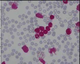
ICCCEBS 2021                                                                               IOP Publishing
Journal of Physics: Conference Series           1916 (2021) 012220   doi:10.1088/1742-6596/1916/1/012220

             Figure 2. Normal cell                                   Figure 3. Leukemia cell

4. Image Segmentation
Segmentation - progression of sectionalization of the image into its numerous segments. segmentation was
classically used for locating matters and borders in images. We have used K means clustering algorithm.
We have used color images for segmentation. The ROI (Region of Interest) is WBC’s as those are the
ones affected in case of leukemia. The input image is changed into HSV color space.

                                  Figure 4. Hue (Cancer and Normal cell)

                                    Figure 5. Saturation (Cancer and Normal cell)

                                                      3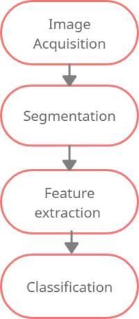
ICCCEBS 2021                                                                                  IOP Publishing
Journal of Physics: Conference Series             1916 (2021) 012220    doi:10.1088/1742-6596/1916/1/012220

                                        Figure 6.Value (Cancer and Normal cell)

                       Figure 7. .Detected WBC’sDilation and Erosion:

In any image Dilation adds extra pixels to the object boundaries, whereas Erosion helps remove the pixel
values from the boundaries of the objects. Depending on the size of the element, pixels will be added or
removed from the object.

                                        Figure 8. Output after dilation and erosion
5. Feature Extraction and Classification
It is the part of dimensionality reduction procedure, in which, an early set of the raw data is divided and
condensed to more controllable groups. So when you want to process it will be easier.
Classification is a process relating to categorization, the process in which objects and ideas are recognized,
distinguished.
Feature Extraction and classification are done using VGG Architecture. The shape and color features of the
image were obtained from processed input data and then features obtained were fed as the input of the
classifier system.

                                                       4
ICCCEBS 2021 IOP Publishing
Journal of Physics: Conference Series 1916 (2021) 012220 doi:10.1088/1742-6596/1916/1/012220
The accuracy of developed system is 93%. This system could be of real help as the time taken is reduced
compared to visual inspection and the possible errors due to humans could be avoided.
Figure 9 ,Accuracy plot of the Model
6. Conclusion
In this project we have developed a model to detect Leukemia from microscopic images using python. In
this method, image processing techniques like segmentation, Dilation and erosion are used to obtain the
affected White blood cells. Then the segmented image is used and features like shape and color features
are extracted using the VGG16 architecture and the images are classified based on the acquired features.
The output of the model will show if the input image is cancerous or not.
5ICCCEBS 2021 IOP Publishing
Journal of Physics: Conference Series 1916 (2021) 012220 doi:10.1088/1742-6596/1916/1/012220
References
[1] A. Haldorai and A. Ramu, Security and channel noise management in cognitive radio networks,
Computers & Electrical Engineering, vol. 87, p. 106784, Oct. 2020.
doi:10.1016/j.compeleceng.2020.106784
[2] A. Haldorai and A. Ramu, Canonical Correlation Analysis Based Hyper Basis Feedforward
Neural Network Classification for Urban Sustainability, Neural Processing Letters, Aug. 2020.
doi:10.1007/s11063-020-10327-3N., Ritter, J., Cooper, Segmentation and Border Identification
of Cells in Images of Peripheral Blood Smear Slides , 30th Australasian Computer Science
Conference, Conference in Research and Practice in Information Technology, 62, 2007, pp. 161-
169
[3] D.M.U., Sabino, L.D.F., Costa, L.D.F., E.G., Rizzatti, M.A., Zago, A Texture Approach to
Leukocyte Recognition , Real Time Imaging, 10, 2004, pp. 205-206.
[4] M.C., Colunga, O.S., Siordia, S.J., Maybank, Leukocyte Recognition Using EM-Algorithm ,
MICAI 2009, LNAI 5845, Springer Verlag Berlin Heidelberg, 2009, pp. 545-555.
[5] R. Donida Labati, V. Piuri, F. Scotti, ALL-IDB: the acute lymphoblastic leukemia image
database for image processing , in Proc. of the 2011 IEEE Int. Conf. on Image Processing (ICIP
2011), Brussels.
[6] F. Scotti, Automatic morphological analysis for acute leukemia identification in peripheral
blood microscope images , in Proc. of the 2005 IEEE Int. Conf. on Computational Intelligence
for Measurement Systems and Applications (CIMSA 2005),Giardini Naxos - Taormina, Italy,
pp. 96-101, July 20-22, 2005
[7] V. Piuri, F. Scotti, Morphological classification of blood leucocytes by microscope images , in
Proc. of the 2004 IEEE Int. Conf. on Computational Intelligence for Measurement Systems and
Applications (CIMSA 2004), Boston, MA, USA, pp. 103-108,July 12-14, 2004.
[8] Ruggero Donida Labati, Vincenzo Piuri, Fabio Scotti, The Acute Lymphoblastic Leukemia
Image Database for Image Processing, Universita Degli Studi Di Milano.
[9] Behrooz Ghandeharian, Hadi Sadoghi Yazdi and Faranak Homayouni, Modified Adaptive
Centre Eighted Median Filter for Uppressing Impulsive Noise in Images , IJRRAS, Vol,1,
Issue.3, December 2009.
[10] MedlinePlus, The U.S. National Library of Medicine
[Online].http://www.nlm.nih.gov/medlineplus/ency/article/003643.htm
[11] G. Naxos and F. Scotti, CIMSA 2005-IEEE International Conference on Computational
Intelligence for Measurement Systems and Applications Automatic Morphological Analysis for
Acute Leukemia Identification in Peripheral Blood Microscope Images, no. July, pp. 20–22,
2005,
[12] F. Scotti, Robust segmentation and measurements techniques of white cells in blood
microscope images, April, pp. 43–48, 2006.
[13] T. Bergen, D. Steckhan, T. Wittenberg, and T. Zerfaß, Segmentation of leukocytes and
erythrocytes in blood smear images, Proc. 30th Annu. Int. Conf. IEEE Eng. Med. Biol. Soc
EMBS’08 - Personalized Healthc. through Technol. pp. 3075–3078, 2008
6You can also read