Marking a surgical margin for excision of a keratinocyte cancer - RACGP
←
→
Page content transcription
If your browser does not render page correctly, please read the page content below
Clinical

Marking a surgical margin for
excision of a keratinocyte cancer

Chloe A Mutimer, Anthony J Dicker                              THE RECENTLY REVISED Australian                 1. Wipe the skin with an alcohol wipe.
                                                               guidelines for managing keratinocyte               Rubbing a BCC can cause a flush of the
                                                               cancers (formally known as non-melanoma            lesion to assist in identifying the clinical
Background
The size of the measured margin for
                                                               skin cancers) includes a section on suggested      margins.
excision of a keratinocyte cancer is often                     clinical margins for excising these tumours.1   2. Stretching the skin can greatly
discussed; however, a technique for                            While the size of the measured margin is           assist in identifying the edge of
marking the skin is rarely described.                          discussed, a technique for marking the             a lesion, particularly for a BCC.
                                                               skin is rarely described. Here the authors         Dermatoscopic examination of the
Objective
The aim of this article is to describe
                                                               describe their method for marking out a            lesion before marking with a Codman
a method for marking a lesion                                  surgical lesion for excision, which has been       marker can help identify extensions
for excision.                                                  used and supported in several studies.2–6          of tumour.
                                                                                                               3. Using bright lighting, stretch and
Discussion
                                                                                                                  magnification, dot around the outer
The key to surgical excision of a
keratinocyte cancer is the assessment
                                                               Equipment                                          margin of the lesion using the Codman
of the tumour border in good light under                       The following equipment is recommended.            marker, placing the dots on the adjacent
loupe magnification and delineation                            • Alcohol wipe: Cleaning the skin before           normal appearing skin (Figure 1).
using a skin marker. The surgical margin                         any surgical drawing makes it easier          4. Connect the dots to create one
is then added around the lesion. Often                           to apply marks to the skin. An alcohol-          continuous line around the lesion
an elliptical excision is performed, with
                                                                 based solution will evaporate within             border. Fill in and thicken the line
lesions excised adhering to the drawn
                                                                 seconds, while an aqueous-based wash             so that it has a diameter of 2–3 mm
line and then closed appropriately.
                                                                 may cause the pen markings to run.               (Figure 2). Add extra circles so that
                                                               • Bright light: Good illumination of the           the distance from the inner edge of
                                                                 lesion is essential to define the clinical       the inner circle and the outer edge of
                                                                 edges of a lesion.                               the outer circle is the required clinical
                                                               • Magnification: A simple surgical loupe           margin. Alternatively, an outer line with
                                                                 enhances the ability to correctly identify       a margin of 2–3 mm from the lesion’s
                                                                 the clinical edge of the lesion.                 border can be drawn.
                                                               • Dermatoscope: This is an extension of         5. Examine and palpate the area to
                                                                 the role of magnification and can help           identify the natural direction of closure.
                                                                 further define the clinical edge.7               The direction of closure may be
                                                               • Codman marker: These surgical pens are           indicated by Langer’s lines, biodynamic
                                                                 available in sterilised packs with a ruler.      excisional skin tension (BEST) lines,
                                                                                                                  normal skin creases or junctions of
                                                                                                                  cosmetic units (Figure 3).
                                                               Technique                                       6. When excising the lesion, cut on the
                                                               The following outlines an example for              outside of the pen markings with the
                                                               basal cell carcinoma (BCC) removal;                lesion itself excised as a circle, then
                                                               margins can be adjusted for squamous               the ellipses formed excised afterwards
                                                               cell carcinoma (SCC) removal.                      (Figure 4).

© The Royal Australian College of General Practitioners 2021                                                       Reprinted from AJGP Vol. 50, No. 6, June 2021   385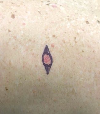
Clinical Marking a surgical margin for excision of a keratinocyte cancer
7. The method of wound closure depends tumour being evident at the edge of the performed by dermatologists or plastic
on the size of the lesion, with direct histological specimen. surgeons. There may be a bias for the
closure the preferred method. Flap Recommendations for surgical margins population treated in these studies, as
repairs or skin grafts are options for are shown in Table 1. In relation to SCC, patients referred to these centres are more
larger wounds. surgical margins of 4 mm are generally likely to have high-risk or difficult-to-treat
recommended for low-risk lesions and lesions, compared with those treated in
6 mm for high-risk lesions.9 An adequate primary centres.
Surgical margins surgical margin for BCCs has been widely Ultimately, selection of an appropriate
The surgical margin should aim to debated. Cancer Council Australia’s surgical margin should aim to achieve
meet the widely accepted incomplete clinical guidelines suggest 2–3 mm a balance between rates of incomplete
excision rate, in which histopathological surgical margins for low-risk tumours and excision of the tumour and normal tissue
involvement of margins is seen in 5 mm for high-risk tumours.1 sacrifice, which can affect functional and
of excisions.8 Margin involvement is Most research has occurred in tertiary cosmetic outcomes, a factor that is often
judged by histological assessment, with referral centres, where excisions were important to patients.
When to consider referral
Most BCCs can be managed in a primary
care centre. However, it is recommended
that referral to a dermatologist or
plastic surgeon for surgical excision is
considered on the basis of individual
patient factors such as tumour location
(head and neck), histological subtype
(infiltrating or sclerosing), size (>2 cm
in diameter) and patient preference.
Other treatments – such as Mohs
micrographic surgery, curettage and
cautery, cryotherapy, topical therapies
(such as imiquimod) or radiotherapy –
can be considered on a case-by-case
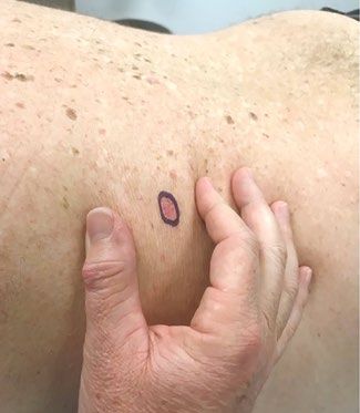
Figure 1. Basal cell carcinoma with outer edges Figure 2. Dots around the margin connected
of lesion marked with a Codman marker to form a continuous line, measured to have basis and compared with surgery, with
a diameter of 2–3 mm the aim of achieving the best outcome
for the patient.
High-risk features of SCC include size
>20 mm, tumour depth >4 mm, recurrent
lesion, high-risk anatomic location (head
and neck), perineural or lymphovascular
invasion, poorly differentiated subtype
or immunosuppression.1 Patients with
high-risk features should be referred to
a specialist or multidisciplinary team for
management. Other management options
include Mohs micrographic surgery,
curettage and cautery, cryotherapy, topical
medications and photodynamic therapy.10
If a tumour is not appropriate for excision
because of patient factors (eg not being fit
for surgery) or further locoregional control
is sought, referral to a radiation oncologist
can also be considered. Referral for
medical oncology review should be sought
Figure 3. Skin placed under tension to Figure 4. Ellipse drawn around the lesion
determine the direction of closure for elliptical excision; the outer edge of the in the case of metastatic SCC.
markings is the line of excision The Australian guidelines suggest
that recurrent or incompletely excised
386 Reprinted from AJGP Vol. 50, No. 6, June 2021 © The Royal Australian College of General Practitioners 2021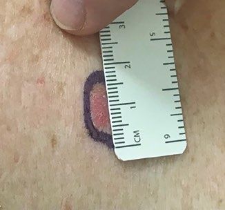
Marking a surgical margin for excision of a keratinocyte cancer Clinical
excision margin. Clin Otolaryngol Allied Sci
Table 1. Suggested margins depending on features of the lesion1 2000;25(5):370–73. doi: 10.1046/j.1365-
2273.2000.00376.x.
Margins for basal cell Margins for squamous cell 3. Kimyai-Asadi A, Alam M, Goldberg LH,
carcinoma excision carcinoma excision Peterson SR, Silapunt S, Jih MH. Efficacy of
narrow-margin excision of well-demarcated
Low-risk lesion 2–3 mm 4 mm primary facial basal cell carcinomas. J Am Acad
Dermatol 2005;53(3):464–68. doi: 10.1016/j.
jaad.2005.03.038.
High-risk lesion >5 mm 6 mm
4. Bisson MA, Dunkin CS, Suvarna SK, Griffiths RW.
Do plastic surgeons resect basal cell carcinomas
too widely? A prospective study comparing
surgical and histological margins. Br J Plast Surg
lesions be referred for specialist • Bright light and good magnification are 2002;55(4):293–97. doi: 10.1054/bjps.2002.3829.
5. Thomas DJ, King AR, Peat BG. Excision margins
management.1 In regard to incomplete important for determining the edges of for nonmelanotic skin cancer. Plast Reconstr
excision, management options include the tumour. Surg 2003;112(1):57–63. doi: 10.1097/01.
further re-excision, topical treatments or • Referral should be considered based
6.
PRS.0000067479.77859.31.
Griffiths R, Suvarna SK, Stone J. Basal cell
monitoring depending on a number of on individual factors, such as tumour carcinoma histological clearance margins:
lesion factors including whether the lateral location, size and patient preference. An analysis of 1539 conventionally excised
tumours. Wider still and deeper? J Plast Reconstr
or deep margins were involved.11,12 Aesthet Surg 2007;60(1):41–47. doi: 10.1016/j.
bjps.2006.06.009.
Authors 7. Lallas A, Apalla Z, Ioannides D, et al. Dermoscopy
Chloe A Mutimer BBMed, MD (Distinct), Medical in the diagnosis and management of basal cell
Further management Registrar, Alfred Hospital, Vic; Australian Skin Cancer carcinoma. Future Oncol 2015;11(22):2975–84.
All patients treated for keratinocyte Clinics, Vic doi: 10.2217/fon.15.193.
Anthony J Dicker MBBS, PhD, Skin Cancer Physician, 8. Wolf DJ, Zitelli JA. Surgical margins for basal cell
carcinomas require follow-up. This can Australian Skin Cancer Clinics, Vic; University of carcinoma. Arch Dermatol 1987;123(3):340–44.
either be undertaken by their general Queensland, Qld 9. Nahhas AF, Scarbrough CA, Trotter S. A review
practitioner or non-GP specialist, and Competing interests: None. of the global guidelines on surgical margins
Funding: None. for nonmelanoma skin cancers. J Clin Aesthet
is needed to assess for evidence of Dermatol 2017;10(4):37–46.
Provenance and peer review: Not commissioned,
recurrence, metastasis and new primary externally peer reviewed. 10. Kallini JR, Hamed N, Khachemoune A. Squamous
cell carcinoma of the skin: Epidemiology,
skin cancers. Patient education regarding Correspondence to:
classification, management, and novel trends. Int J
c.mutimer@alfred.org.au
the prevention of skin cancer is also crucial. Dermatol 2015;54(2):130–40. doi: 10.1111/ijd.12553.
11. Gulleth Y, Goldberg N, Silverman RP, Gastman BR.
References What is the best surgical margin for a basal cell
1. Cancer Council Australia Keratinocyte Cancers carcinoma: A meta-analysis of the literature. Plast
Key points Guideline Working Party. Draft clinical practice Reconstr Surg 2010;126(4):1222–31. doi: 10.1097/
guidelines for keratinocyte cancer. Sydney, NSW: PRS.0b013e3181ea450d.
• The keratinocyte cancer guidelines Cancer Council Australia, 2019. Available at https:// 12. Bovill ES, Banwell PE. Re-excision of incompletely
provide good advice for clinicians wiki.cancer.org.au/australia/Guidelines:Keratinocyte_ excised cutaneous squamous cell carcinoma:
carcinoma [Accessed 5 February 2021]. Histological findings influence prognosis. J Plast
undertaking surgical excision of 2. Lalloo MT, Sood S. Head and neck basal cell Reconstr Aesthet Surg 2012;65(10):1390–95.
skin tumours. carcinoma: Treatment using a 2-mm clinical doi: 10.1016/j.bjps.2012.04.031.
© The Royal Australian College of General Practitioners 2021 Reprinted from AJGP Vol. 50, No. 6, June 2021 387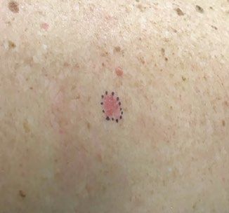
You can also read