Case Report Correction of Diastasis Rectus Abdominis with Tacking the Rectus Sheath and Resection of Excess Skin for Cosmesis
←
→
Page content transcription
If your browser does not render page correctly, please read the page content below
Hindawi
Case Reports in Medicine
Volume 2020, Article ID 7635801, 4 pages
https://doi.org/10.1155/2020/7635801

Case Report
Correction of Diastasis Rectus Abdominis with Tacking the Rectus
Sheath and Resection of Excess Skin for Cosmesis

          Kento Takaya ,1 Noriko Aramaki-Hattori ,1 Hanayo Yabuki,1 Norihito Wada,2
          Shigeki Sakai,1 Keisuke Okabe,1 and Kazuo Kishi1
          1
              Department of Plastic and Reconstructive Surgery, Keio University School of Medicine, Tokyo, Japan
          2
              Department of Surgery, Keio University School of Medicine, Tokyo, Japan

          Correspondence should be addressed to Kento Takaya; kento-takaya312@keio.jp

          Received 5 April 2020; Accepted 26 May 2020; Published 16 June 2020

          Academic Editor: Mark E. Shaffrey

          Copyright © 2020 Kento Takaya et al. This is an open access article distributed under the Creative Commons Attribution License,
          which permits unrestricted use, distribution, and reproduction in any medium, provided the original work is properly cited.
          Introduction. We report a case of diastasis rectus abdominis (DRA), in which the improvement of the appearance was obtained by
          performing extra skin resection. Case Report. A 30-year-old woman presented persistent abdominal bulging after her second
          delivery. She was diagnosed as DRA by computed tomography. We underwent a surgery that tacking the anterior layer of the
          rectus sheath and resecting excess skin. Results. There has been no clinical evidence of recurrence, and the patient satisfies her
          abdominal appearance. Conclusion. Because DRA is not a true hernia, surgery for DRA should be performed in understanding
          how patients want to improve their aesthetic appearance.

1. Introduction                                                              A 5 cm vertical surgical scar was noted to extend distally
                                                                         from the umbilicus. The abdomen bulged outward from the
Diastasis rectus abdominis (DRA) is a state in which the                 level of the lower sternum to below the umbilical fossa
abdominal wall is stretched due to an increase in abdominal              (Figure 1), and a 15 cm separation of rectus abdominis was
pressure, such as that occurs during pregnancy or with                   palpated both above and below the umbilical fossa. Com-
obesity, resulting in abdominal bulging [1]. Because DRA is              puted tomography showed the midline separation of the
not a true hernia and is not associated with strangulation, it           rectus abdominis muscle around the navel. The rectus
does not necessarily require surgical repair, and there are no           abdominis fascia was maintained, and there was no extru-
defined indications for surgery [2].                                      sion of abdominal contents. The inter-recti distance (IRD)
                                                                         was 36 mm at 3 cm above the superior border of the um-
2. Case Presentation                                                     bilicus, 40 mm at the center of the umbilicus, and 36 mm at
                                                                         2 cm below the inferior border of the umbilicus (Figure 2).
A 30-year-old woman noted an increase in weight of 20 kg                     Surgery was performed under general anesthesia. An
during her second pregnancy, which was delivered by ce-                  abdominal midline incision was made on the cesarean scar,
sarean. Her postpartum weight decreased by 17 kg, but she                and the linea alba was noted to be thinned and being pulled
noted persistent abdominal bulging. Computed tomography                  to the left and right sides of the abdomen. The rectus
showed a separation of the rectus abdominis at the site of the           abdominis muscle was separated horizontally. The anterior
abdominal wall scar. The patient presented to us desiring                rectus sheath was sutured in the midline using nonab-
cosmetic improvement. Her history was significant for a                   sorbable suture without the performance of the midline
20 kg weight gain with her first pregnancy, but she had no                incision on the anterior sheath, and a high-density poly-
subsequent abdominal distension. Her medical history was                 ethylene mesh was applied to the linea alba and rectus
otherwise unremarkable.                                                  abdominis muscle. Attention was then turned to the excess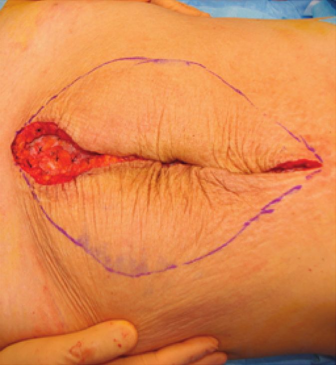
2 Case Reports in Medicine
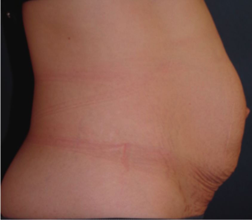
(a) (b)
Figure 1: A 5 cm vertical surgical scar is present distal to the umbilicus. Significant midline abdominal bulging is noted to extend beyond the
level of the scar.
(a) (b)
(c)
Figure 2: Preoperative abdominal computed tomography: (a) 3 cm above the superior border of the umbilicus; (b) center of the umbilicus;
(c) 2 cm below the inferior border of the umbilicus. The inter-rectus distance is indicated by a white line.
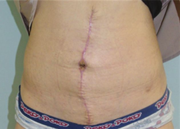
skin and subcutaneous adipose tissue, including the pig- 3. Discussion
mented portion of skin. A spindle-shaped incision was used
to excise this tissue, with the spindle measuring 15 cm in the Surgical repair for DRA is widely performed in Europe and
horizontal direction (Figure 3). the United States for cosmetic purposes, but it is a little-
A drain was placed over the mesh. The superficial fascia known clinical condition in Japan [3]. Both laparotomy and
was sutured, and skin closure was performed, using size zero laparoscopic methods are used, and various techniques, such
polydioxanone suture. Adhesive skin closure strips were as the use of mesh and layered suturing, are employed [4].
applied to the incision. The patient’s operative course was Open surgery is often performed by suturing the anterior or
uneventful. Computed tomography performed 2 months posterior sheath of the rectus abdominis muscle with both
after surgery showed no recurrence of the swelling of her an absorbable and nonabsorbable suture to form a fistula.
abdomen. The patient is satisfied with her abdominal ap- Indwelling mesh may be placed at the surgeon’s discretion
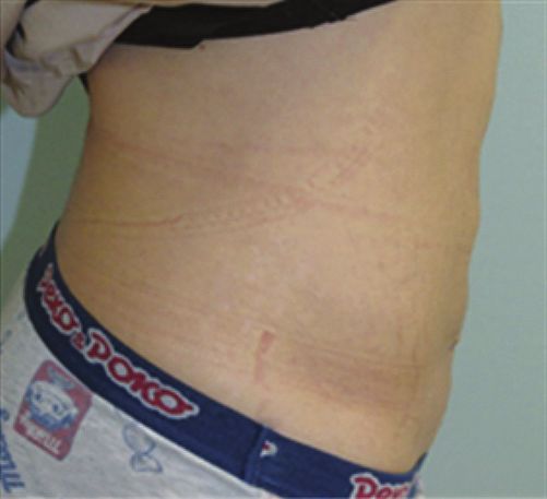
pearance (Figure 4). There has been no clinical evidence of [5]. In laparoscopic surgery, as in the open technique, the
recurrence and no complications for one year after surgery. posterior surface of the linea alba is sutured to form pleats;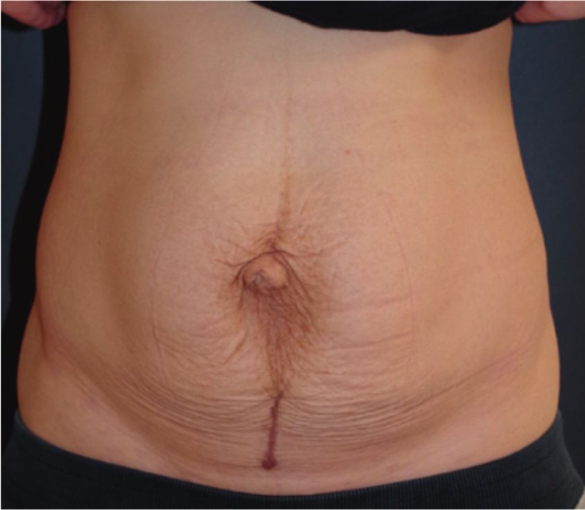
Case Reports in Medicine 3
(a) (b) (c)
(d) (e)
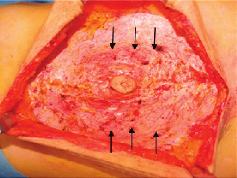
Figure 3: Intraoperative photographs. (a) The rectus abdominis muscle is displaced to the left and right. The peritoneum is intact. (b) The
anterior rectus muscle is sutured. (c) A mesh is applied to the rectus abdominal muscle. (d) Excess skin is removed in a spindle shape. (e)
Closure of the incision.
(a) (b)
(c) (d) (e)
Figure 4: (a–c) Postoperative abdominal computed tomography. (a) 3 cm above the superior border of the umbilicus; (b) center of the
umbilicus; (c) 2 cm below the inferior border of the umbilicus. There is no apparent recurrence of the hernia. (d-e) Appearance three months
after surgery. The patient’s abdominal bulge has resolved.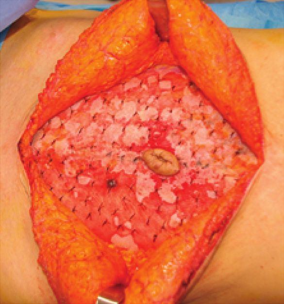
4                                                                                                               Case Reports in Medicine

mesh is placed in almost all patients. A report comparing                     nulliparous women,” Clinical Anatomy, vol. 22, no. 6,
open with laparoscopic surgery notes that the latter results in               pp. 706–711, 2009.
smaller surgical scars does not require placement of an                 [2]   F. X. Nahas, “An aesthetic classification of the abdomen based
indwelling drain and reduces perioperative complications;                     on the myoaponeurotic layer,” Plastic and Reconstructive
however, this report does not have a high level of evidence.                  Surgery, vol. 108, no. 6, pp. 1787–1795, 2001.
                                                                        [3]   F. Hickey, J. G. Finch, and A. Khanna, “A systematic review on
    When planning surgery, attention is focused on
                                                                              the outcomes of correction of diastasis of the recti,” Hernia,
repairing the fascia, since the cause of the problem is                       vol. 15, no. 6, pp. 607–614, 2011.
stretching and thinning of the anterior layer of the rectus             [4]   C. Palanivelu, M. Rangarajan, P. A. Jategaonkar, V. Amar,
sheath and the linea alba. Subcutaneous liposuction is                        K. S. Gokul, and B. Srikanth, “Laparoscopic repair of diastasis
sometimes combined with this repair [6]. In our patient, we                   recti using the “Venetian blinds” technique of plication with
repaired the abdominal wall, and then, we tightened the                       prosthetic reinforcement: a retrospective study,” Hernia,
entire abdominal area by removing excess skin and sub-                        vol. 13, no. 3, pp. 287–292, 2009.
cutaneous fat, allowing for an aesthetically pleasing result.           [5]   L. G. Angiò, E. Piazzese, V. Pacilè et al., “The surgical
    Reported complications of abdominoplasty include                          treatment of the diastasis recti abdominis: an original tech-
hematoma and seroma formation, wound infection, skin                          nique of prosthesis repair of the abdominal wall,” Giornale di
                                                                              Chirurgia, vol. 28, no. 5, pp. 187–198, 2007.
flap necrosis, and hypertrophic scarring [7]. Generally, the
                                                                        [6]   M. L. Zukowski, K. Ash, D. Spencer, M. Malanoski, and
recurrence rate is low and complications are rare, but any                    G. Moore, “Endoscopic intracorporal abdominoplasty: a re-
surgery requires a sufficient explanation of the risks and                      view of 85 cases,” Plastic and Reconstructive Surgery, vol. 102,
benefits to the patient [8].                                                   no. 2, pp. 516–527, 1998.
    Additionally, our method has a limitation. It is a long             [7]   J. Akram and S. H. Matzen, “Rectus abdominis diastasis,”
vertical scar that remains in the midline of the abdomen. In                  Journal of Plastic Surgery and Hand Surgery, vol. 48, no. 3,
this case, a scar after the caesarean section was originally                  pp. 163–169, 2014.
found under the umbilicus, and a remarkable skin laxity was             [8]   A. Michalska, W. Rokita, D. Wolder, J. Pogorzelska, and
observed from the upper part of the umbilicus to the lower                    K. Kaczmarczyk, “Diastasis recti abdominis: a review of
abdomen, so a long scar was needed to remove it. However,                     treatment methods,” Ginekologia Polska, vol. 89, no. 2,
new scarring from the surgical procedure should be mini-                      pp. 97–101, 2018.
                                                                        [9]   R. W. Dabb, W. W. Hall, M. Baroody, and A. A. Saba,
mized for further cosmetically superior results in future
                                                                              “Circumferential suction lipectomy of the trunk with anterior
cases. Laparoscopic surgery is an alternative to minimize                     rectus fascia plication through a periumbilical incision: an
postsurgical scarring, but at the risk of postoperative                       alternative to conventional abdominoplasty,” Plastic and
complication such as organ damage and adhesive ileus. If                      Reconstructive Surgery, vol. 113, no. 2, pp. 727–732, 2004.
skin is less lax, we may need to consider a previously re-             [10]   J. C. C. Restrepo and J. A. M. Ahmed, “New technique of
ported approach to the rectus anterior sheath through a                       plication for miniabdominoplasty,” Plastic and Reconstructive
small incision around the navel [9, 10].                                      Surgery, vol. 109, no. 3, pp. 1170–1177, 2002.
    We repaired the abdominal wall in this patient with DRA
by tacking the anterior layer of the rectus sheath and by
placing mesh. We also resected the excess skin to improve
cosmesis and achieve a high level of patient satisfaction. We
think that it is necessary to consider the possibility of DRA
when planning surgery for a patient with abdominal bulging
extending beyond an operative scar.

Data Availability
Data sharing not applicable to this article as no datasets were
generated or analyzed during the current study.

Consent
The patient has given her permission for publication of this
report, including images.

Conflicts of Interest
The authors declare that they have no conflicts of interest.

References
 [1] G. M. Beer, A. Schuster, B. Seifert, M. Manestar, D. Mihic-
     Probst, and S. A. Weber, “The normal width of the linea alba in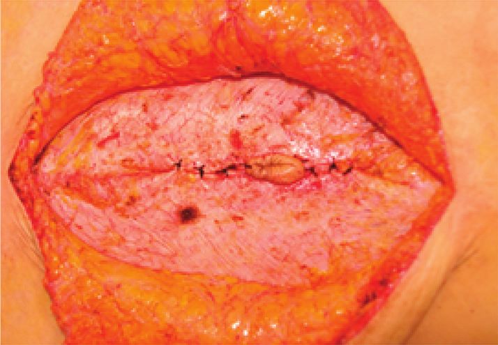

You can also read