TEMPORAL SUBLUXATION OF A SCLERAL-FIXATED IOL
←
→
Page content transcription
If your browser does not render page correctly, please read the page content below
s   CATARACT SURGERY CASE FILES

            TEMPORAL SUBLUXATION OF
            A SCLERAL-FIXATED IOL
            What is the best strategy for management when the suture securing the inferior haptic breaks years after
            cataract surgery?
             BY BRANDON D. AYRES, MD; ASHLEY KHALILI, MD; SOON-PHAIK CHEE, FRCS(G), FRCS(ED), MMED(S’PORE); AND ZEBA A. SYED, MD

             CASE PRESENTATION
               A 58-year-old woman presents for a consultation. The patient reports                 a fall with no head trauma. Ten years ago, the left eye underwent scleral
            that she has experienced 3 months of blurry vision in the left eye following            fixation of a PMMA IOL (CZ70BD, Alcon) with 10-0 polypropylene (Prolene,
                                                                                                    Ethicon) sutures and a pupilloplasty. The patient also underwent a pars plana
                                                                                                    vitrectomy many years ago for a retinal detachment.
                                                                                                       On examination, her BCVA is 20/40 OS. The lids and lashes are normal.
                                                                                                    The ends of a 10-0 polypropylene suture are seen to be buried under the
                                                                                                    conjunctiva superiorly and inferiorly. The cornea is clear, and the anterior
                                                                                                    chamber is deep and quiet. The superotemporal pupilloplasty suture is intact,
                                                                                                    and temporal transillumination defects are visible. The examination findings
                                                                                                    are consistent with a temporally subluxated IOL due to the release of the
                                                                                                    inferior polypropylene suture from the inferior haptic. When the patient is
                                                                                                    supine, the view of the IOL is lost with posterior hinging owing to a release of
                                                                                                    inferior fixation (Figure 1). The retinal examination is normal with 360º laser
                                                                                                    scars from prior surgery.
                                                                                                       How would you address the dislocated IOL?

              Figure 1. Preoperative appearance of the left eye while the patient is supine. Note                 —Case prepared by Brandon D. Ayres, MD, and Ashley Khalili, MD
              the lack of visibility of the IOL and the superotemporal iris suture.

                                                                              however, is likely to give way during                 pupilloplasty suture would be released.
                                                                              surgery, risking a dropped IOL in this                With iris hooks exposing the attached
                                                                              vitrectomized eye. My preference,                     haptic, the sclera would be depressed,
                                                                              therefore, would be to perform an                     bringing the suture into view. The
                                                                              IOL exchange and intrascleral haptic                  haptic would be grasped with
              SOON-PHAIK CHEE, FRCS(G), FRCS(ED),                             fixation and to use the Yamane1                       intraocular forceps, and a 27-gauge
                        MMED(S’PORE)                                          instead of a glued IOL technique.                     needle would be used to nick the
                                                                                 Under peribulbar anesthesia, the                   suture. The IOL would be brought into
              The options here are resuturing                                 6 and 12 clock positions at the lim-                  the anterior chamber and explanted.
            the inferior haptic to the sclera                                 bus and 2 mm posteriorly on the                          After removal of the iris hooks,
            with a stronger suture through                                    conjunctiva would be marked. With                     a superior peripheral iridectomy
            the eyelet (technically difficult)                                anterior chamber infusion running,                    would be created. A three-piece,
            or securing the haptic with a belt                                paracenteses would be created on                      round anterior–edged IOL would be
            loop (PTFE [off-label use] or flanged                             either side of the sutured haptic and                 inserted into the anterior chamber
            polypropylene). The existing suture,                              at the 1 and 4:30 clock positions. The                under a dispersive OVD. The incision

    26 CATARACT & REFRACTIVE SURGERY TODAY | AUGUST 2021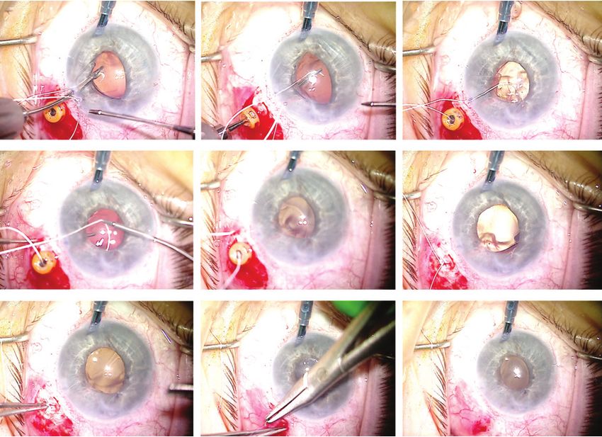
CATARACT SURGERY CASE FILES
s
would be sutured. Starting 2 mm counterclockwise to the IOL, an OVD cannula would be inserted through the
scleral fixation marks, bent 27-gauge needles on insulin pars plana to elevate the IOL and rest it on the iris.
syringes filled with balanced salt solution would be made to After the creation of an adjacent paracentesis, the
traverse the sclera and enter the eye at the fixation marks. haptic of the CZ70BD would be externalized, and a
Beginning with the trailing haptic, the haptics would be 5-0 polypropylene suture would be threaded through the
threaded into the needle. The haptics would be retrieved, eyelet. Low-temperature cautery would be used to make
flanged, and buried after IOL centration is confirmed. a flange at the suture’s tail, posterior to the IOL. The
A four-throw pupilloplasty would be performed using externalized haptic can then be returned into the anterior
10-0 polypropylene. The OVD would be removed, infusion chamber. Next, a 27-gauge needle would be inserted
halted, and the incisions hydrated. 2.5 mm posterior to the inferior limbus, and intraocular
forceps (MicroSurgical Technology) would be used to
thread the free end of the 5-0 polypropylene suture into
the lumen. Once the needle is externalized, the suture can
be shortened to center the IOL and cauterized to create a
flange that remains subconjunctival.
It is important to recognize that the superior
10-0 polypropylene suture is at similar risk of future
ZEBA A. SYED, MD disinsertion. I would therefore consider refixating that
haptic using the same approach.
One technique that I find useful in this scenario is a
modification of the approach first described by Canabrava
and colleagues.2 This particular case offers two unique
considerations. First, initial IOL fixation was with a
10-0 polypropylene suture, which is at risk of degradation
years later. Second, the patient has a history of pars plana
vitrectomy, obviating the need for concurrent vitrectomy at
the time of haptic fixation. WHAT WE DID: BRANDON D. AYRES, MD,
To start this case, a single pars plana port would be AND ASHLEY KHALILI, MD
placed, and a paracentesis would be created for an
anterior chamber maintainer. To retrieve the existing Surgical options for this patient are an IOL exchange
or refixation of the current lens. An important point of
consideration is the need for a large wound, approximately
Figures 2 through 5 courtesy of Brandon D. Ayres, MD
A B 6 to 7 mm, if the PMMA IOL is to be removed and
replaced. Additionally, the pupilloplasty suture may limit
full dilation of the pupil and may have to be replaced to
improve visualization during surgery.
The risks, benefits, and alternatives to surgery were
discussed extensively with the patient, and she elected
to undergo IOL refixation. To avoid the need for a large
corneal wound, a surgical plan was made to refixate the
C D haptics using a PTFE suture (Gore-Tex CV-8, W.L. Gore &
Associates; off-label use). The patient was also made aware
of the plan for a pupilloplasty as needed.
A peritomy was created inferiorly, and a centration
mark was made 3 mm posterior to the limbus with a
marking pen. Two circumlimbal sclerotomies were made
4 mm apart and centered over the 3-mm centration mark.
With a Charles lens used for posterior visualization, the
Figure 2. Retrieval of the released inferior haptic of the CZ70BD lens. An anterior chamber
maintainer is placed nasally. A trocar and a sclerotomy (indicated with an *) are placed
inferior haptic was grasped with microforceps, and the
3 mm posterior to the limbus and 4 mm apart in a circumlimbal fashion after a peritomy is haptic was elevated into the anterior chamber. The haptic
created (A). With posterior visualization using a Charles lens, the inferior haptic is grasped was then externalized through an inferior paracentesis,
with a pair of microholders (B). The haptic is elevated into the anterior chamber and then and a CV-8 PTFE suture strand was laced through the
externalized through an inferior paracentesis (C). A CV-8 PTFE suture strand is laced through externalized haptic eyelet (Figure 2). The haptic was
the externalized haptic eyelet (D). then rotated back into the eye, and the suture ends
AUGUST 2021 | CATARACT & REFRACTIVE SURGERY TODAY 27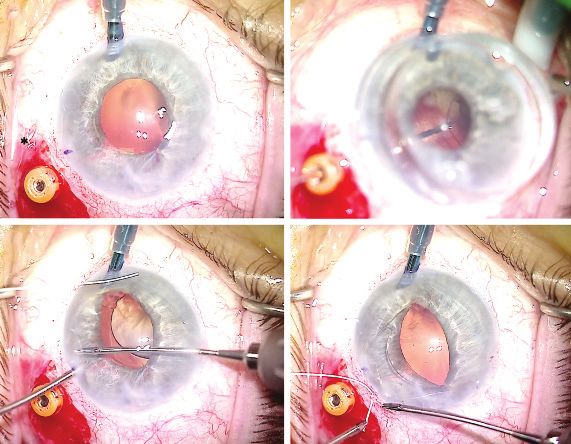
s CATARACT SURGERY CASE FILES
A B C A B C
D E F D E F
G H I G H I
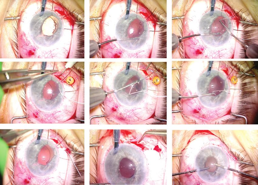
Figure 3. Scleral fixation with a CV-8 PTFE suture using a handshake technique. The Figure 4. Refixation of the superior haptic. Iris hooks are placed to improve visualization (A).
suture strand is brought into the anterior chamber with microforceps via the inferior The superior haptic is released from the polypropylene suture (B). A PTFE suture is introduced
paracentesis (A). The suture end is grasped behind the optic by forceps introduced through into the anterior chamber via a superior paracentesis and laced through the eyelet (C). A
the trocar (B) and then externalized (C). Next, the other end of the suture is introduced into peritomy is created, and two sclerotomies are created 4 mm apart in a circumlimbal direction
the anterior chamber and passed from superior limbal approaching forceps to microforceps and centered over a centration mark located 3 mm posterior to the limbus (D). One end of the
placed in the distal sclerotomy (D). With removal of the microforceps, the suture end is suture is handed off through the anterior chamber from one microforceps to another, which
externalized through the sclera (E). The trocar is removed, and the suture ends are tied (F). was introduced through the sclerotomy (E). The suture is then externalized through the scleral
The knot is buried into the sclerotomy (G). The peritomy is closed (H and I). incision using the microforceps. The same steps are repeated for the other suture end via the
proximal trocar (F). The trocar is removed, and the knot is tied and buried (G). The iris hooks are
removed, and the peritomy is closed (H). The anterior chamber is cleared of OVD (I).
cornea had mild peripheral edema, SOON-PHAIK CHEE, FRCS(G), FRCS(ED),
and the anterior chamber was deep MMED(S’PORE)
n S ingapore National Eye Centre
with mild cell. The PTFE suture was
n Department of Ophthalmology, Yong Loo Lin School
well covered superiorly and inferiorly
by conjunctiva. The IOL was in of Medicine, National University of Singapore
n C ataract Research Team, Singapore Eye
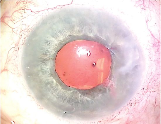
position and well centered (Figure 5).
The patient began a postoperative Research Institute
n D epartment of Ophthalmology & Visual Sciences
regimen of topical steroids and
antibiotics. One month after Academic Clinical Program, Duke-NUS Medical
surgery, the patient reported an School, Singapore
n c hee.soon.phaik@singhealth.com.sg
improvement in her vision. Her
n F inancial disclosure: None
visual acuity was 20/40, the corneal
Figure 5. After surgery, the IOL is in position and exhibits edema had resolved, and the IOL
good centration and no tilt. ASHLEY KHALILI, MD
was stable. n
n Cornea fellow, Wills Eye Hospital, Philadelphia
1. Yamane S, Sato S, Maruyama-Inoue M, Kadonosono K. Flanged intrascleral n a khalili@willseye.org
were each externalized through the intraocular lens fixation with double-needle technique. Ophthalmology.
n F inancial disclosure: None
sclerotomies with microforceps using 2017;124(8):1136-1142.
2. Canabrava S, Canêdo Domingos Lima AC, Ribeiro G. Four-flanged intrascleral
a handshake technique (Figure 3). intraocular lens fixation technique: no flaps, no knots, no glue. Cornea.
The superior haptic was released 2020;39(4):527-528. ZEBA A. SYED, MD
from polypropylene fixation and n Codirector, Cornea Fellowship Program, Wills Eye
fixated with a PTFE suture using the Hospital, Philadelphia
same technique (Figure 4). Iris hooks SECTION EDITOR BRANDON D. AYRES, MD n A ssistant Professor of Ophthalmology, Sidney
were placed superiorly to improve n Surgeon on the Cornea Service, Wills Eye Kimmel Medical College, Thomas Jefferson
visualization and avoid the need to Hospital, Philadelphia University, Philadelphia
replace the pupilloplasty sutures. n b ayres@willseye.org n z syed@willseye.org
One day after surgery, the patient's n F inancial disclosure: Consultant (Alcon, Carl Zeiss n F inancial disclosure: Research support (Dompé
visual acuity was 20/80 OS. The Meditec, MicroSurgical Technology) Farmaceutici); Speakers bureau (Bio-Tissue)
28 CATARACT & REFRACTIVE SURGERY TODAY | AUGUST 2021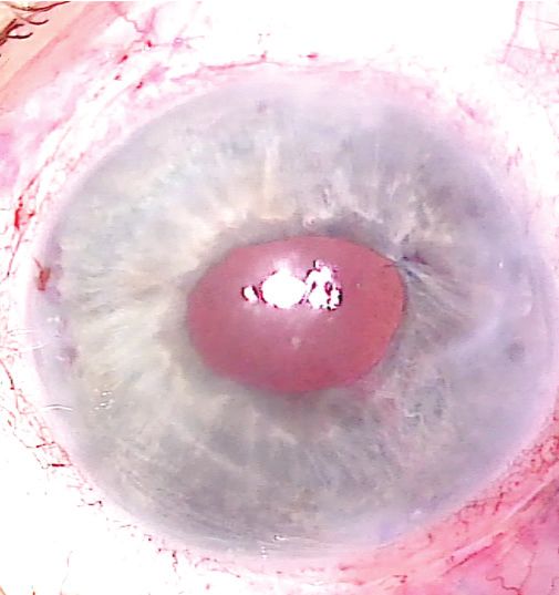


You can also read