DENSE BONE ISLAND OF THE JAW: A CASE REPORT - Exodontia.info
←
→
Page content transcription
If your browser does not render page correctly, please read the page content below
case report

DENSE BONE ISLAND OF THE JAW:
A CASE REPORT
G. CERULLI MARIANI*, F. FAVARETTI*, L. LAMAZZA**, A. DE BIASE**
* Freelancer Rome
** Department of Oral Science, Sapienza University of Rome, Italy

  SUMMARY                                                         RIASSUNTO
  Dense bone island of the jaw: a case report                     Dense bone island della mandibola: descrizione di un
  The “Dense Bone Island” (DBI) is a radiopaque lesion re-        caso
  ferred in leterature as idiopathic osteosclerosis, enostosis,   La Dense Bone Island (DBI) è una lesione radiopaca de-
  focal osteosclerosis, periapical osteopetrosis, and bone        scritta in letteratura come osteosclerosi idiopatica, eno-
  scar. The DBI are accidentally found in routinary Xray of       stosi, osteosclerosi focale, osteopetrosi periapicale, cica-
  bone structures. In the maxillary bones, often localized in     trice ossea. La DBI è spesso riscontro casuale in radio-
  the mandible, especially in the molar region, with a report-    grafie di routine eseguite per altri motivi. Si localizza prin-
  ed incidence ranging from 2.3 to 9.7%. DBI does not             cipalmente a livello della mandibola in regione molare con
  seem to develop until the first phase of adolescence and        incidenza compresa tra il 2,3 e il 9.7%. Sembra che il suo
  it is usually found in adolescents and in young adults. In      sviluppo inizi dopo la prima fase dell’adolescenza. Il ri-
  40% of cases DBI seems to increase in size after a 10           scontro diagnostico avviene normalmente in soggetti ado-
  year follow-up, because the DBI found in jaws and in long       lescenti o giovani adulti. Nel 40% dei casi la DBI sembra
  bones seem to increase proportionally to the bone growth.       aumentare di volume dopo circa 10 anni, probabilmente
  A case of a 26 years old patient and the surgical treatment     perché si accresce proporzionalmente alla crescita ossea.
  is presented. This is the first reported case where com-        Viene presentato un caso di una paziente di 26 anni sot-
  plete X-ray Orthopantomography follow-up showed the             toposta a trattamento chirurgico. Il caso presentato è il pri-
  evolution of the lesion since its onset.                        mo caso in cui la lesione è stata documentata radiografi-
                                                                  camente dal momento della sua insorgenza fino al tratta-
                                                                  mento chirurgico.

  Key words: enostosis, radiopaque lesion, bone island.           Parole chiave: enostosi, lesioni radiopache, bone island.

                                                                   ed from the surrounding normal bone, and smooth
     Introduction                                                  or irregular in outline.
                                                                   Histopathologically, DBIs are composed of dense
                                                                   calcified tissue without marrow spaces and gener-
A “Dense Bone Island” (DBI) is a localized, well-                  ally no inflammatory cell infiltration (3, 4). Most
defined, radiopaque mass in the jaw with a round,                  of the DBIs described in literature are smaller than
elliptical or irregular shape and a variable size (1).             2 cm (2, 3); this leads to hypothesize that they may
Most of these lesions are asymptomatic, and rep-                   not reach the sufficient size to cause jaw expansion.
resented casual finds in routine X-rays.
Although DBI has been reported with a variety of
names including enostosis, bone scar, focal os-
teosclerosis, idiopathic osteosclerosis and periapi-                     Case report
cal osteopetrosis, their cause and classification are
controversial (2).                                                 Twenty-six-year-old female patient, negative
On radiographic evaluation, they are well separat-                 anamnesis for systemic diseases, showed no rele-

                    ORAL & Implantology - Anno I - N. 2/2008                                                                        87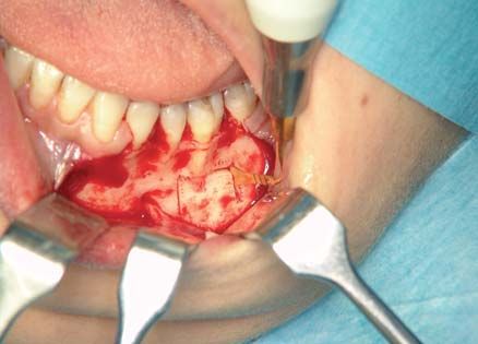
vant alterations at the intra- oral examination. The
case report
radiographic examinations (Orthopantomography,
CT Dentascan), showed a 1cm osteocondensing
lesion, localized under the left first lower molar
(Figg. 1, 2).
The patient had radiographic examinations that
showed the evolution of the lesion since its onset
(Figg. 3, 4, 5).
In consideration of its remarkable increase in the
last 10 years, and of an intermittent painful symp-
tomatology, was decided to perform a surgical Figure 3
OPT Rx at 6 years of age with mixed dentition, ab-
enucleation of the lesion. sence of endosteal lesions.
Surgical treatment was performed under general
anesthesia using ultrasonic device. DBI was re-
moved preserving surrounding bone tissue and
alveolar nerve (Figg. 6, 7). Notwithstanding the
lack of cleavage plane between lesion and sur-
Figure 4
OPT Rx at 12 years of age, with permanent denti-
tion, absence of endosteal lesions.
Figure 1
Preoperative ortophantomography.
Figure 5
OPT Rx at 16 years of age, initial onset of endosteal
lesion near the left first lower molar.
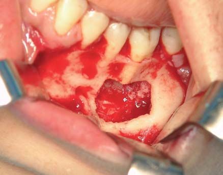
rounding bone, precision and selective cut of ul-
trasound device allowed a conservative excision
(Fig. 8). Microscopic examination showed cortical
lamellar bone tissue partly sclerotized in the site of
previous reworking, locally in continuity with
Figure 2 porosus bone whose medulla spaces were occu-
Detail of CT Dentascan in axial projection.
pied by vascularized loose fibrous tissue (Fig. 9).
88 ORAL & Implantology - Anno I - N. 2/2008
case report

 Figure 6                                                                                    Figure 8
 Osteotomy performed by ultrasound device.                                                   Removed lesion.

                                                           Figure 9
                                                           Histological section. Cortical lamellar bone tissue
 Figure 7
                                                           (HE, 10X).
 Intraoral view showing precise and selective cut
 with complete excision of lesion.

The diagnosis made, in consideration of both the
histological and radiographic examination of the
lesion, was of solitary enostosis, known as “bone
island”.
No recurrence was showed in one year follow-up
radiograph (Fig. 10).

    Discussion
                                                           Figure 10
DBI are generally modest in size and do not                One year follow-up showing a prosthetic rehabilita-
                                                           tion and good bone healing.
change overtime (5). Mirra (6), presented the case

                ORAL & Implantology - Anno I - N. 2/2008                                                         89
of a 5-year-old child with a lesion of 1.1 × 1.7 cm secondary infection, through the element in-
case report
in the distal portion femur, initially considered as volved, the surgical procedures must be carefully
a DBI, whereas 6 months later the lesion had carried out in order not to cause lesions to the sur-
reached the size of 4.2 × 3.0 cm, and a biopsy rounding areas.
showed evidence of osteosarcoma. The author
suggests to perform an open surgery biopsy of
DBI when the size of the lesion increases by 25%
in 6 months or by 50% in one year.
References
DBI may be differentiated from more aggressive
1. Petrikowski CG, Peters E. Longitudinal radiographic
or malignant bone lesions by one of the following: assessment of dense bone island of the jaws. Oral Sur-
absence of a primitive tumor, slow growth over a gery Oral Med Oral Path Oral Radiol and Endod 1997;
period of years, a clearly demarcated margin with 83: 627-634.
thorny radiation from the sclerotic lesion and the 2. Kawai T, Muratami S. Gigantic dense bone island of
absence of pain. the jaw. Oral Surg Oral Med Oral Pathol Oral Radiol
Endod 1996; 82: 108-15.
Petrikowski and Peters (1) noted that, for practical
3. Eversole LR, Stone CE, Strub D. Focal sclerosing
purpose, the calcifying encondroma, medullary osteomyelitis/focal periapical osteopetrosis: radiogra-
bone infarct, healing non-ossifying fibroma, os- phic patterns. Oral Surg 1984; 58: 456-60.
teosarcoma and osteoid osteoma can be eliminate 4. Nakano K, Ogawa T, Sobue S, Ooshima T. Dense bo-
from the differential diagnosis of DBI. ne island: clinical features and possible complications.
Always according to Petrikowski and Peters (1), Int J Paed Dent 2002; 12: 433-437.
5. Greenspan A, Standalnik RC. Bone island: scintigra-
the age in which a DBI is found ranges between
phic findings and their clinical application. Canadian
9.4 and 14.0 years, whereas according to other Association of radiology journal 1995; 46: 368-379.
studies, DBI seems to be more frequent in the third 6. Mirra JM. Enostosis. In: Bone tumor. Philadelphia:
decade of life (7, 8). Lea & Febiger, 1989: 182-191.
Various therapeutic choices apply. If the lesion is 7. Yonetsu K, Yuasa K, Kanda S. Idiopatic osteosclerosis
an isolated radiopaque area without any connec- of the jaws. Panoramic radiographic and computed to-
mographic findings. Oral Surg Oral Med Oral Pathol
tion with the teeth, and no painful symptomatol-
1997; 8: 517-521.
ogy occurs, it is preferable not to surgically inter- 8. Geist JR, Katz JO. The frequency and distribution of
vene. idiopathic osteosclerosis. Oral Surg Oral Med Oral Pa-
When, the thick radiopaque area is associated to a thol 1990; 69: 388-393.
Correspondence to:
Dott. Alberto De Biase
E-mail: alberto.debiase@uniroma1.it
90 ORAL & Implantology - Anno I - N. 2/2008You can also read