Amalgam tattoo removal with diode laser - ZWP online
←
→
Page content transcription
If your browser does not render page correctly, please read the page content below
| case report

           Amalgam tattoo removal
           with diode laser
           Dr Maziar Mir, Prof. Norbert Gutknecht, Dr Masoud Mojahedi, Germany; Dr Jan Tunér, Sweden;
           Prof. Ramin Mosharraf, Sevda Noori & Dr Masoud Shabani, Iran

           Introduction                                                               The clinical appearance of the lesion and the presence
                                                                                      of radiopaque amalgam fragments in radiographs are
           An amalgam tattoo belongs to the group of common pig-                      enough to diagnose an amalgam tattoo. If no metallic
           mented lesions in the oral cavity. Such tattoos are created                fragments can be identified in a radiograph, a biopsy
           through emergence of amalgam particles into the dam-                       of the lesion is recommended to rule out melanocytic
           aged oral mucosa during placement of amalgam fillings                      neoplasia. Unless for aesthetic reasons, treatment of an
           or a discolouring of the soft tissue close to the amalgam                  amalgam tattoo is not necessary.3, 4 Owing to the sever-
           filling over time. Clinically, amalgam tattoos appear as blu-              ity of melanoma, clinicians must differentiate between be-
           ish-black or bluish-grey lesions on the oral mucosa, most                  nign lesions and serious lesions, most notably melanoma.
           commonly on the gingival surfaces, alveolar mucosa and                     Based on the pigment origin, a bluish-black or bluish-grey
           buccal mucosa. The incidence of amalgam tattoos has                        pigmented lesion may be classified as an endogenous or
           been reported as up to 8 per cent of samples surveyed.1, 2                 exogenous pigmented lesion. Based on the involved ar-
                                                                                      eas, such lesions are further differentiated into localised
                                                                                      and diffuse pigmentations.

                                                                                      Localised bluish-black or bluish-grey pigmented le-
                                                                                      sions include:
                                                                                      1. pigmentation due to metals like amalgam, graphite,
                                                                                         lead (in the oral cavity, amalgam tattoos usually occur
                                                                                         near restorations);
                                                                                      2. melanotic macule (small in size, mostly occurs on lips,
                                                                                         due to increase in melanin synthesis);
                                                                                      3. naevus (increased proliferation of melanocytes, usually
                                                                                         innate/congenital);
                                                                                      4. malignant melanoma (dark, irregular borders, asym-
                                                                                         metrical and rapid in growth); and
Fig. 1                                                                                5. post-inflammatory pigmentation due to healing of le-
                                                                                         sions such as lichen planus, pemphigus and pemphi-
                                                                                         goid disease.

                                                                                      Diffuse pigmentations include:
                                                                                      1. physiological pigmentation (present from birth);
                                                                                      2. smoker’s melanosis (due to history of smoking);
                                                                                      3. endocrine disorders like Addison’s disease/Cushing’s
                                                                                         syndrome (based on systemic signs and symptoms);
                                                                                      4. HIV-associated melanosis (based on ELISA test for
                                                                                         HIV); and
                                                                                      5. pigmentations associated with syndromes, such as
                                                                                         McCune–Albright and Peutz–Jeghers (requires general
                                                                                         examination for other associated features).

Fig. 2
                                                                                      All of the above-mentioned lesions have to be considered
           Fig. 1: Clinical appearance of the amalgam tattoo at the alveolar ridge.   in differential diagnosis when considering the treatment
           Fig. 2: Radiograph showing some amalgam particles at the lesion site.      of such darkly pigmented areas in the oral cavity.5

06         1 2019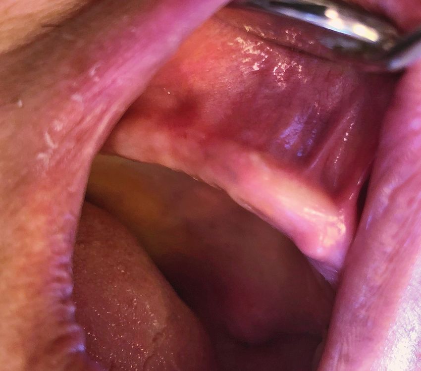
AD
Once amalgam tattoo diagnosis has lar joint disorder or myofascial distur-
been confirmed, treatment may be per- bances, and no functional or parafunc-
formed with a dental laser, surgical blade tional habits, but poor oral hygiene and a
or placement of subepithelial connective fully edentulous maxilla.
tissue.6, 7
Clinical findings
Case presentation The clinical examination showed a dark
pigmentation on the maxillary alveolar
A 60-year-old female patient with a dark ridge that was firm and well demarcated
pigment at the edentulous alveolar ridge and caused no pain (Fig. 1).
was referred for treatment of the lesion
as a pre-prosthodontic preparation pro- Radiographic examination
cedure. Radiographic examination revealed the
appearance of radiopaque particles at
Medical history the area of the dark pigmentation (Fig. 2).
The patient’s medical history showed no
systemic medical problems, no allergic Diagnosis
reaction, no medications or recreational An amalgam tattoo lesion was thus diag-
drugs and no history of past surgical pro- nosed and removal by diode laser was
cedures; thus, the patient did not need decided on.
to be referred for medical consultation.
Amalgam tattoo removal
Dental history with diode laser
Oral and maxillofacial examination of the After the patient had completed the con-
patient revealed no temporomandibu- sent form, the operation area was anaes-
Fig. 3
Fig. 4
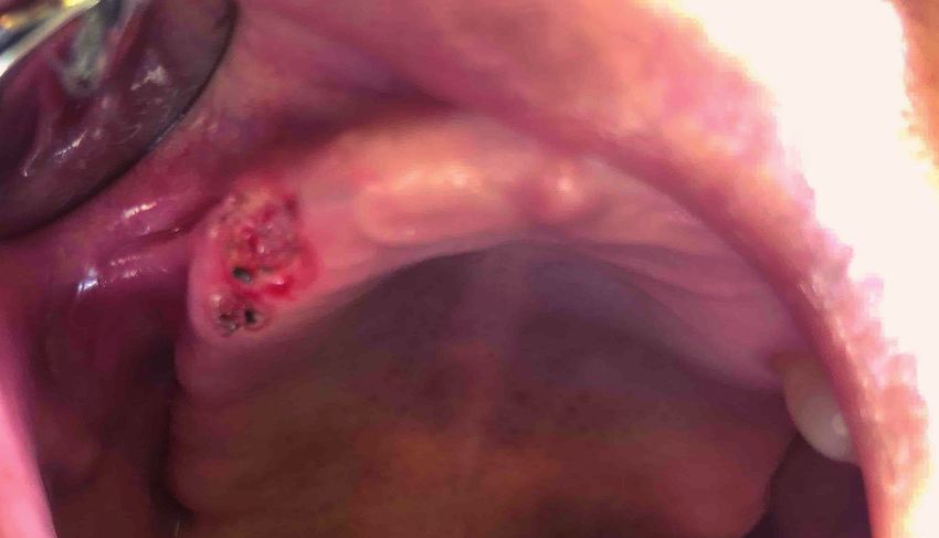
Fig. 3: Situation immediately after amalgam tattoo removal. Fig. 4: Follow-up examination one day after amalgam
tattoo removal.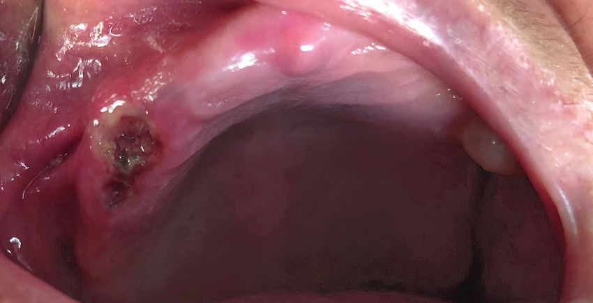
| case report
Fig. 5 Fig. 6
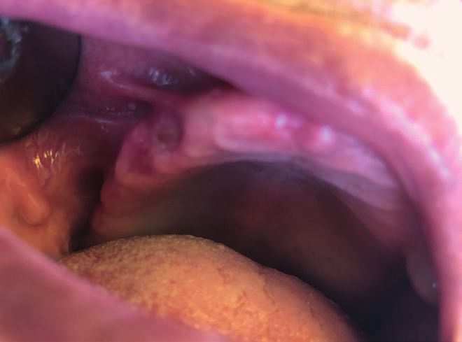
Fig. 5: Situation at the one-week follow-up. Fig. 6: One month after amalgam tattoo removal.
thetised through infiltration with 2 % lidocaine (1:80,000 month follow-up, a successful treatment could be clini-
adrenaline; 1.8 ml; Darou Pakhsh Pharmaceutical). cally observed (Fig. 6).
In the next step, the controlled area was defined and laser Discussion
warning signs were properly displayed to secure the op-
erating room. After the eye protection of the patient, the Diode lasers are used extensively in many dental prac-
patient’s guardian and the assistant had been checked, tices.8 Laser–tissue interaction with high-power diode
the patient information (examination sheet and radio- lasers is based on photothermal effects.9 Q-switched
graph, consent form, etc.) was reviewed. alexandrite (755 nm), diode (980 nm) and Er,Cr:YSGG
(2,780 nm) lasers have been used for amalgam re-
Mouth rinsing was done with a 0.2 % chlorhexidine oral moval.10–12
rinse (Shahre Daru Laboratories) for about one minute.
Subsequently, the amalgam tattoo was removed with a In comparison with conventional excisional biopsy pro-
high-power diode laser (Gigaa Laser). The laser param- cedures (scalpel and suturing), laser-assisted amalgam
eters applied for the amalgam tattoo removal were as tattoo removal can be performed very quickly, with no
follows: wavelength of 980 nm, power of 1.5 W, fibre of bleeding, little or no pain, less or no oedema, and a re-
400 µ, initiated fibre, continuous wave and contact mode. duced or no need for analgesics. Owing to the close-
The laser settings were registered in the patient docu- ness of the lesion to the alveolar bone and the preven-
ments. tion of heat transfer to the alveolar bone, this procedure
is traditionally classified as an advanced laser proce-
Post-procedural education dure.
The patient was advised on keeping the area clean,
avoiding food and liquids that might have caused pain Conclusion
or irritation of the sensitive tissue, and taking over-the-
counter analgesics as needed. The application of a diode laser according to the laser
protocol used in this case example proved to be a suc-
Final result cessful treatment choice for amalgam tattoo removal.
Excellent amalgam tattoo removal treatment was
achieved with no bleeding, carbonisation or char. The
patient did not experience any discomfort and was sat-
isfied. The amalgam particles were removed after the
soft-tissue removal (Fig. 3). contact
Follow-up Dr Masoud Shabani Author details
The first visit after treatment was scheduled for one Department of Community Dentistry
day after the procedure (Fig. 4). The healing process School of Dentistry
was found to be as expected, with healing progressing Ardabil University of Medical Sciences
well and without any swelling or pain. The next visit was Ardabil, Iran
planned for one week later (Fig. 5). Finally, at the one- m.shabani@arums.ac.ir
08 1 2019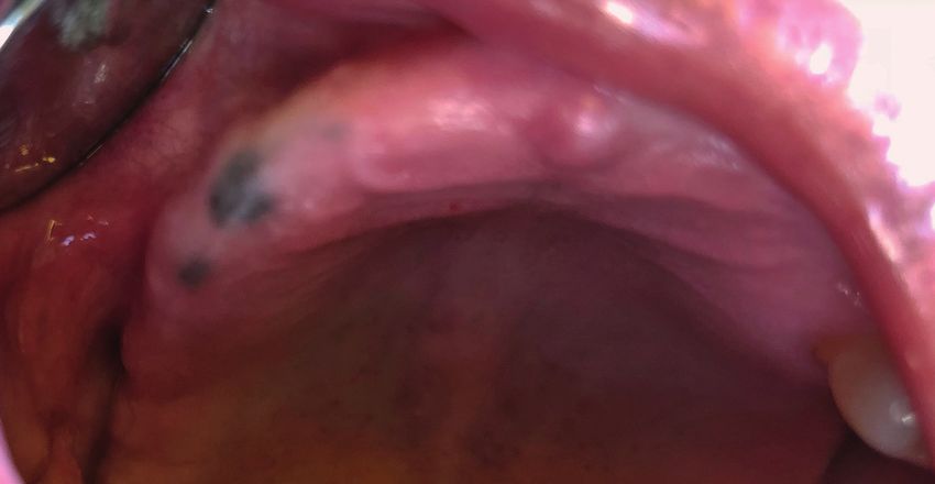

You can also read