Radiofrequency ablation, A new paradigm for the treatment of adenomyosis: Case series - Sohag Medical Journal
←
→
Page content transcription
If your browser does not render page correctly, please read the page content below
Sohag University Sohag Medical Journal Faculty of Medicine
ــــــــــــــــــــــــــــــــــــــــــــــــــــــــــــــــــــــــــــــــــــــــــــــــــــــــــــــــــــــــــــــــــــــــــــــــــــــــــــــــــــــــــــــــــــــــــــــــــــــــ
Radiofrequency ablation, A new paradigm for the
treatment of adenomyosis: Case series
Elsemary M.A, Sabri M.M, Abdelmonem A. M, Ibrahim M.,
Hisham A.Alghany Algahlan.
Obesterics and gynacology faculty of medicine sohag university
Abstract
Adenomyosis is a challenging clinical condition that is commonly being diagnosed in
women of reproductive age. To date, many aspects of the disease have not been fully
understood, making management increasingly difficult. Heavy menstrual bleeding and
dysmenorrhea are the typical symptoms of adenomyosis, occurring in approximately 60
and 25 percent of women, respectively. The diagnosis of adenomyosis is based mainly on
transvaginal ultrasonography and magnetic resonance imaging (MRI).A thickness of the
junctional zone of at least 12 mm is the most frequent MRI criterion in establishing the
presence of adenomyosis. Adenomyosis can appear as a diffuse or focal form. Although
hysterectomy is a definitive treatment option, minimally invasive treatment methods have
been developed as more women desire uterine preservation for future fertility or to avoid
major surgery. Several uterine-sparing treatment options are now available, including
medication, hysteroscopic resection or ablation, conservative surgical methods, high-
intensity focused ultrasound, uterine artery embolization and radiofrequency ablation
each with its own risks and benefits.
Keywords: Adenomyosis, Pelvic pain, Dysmenorrhea, Radiofrequency
Introduction:
Adenomyosis is a heterogeneous adenomyoma(more or less clear borders
gynecological condition commonly with mainly solid characteristics) or
encountered in our clinical practice. It is cystic adenomyosis (2).
caused by the benign invasion of ectopic Transvaginal ultrasonography (TVUS)
endometrial glands and stroma in the and magnetic resonance imaging (MRI)
myometrium. Patients with adenomyosis are the main radiologic tools for the
can have a wide range of clinical diagnosis of adenomyosis. MRI has a
presentations. The most common diagnostic accuracy of 85 %, with
presenting features of adenomyosis additional value in confirming the
include heavy menstrual bleeding, diagnosis and determining disease
secondary dysmenorrhoea and an characteristics and extent and other
enlarged uterus (which may produce uterine lesions (3).
pressure symptoms on the bladder or the Radiofrequency ablation (RFA) of
bowel). However, patients can also be tumors results from the heat that is
asymptomatic (1). generated from the medium frequency
Adenomyosis can be diffuse, where alternating current. The heat causes
islands of adenomyosis may be found coagulation necrosis in targeted tumors
throughout the myometrium, or it can be and obliteration of interstitial vessels. It
localized which is subdivided into was first used via a laparoscopic
14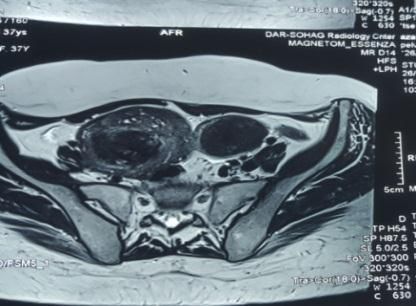
SOHAG MEDICAL JOURNAL Radiofrequency ablation, A new paradigm for the treatment
Vol. 24 No. 1 Jan 2020 Elsemary M.A
approach for treating uterine fibroids and MRI revealed ill-defined hypointense
subsequently with ultrasound guidance. lesion about 3.5x2.8x3 cm in continuity
Ultrasound-guided RFA can be with junctional zone and showing multi
performed as an outpatient procedure minor cystic changes and focal bulge
under sedation and many studies have within the endometrial cavity for follow
reported it as a safe and effective up. Bleeding was relieved completely
treatment for uterine fibroids (4). and patient resuming normal cycles,
Case (1): partial relieving of pain was noticed.
A 35 years old patient , nulligravida
married since 10 years , before and after
marriage the patient gave a history of
severe and worsening dysmenorrhea
with cramps and irregular menstrual
cycles (oligomenorrhea) which were
insufficiently relieved by NSAIDs, one
year after regular marital life she sought
medical advice and all infertility workup
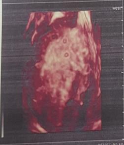
Pre-RFA 2D TVS Pre-RFA 3D TVS
was done and all were normal.
Two years after marriage the patient
starts to complaining of menorrhagia
(period 8 days with blood clots), After 5
years here husband marry again and now
He has two kids.
At the same time, she also sought
medical advice and D & C was done for
marked bleeding which was relieved for
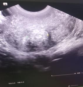
one year and then returned back for Post_RFA 3 M. Post_RFA 6 M.
which she sought medical advice many
times and many gynecologists told here
that she has a fibroid uterus (wrong
diagnosis) and advice here to do a
hysterectomy. Nine months ago 3D
transvaginal ultrasound was done It
reveals anterior uterine wall localized
adenomyosis about 5 x5 cm.
Routine investigations were done which
revealed that Hb was 7.5 mg/d, cross-
matched blood (3 units) was received,
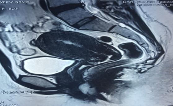
MRI pelvis showing ant. Wall localized
then ultrasound-guided Radiofrequency
adenomyosis
ablation was done in Sohage University
Hospital after agreement of department Case (2):
committee, Three and Six months follow Thirty-seven years old, unmarried
up with ultrasound and MRI respectively patient with a history of dysmenorrhea 6
revealed decrease in size of months after menarche, which was
adenomyoma to be 3x3.3 cm then increased gradually in strength in the
2.87x3 cm. Six months Follow up by
15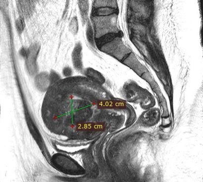
SOHAG MEDICAL JOURNAL             Radiofrequency ablation, A new paradigm for the treatment
Vol. 24 No. 1 Jan 2020                              Elsemary M.A

following years and was relieved
partially by analgesics.
Two years ago the patient starts
complaining of heavy menstrual
bleeding (12 days with blood clots)
besides the worsening dysmenorrhea.
Abdominal ultrasound was done which
revealed abnormally enlarged and
distorted contour of the uterus with
lateral subserous 5x5.5 cm leiomyoma             Post-RFA MRI
and a suspicious posterior uterine wall          Case(3):
ill-defined lesion which necessitate the         A 53 years old patient, Grandmultipara,
need for MRI pelvis which was done and           complaining of heavy menstrual
revealed that the lesions were localized         bleeding for 2 years. pelvic and
adenomyosis about 53x58 mm with                  ultrasound examination revealed a uterus
multiple cystic changes of adjacent              ± 10 wks.MRI was done and revealed
myometrial tissue and a subserous                enlarged uterus with ill-defined diffuse
leiomyoma 52x55                                  thickening of the inner myometrium with
mm.                                              few foci and thickened junctional zone
Two months ago ultrasound-guided                 2.5 cm.
Radiofrequency ablation was done for             Six months ago ultrasound-guided
both lesions.                                    Radiofrequency ablation was done for
Follow up MRI was done, It revealed              fundus, anterior and posterior uterine
that the size and extension were                 walls(5      deployments      of      the
decreased, looks more heterogenous               radiofrequency needle).
signal with cystic changes impressive of         Vaginal bleeding occurs once 7 days
degeneration as well as reduced the              after the procedures and there is no
associated microcystic area of localized         vaginal bleeding till now suggesting that
adenomyosis which was decreased to               diffuse adenomyosis was the cause for
2.1x3.7 cm and the size of leiomyoma             continuing menses till that age of 53
was reduced to 3.8x4.3 cm.bleeding was           years. Follow up MRI was done 6
completely resolved and the patient              months after the procedure, It reveals
resuming her normal cycles. Pain is              diffuse    heterogeneous     hypointense
relieved partially.                              thickening of the junctional zone with
 Follow up is needed for that case to            maximum transverse thickness 14 mm.
know the effect of the procedure on pain.

Pre-RFA MRI
                                                 Pre-RFA MRI

                                            16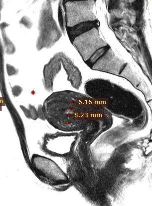
Sohag University Sohag Medical Journal Faculty of Medicine
ــــــــــــــــــــــــــــــــــــــــــــــــــــــــــــــــــــــــــــــــــــــــــــــــــــــــــــــــــــــــــــــــــــــــــــــــــــــــــــــــــــــــــــــــــــــــــــــــــــــــ
Post-RFA MRI
Conclusion:
Ultrasound-guided RFA might be a safe 2. Wang JH, Wu RJ, Xu KH, Lin J.
and effective minimally invasive Single large cystic adenomyoma of
alternative in the treatment of the uterus after corneal pregnancy and
symptomatic adenomyosis in which curettage. Fertil Steril 2007;88:965–7.
there is reduced uterine adenomyosis–
related volume, and significant relief of 3. Dueholm M, Lundorf E, Hansen ES,
symptoms. However large studies and Sorensen JS, Ledertoug S, Olesen F.
longer follow-ups with large data to Magnetic resonance imaging and
confirm the exact recurrence rate, long transvaginal ultrasonography for the
term complications, and effect on diagnosis of adenomyosis. Fertil
pregnancy are needed. Steril. 2001;76:588–594.
References: 4. Bongers M, Brlmann H, Gupta J,
1. Brucker SY, Huebner M, Wallwiener Garza-LealJG,Toub
M, Stewart EA, Ebersole S, D. Transcervical,intrauterine
Schoenfisch B, et al. Clinical ultrasound-guided radiofrequency
characteristics-indicating ablation of uterine fibroids with the
adenomyosis coexisting with VizAblate® System: three- and six-
leiomyomas: a retrospective, month endpoint results from the
questionnaire-based study. Fertil FAST-EU study. Gynecol
Steril 2014; 101: 237–41. Surg 2015; 12: 61–70.
17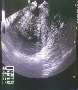
You can also read