THE BENEFITS OF IDENTIFYING PARACENTRAL ACUTE MIDDLE MACULOPATHY
←
→
Page content transcription
If your browser does not render page correctly, please read the page content below
s IMAGING
THE BENEFITS OF IDENTIFYING
PARACENTRAL ACUTE MIDDLE
MACULOPATHY
This important OCT sign can be the sole indicator of a significant retinal vascular event with systemic implications.
BY MANAB J. BARMAN, MD; RAGHUDEV BHATTACHARJEE, MD; SAURABH DESHMUKH, MD; AND AWANEESH UPADHYAY, MD
P
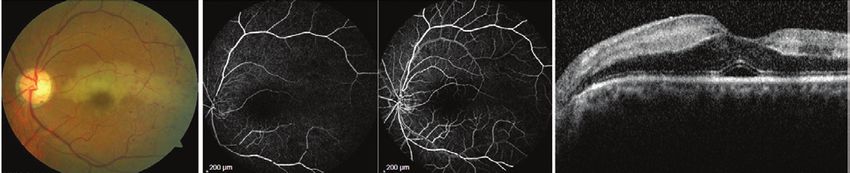
aracentral acute middle maculopathy (PAMM), first CASE NO. 1
described by Sarraf et al in 2013, typically manifests as a A 38-year-old man presented with complaints of blurred
distinct paracentral scotoma with or without diminution vision in the left eye for 7 days. His BCVA was 20/20+. Amsler
of vision.1 Fundus examination shows a dark gray paracen- grid testing identified a small paracentral scotoma inferior to
tral lesion that points toward the center of the fovea.2 The the fixation point, confirmed with visual field testing. Fundus
condition can present in conjunction with a number of retinal examination revealed a yellowish-white, well-demarcated
vascular diseases.3,4 lesion superior to the fovea (Figure 1A). Multicolor imag-
Although PAMM was originally described as a variant of ing showed a corresponding lesion in green (Figure 1B).
acute macular neuropathy (AMN), the two are now regarded Fluorescein angiography (FA) did not show any filling defect.
as distinct entities.5 The retinal ischemic cascade of PAMM in OCT revealed a hyperreflective band involving the inner
its mildest form (known as perivenular PAMM) involves the plexiform layer (IPL) and OPL, indicating PAMM, possibly
venular end of the deep capillary plexus (DCP). With increas- secondary to cilioretinal artery insufficiency. The EZ was
ing severity it may progress to diffusely involve the inner intact (Figure 1C). Systemic workup showed dyslipidemia.
nuclear layer (INL) or even to infarct the inner retina. The lesion persisted at the 6-month follow-up.
AMN, by contrast, displays hyperreflectivity of the outer
A B C
plexiform layer (OPL) and outer nuclear layer (ONL) and
may be associated with disruption of the ellipsoid zone (EZ).6
OCT angiography (OCTA) shows reduced flow in the
intermediate retinal capillary plexus (ICP) and DCP in
PAMM, whereas AMN is associated with reduced flow in the
Figure 1. The color fundus photo shows a grayish-white, well-demarcated lesion superior
DCP only.7,8 to the fovea (A). Multicolor imaging depicts the lesion in green (B). SD-OCT shows a
New imaging modalities such as OCTA have added sub- hyperreflective band in the IPL and OPL (C).
stantial knowledge to the pathogenesis of PAMM, but the
condition’s clinical course and treatment outcome are still CASE NO. 2
under investigation. In a single-center retrospective observa- A 52-year-old man presented with blurred vision in the
tional study, we analyzed seven eyes of seven patients with right eye for 15 days despite a BCVA of 20/20. He had a med-
PAMM of varied etiology. The study was conducted follow- ical history of hypercholesterolemia. Fundus examination
ing institutional review board guidelines and adhering to the showed a well-defined, grayish, wedge-shaped lesion superior
tenets of the Declaration of Helsinki. to the fovea (Figure 2A). Multicolor imaging depicted the
52 RETINA TODAY | APRIL 2021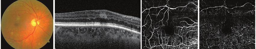
IMAGING
s
A B C
Figure 2. The color fundus photo shows PAMM superior to the fovea (A). Multicolor imaging depicts PAMM in green (B). Thinning of INL is noted on SD-OCT (C).
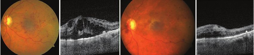
lesion in green (Figure 2B). FA was inconclusive, but OCT at significant macular edema and a grayish lesion superior to
the level of the lesion revealed thinning of the INL, possibly the center of the fovea (Figure 4A). OCT revealed a hyper-
secondary to branch retinal artery insufficiency (Figure 2C). reflective band at the level of the OPL (Figure 4B). OCTA
showed an area of capillary abnormality in the DCP superior
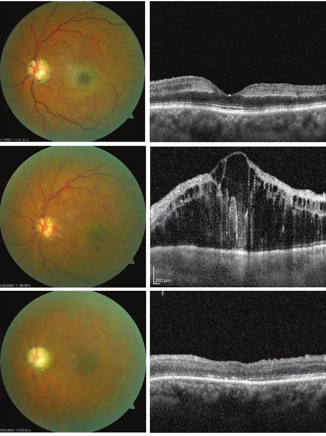
CASE NO. 3 to the foveal center (Figure 4C and 4D). After 3 months, the
A 46-year-old hypertensive man presented with blurred retinal condition was stable with the persistence of PAMM.
vision in the right eye for 15 days. BCVA was 20/40 OD.
Fundus examination revealed evidence of a nonisch- CASE NO. 5
emic central retinal vein occlusion (CRVO) and a well- A 62-year-old man presented with blurred vision in the
defined grayish‑white lesion inferotemporal to the fovea left eye for 2 months. He had hypertension and diabetes and
(Figure 3A). FA showed inferior extension of the foveal was being treated for both. BCVA was 20/80 OS. Diagnosis of
avascular zone. OCT showed cystic changes with a hyper- branch retinal vein occlusion (BRVO) with CME was made
reflective band at the level of the IPL (Figure 3B). OCTA based on the clinical picture and OCT findings (Figure 5A
revealed capillary abnormalities in both the DCP and SCP and 5B). FA was deferred. The patient received three intra-
inferior to the fovea (Figures 3C and 3D). The patient vitreal injections of ranibizumab at monthly intervals. After
received three intravitreal injections of ranibizumab the first injection, PAMM was detected superior to the fovea
(Lucentis, Genentech) at monthly intervals. After 6 months, and confirmed with OCT (Figure 5C). At 4-month follow-up,
fundus examination revealed the persistence of PAMM PAMM persisted with CME (Figure 5D).
with cystoid macular edema (CME).
A B C D
A B C D
Figure 5. Fundus photo and OCT imaging are suggestive of BRVO with macular edema (A and
Figure 3. The color fundus photo suggests nonischemic CRVO with PAMM inferotemporal to B). PAMM was detected superior to the fovea 1 month after anti-VEGF injection (C and D).
the fovea (A). SD-OCT shows cystic changes in the center with a hyperreflective band at the
level of IPL (B). OCTA shows capillary abnormalities in both the DCP and SCP (C and D). CASE NO. 6
A 57-year-old man with hypertension presented with
CASE NO. 4 blurred vision in the left eye for 2 weeks. BCVA was 20/40 OS.
A 60-year-old woman with diabetes presented with mild Fundus examination revealed a grayish-white lesion at the
blurred vision in the right eye for 3 months. Her BCVA was distribution of the superior branch retinal artery with evidence
20/20+ OD. Fundus examination revealed mild nonpro- of nonischemic CRVO (Figure 6A). FA showed delayed filling
liferative diabetic retinopathy (NPDR) without clinically of the branch retinal artery (Figure 6B and 6C). OCT revealed
A B C D
Figure 4. The fundus photo suggests diabetic retinopathy with PAMM superior to the center of the fovea (A). SD-OCT shows a hyperreflective band at the OPL (B). OCTA shows an area of
capillary abnormality in the DCP superior to the foveal center (C and D).
APRIL 2021 | RETINA TODAY 53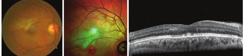
s   IMAGING

             A                    B                   C                  D                                and other clinical findings, for example dyslipidemia in
                                                                                                          Case No. 1 and 2.16 Microcholesterol embolus may lead to
                                                                                                          the occlusion of DCP in such cases.
                                                                                                             CME with PAMM can also occur secondary to CRVO, and
                                                                                                          research shows that intravitreal injections do not help to
            Figure 6. Fundus photo shows PAMM (A). Delayed filling of the branch retinal artery is seen
            on FA (B and C). SD-OCT shows a hyperreflective band at the IPL and OPL (D).                  resolve PAMM in such cases. 4 Not surprisingly, in Case No. 3,
                                                                                                          both CME and PAMM persisted after intravitreal injections
            a hyperreflective band in the inner and middle retinal layers                                 of an anti-VEGF agent. PAMM secondary to diabetic retinop-
            (Figure 6D). A diagnosis of nonischemic CRVO combined with                                    athy, as seen in Case No. 4, can present without CME.3
            branch retinal artery occlusion (BRAO)-associated PAMM was                                       PAMM can develop secondary to BRVO during follow-up
            made. Color Doppler imaging of the carotid and ophthalmic                                     after initiation of anti-VEGF treatment, as in Case No. 5. Pichi
            arteries did not reveal any underlying pathology.                                             et al described a large series of eyes with vascular occlu-
                                                                                                          sion, in which PAMM was detected at presentation.17 This
             CASE NO. 7                                                                                   research team also looked at the association of PAMM with
               A 64-year-old man with hypertension presented with                                         cilioretinal artery occlusion.17 In isolated retinal artery occlu-
            blurred vision in the left eye for 1 day and BCVA of 20/60 OS.                                sion, initial hyperreflectivity of the inner retinal layers is often
            Fundus examination showed advanced cupping in each eye                                        seen on OCT. In Case No. 6 of our series, PAMM developed
            and a well-defined parafoveal, intraretinal, grayish lesion                                   secondary to BRAO with CRVO, and hyperreflectivity was
            with characteristic OCT features suggestive of PAMM                                           noted in both the inner and middle retinal layers.
            (Figure 7A and 7B). Visual field analysis confirmed glauco-                                      Finally, PAMM also may be associated with glaucoma, as
            matous damage, and the patient was started on medication                                       A                               B
            for primary open-angle glaucoma. The patient presented
            3 days later with deterioration of vision to hand movement
            OS (Figure 7C and 7D). A diagnosis of CRVO with CME and
            possible central retinal artery hypoperfusion was made based
            on the findings.
               The patient was started on monthly injections of ranibi-
            zumab. After 3 months, his VA improved to 20/200, with
            resolution of the macular edema (Figure 7E and 7F).
                                                                                                           C                                        D
             DISCUSSION
               PAMM is considered a manifestation of focal ischemia of
            the deep retinal circulation that may herald the presence
            of a secondary underlying condition. Multicolor imaging
            can help to detect PAMM, as it creates three simultaneous
            reflectance images that demonstrate details at different lay-
            ers of the retina.9,10 Blue, green, and red reflectance show the
            inner, middle, and outer retina, respectively. PAMM usually
            presents with the lesion in green.11
                                                                                                           E                                        F
               OCT is invaluable in confirming a diagnosis of PAMM. On
            OCT, the condition initially manifests as a hyperreflective
            band, followed by thinning of the middle retinal layers.11 En
            face OCT may demonstrate a remarkable perivenular pat-
            tern of PAMM in eyes with retinal vein occlusion even in the
            absence of significant funduscopic findings.12 Bakhoum et al
            described characteristic OCT findings of PAMM suggestive
            of an ischemic cascade, indicating more vulnerability of the
            middle retina at the level of the DCP.13                                                      Figure 7. The fundus photograph of this glaucomatous eye shows a perifoveal intraretinal
               FA is a poor imaging modality to illustrate PAMM,                                          greyish lesion (A), and the corresponding OCT shows hyperreflective band-like lesions in
            whereas OCTA at the level of the DCP detects gross capillary                                  the middle retinal layers of the macula, suggestive of PAMM (B). Fundus photography of the
            loss.6,14 OCTA features are described as arteriolar, globular,                                same eye 3 days later shows dilated tortuous vessels with diffuse intraretinal hemorrhages
            fernlike, and combination pattern.15                                                          (C), and the corresponding OCT shows macular edema (D). After three anti-VEGF injections,
               Our case series highlights associations between PAMM                                       the fundus photograph (E) and OCT (F) show resolution of CRVO and macular edema.

    54 RETINA TODAY | APRIL 2021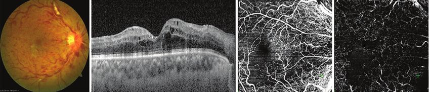
IMAGING

                                                                                                                                      s
seen in our Case No. 7, and in such cases it may be a pre-
monitory sign of CRVO.18

 FINAL THOUGHTS
  PAMM is a sign of deep retinal ischemia, the duration and
severity of which may impact the development of PAMM.
Because of its association with other ocular conditions, the
presence of PAMM without obvious ocular pathology war-
rants a thorough systemic evaluation. n
1. Sarraf D, Rahimy E, Fawzi AA, et al. Paracentral acute middle maculopathy: a new variant of acute macular neuroretinopathy
associated with retinal capillary ischemia. JAMA Ophthalmol. 2013;131(10):1275-1287.
2. Baumüller S, Holz FG. Early spectral-domain optical coherence tomography findings in acute macular neuroretinopathy. Retina.
2012;32(2):409-410.
3. Yu S, Wang F, Pang CE, et al. Multimodal imaging findings in retinal deep capillary ischemia. Retina. 2014;34(4):636-646.
4. Rahimy E, Sarraf D, Dollin ML, et al. Paracentral acute middle maculopathy in nonischemic central retinal vein occlusion. Am J
Ophthalmol. 2014;158(2):372-380.e1.
5. Rahimy E, Kuehlewein L, Sadda SR, Sarraf D. Paracentral acute middle maculopathy: what we knew then and what we know now.
Retina. 2015;35:1921-1930.
6. Scharf J, Freund KB, Sadda S, Sarraf D. Paracentral acute middle maculopathy and the organization of the retinal capillary plexuses
[published online ahead of print 9 August 2020]. Prog Retin Eye Res. 2020:100884.
7. Chu S, Nesper PL, Soetikno BT, et al. Projection-resolved OCT angiography of microvascular changes in paracentral acute middle
maculopathy and acute macular neuroretinopathy. Invest Ophthalmol Vis Sci. 2018;59:2913-2922.
8. Chen YC, Chen SN. Microvascular change in acute macular neuroretinopathy by using optical coherence tomography angiography.
Taiwan J Ophthalmol. 2019;9:118-121.
9. Gramatikov BI. Modern technologies for retinal scanning and imaging: an introduction for the biomedical engineer. Biomed Eng
Online. 2014;13:52.
10. LaRocca F, Nankivil D, Farsiu S, Izatt JA. True color scanning laser ophthalmoscopy and optical coherence tomography handheld
probe. Biomed Opt Expr. 2014;5(9):3204-3216.
11. Shah D, Saurabh K, Roy R. Multimodal imaging in paracentral acute middle maculopathy. Indian J Ophthalmol. 2018;66(8):1186-1188.
12. Ghasemi F K, Phasukkijwatana N, Freund KB, et al. En face optical coherence tomography analysis to assess the spectrum of
perivenular ischemia and paracentral acute middle maculopathy in retinal vein occlusion. Am J Ophthalmol. 2017;177:131-138.
13. Bakhoum MF, Freund KB, Dolz-Marco R, et al. Paracentral acute middle maculopathy and the ischemic cascade associated with
retinal vascular occlusion. Am J Ophthalmol. 2018;195:143-153.
14. Monson BK, Greenberg PB, Greenberg E, et al. High‐speed, ultra‐high‐resolution optical coherence tomography of acute macular
neuroretinopathy. Br J Ophthalmol. 2007;91(1):119-120.
15. Shah A, Rishi P, Chendilnathan C, Kumari S. OCT angiography features of paracentral acute middle maculopathy. Indian J
Ophthalmol. 2019;67(3):417-419.
16. Chen X, Rahimy E, Sergott RC, et al. Spectrum of retinal vascular diseases associated with paracentral acute middle maculopathy.
Am J Ophthalmol. 2015;160(1):26-34.e1.
17. Pichi F, Fragiotta S, Freund KB, et al. Cilioretinal artery hypoperfusion and its association with paracentral acute middle maculopa-
thy. Br J Ophthalmol. 2019;103(8):1137-1145.
18. Aribas YK, Aktas Z, Bayrakceken K, et al. Paracentral acute middle maculopathy in primary congenital glaucoma. Retin Cases Brief
Rep. 2020;14(2):163-165.

MANAB J. BARMAN, MD
n Senior Consultant and Department Head, Vitreo-Retina Service,
  Sri Sankaradeva Nethralaya Hospital, India
n barmanmj@gmail.com

n Financial disclosure: None

RAGHUDEV BHATTACHARJEE, MD
n Fellow, Vitreo-Retina Service, Sri Sankaradeva Nethralaya Hospital, India
n Financial disclosure: None

SAURABH DESHMUKH, MD
n Fellow, Vitreo-Retina Service, Sri Sankaradeva Nethralaya Hospital, India
n Financial disclosure: None

AWANEESH UPADHYAY, MD
n Consultant Vitreoretinal Surgeon, Eye-Q Hospital, India
n Financial disclosure: None

                                                                                                                                            APRIL 2021 | RETINA TODAY 55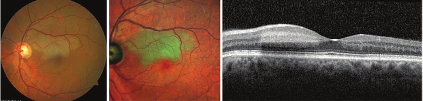


You can also read