Case Report A case of myxoid liposarcoma of the breast
←
→
Page content transcription
If your browser does not render page correctly, please read the page content below
Int J Clin Exp Pathol 2013;6(7):1432-1436
www.ijcep.com /ISSN:1936-2625/IJCEP1305028

Case Report
A case of myxoid liposarcoma of the breast
Tsuyoshi Saito1, Misa Ryu2, Yuki Fukumura1, Miki Asahina1, Atsushi Arakawa1, Katsuya Nakai2, Hiroyoshi
Miura2, Mitsue Saito2, Takashi Yao1
1
 Department of Human Pathology, Juntendo University School of Medicine, Tokyo, Japan; 2Department of Breast
Oncology, Juntendo University School of Medicine, Tokyo, Japan
Received May 20, 2013; Accepted June 4, 2013; Epub June 15, 2013; Published July 1, 2013

Abstract: A 70-year-old woman visited a local hospital complaining of a nodulein the right breast, present since 1
month. She was referred to our hospital for further evaluation. Following mammotome (MMT) biopsy, the nodule
was diagnosed as myxoid/round cell liposarcoma. She underwent total mastectomy of the right breast. Histological
analysis indicated that the tumor was almost entirely composed of proliferating small round mesenchymal cells in
amyxoid matrix background with capillary-like vessels with partial necrosis (
Myxoidliposarcoma of breast
Figure 1. The breast tumor as observed on ultrasonography and magnetic resonance imaging. A, B: Ultrasonogra-
phy revealed a relatively well-demarcated lobulated mass of 51 × 40mm with cystic change. C, D: MRI indicating
as lightly high intensity lesion on a T1-weighted image and a high intensity lesion on a T2-weighted image. E: High-
intensity tumor on diffusion weighted imaging (DWI), reflecting the presence of a lesion with cystic change.
patient underwent total mastectomy of the
right breast.
Because the tumor was pathologically diag-
nosed as amyxoid liposarcoma, and the surgi-
cal margin was histologically considered posi-
tive, adjuvant treatment involvinglocal radiation
therapy (60Gy) was administered. During the
radiation therapy, the patient complained of
dizziness. MRI scan indicated the presence of a
tumor at the left cerebellopontine angle that
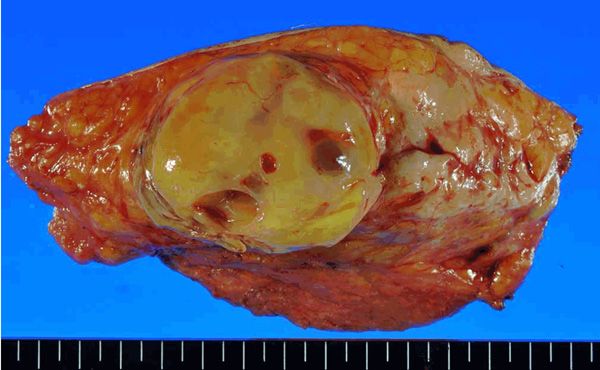
Figure 2. Gross section of the tumor shows a well- had weak low intensity on a T1-weighted image
demarcated yellowish tumor with focal cystic change. and weak high intensity on T2-weighted image.
In view of the clinical history of myxoid liposar-
coma, a metastatic tumor or meningioma were
observed on magnetic resonance imaging also suspected. The tumor was successfully
(MRI) (Figure 1C, 1D). This lesion also showed resected and histologically diagnosed as a
highintensity on diffusion-weighted imaging meningioma with no evidence of metastatic
(DWI), suggesting the presence of cystic change myxoid liposarcoma.
(Figure 1E).
The patientdid not experience any symptoms of
Following MMT biopsy, the tumor was diag- local recurrence and metastases 1 year and 8
nosed as a myxoid/round cell liposarcoma. The months after surgery.
1433 Int J Clin Exp Pathol 2013;6(7):1432-1436Myxoidliposarcoma of breast Figure 3. Histologic features of the breast tumor. A, B: The tumor was histologically almost entirely composed of proliferating small round mesenchymal cells in amyxoid matrix background with capillary-like vessels. C: Frequently seen mitosis (arrows). D: Lipoblasts scattered throughout the tumor. E: p53-positive cells seen only focally (
Myxoidliposarcoma of breast

Table 1. Primer sequences                                                   specimen was con-
                                                                            sidered        positive.
                                                PCR product size            Postoperative radio-
 TLS-CHOP type1, 2-F GACAGCAGAACCAGTACAACAGCAG TLS-CHOP type1: 379bp        therapy was per-
 TLS-CHOP type1, 2-R GCTTTCAGGTGTGGTGATGTATGAAG TLS-CHOP type1: 103bp       formed in this case to
 TLS-CHOP type3-F    gaagtgaccgtggtggcttcaa     208bp                       prevent local recur-
 TLS-CHOP type3-R    ggcaagctggtctgatgcct                                   rence. Adverse prog-
 TLS-CHOP type4-F    cctcagggctatggacagcagaa    208bp                       nostic factors in
 TLS-CHOP type4-R    ggctggaacaagctccatgtagc                                localized soft tissue
 TLS-CHOP type5-F    gcagtcctcctaccctggct       106+αbp                     myxoid liposarcoma
                                                                            include a high-histo-
 TLS-CHOP type5-R    gctgctttcaggtgtggtgat
                                                                            logical grade >10%
 TLS-CHOP type8-F    cccctaaaccagatggccca       190bp
                                                                            round cell compo-
 TLS-CHOP type8-R    ggcaagctggtctgatgcct
                                                                            nent, necrosis >5%,
 TLS-CHOP type9-F    acggacacttcaggctatgg       159bp                       and overexpression
 TLS-CHOP type9-R    ctggaatacagccacatctgtt                                 of p53 >10% of posi-
 EWS-CHOP-F          tggatcctacagccaagctc       EWS-CHOP type1: 111bp       tive cells [17]. In addi-
 EWS-CHOP-R          gctgctttcaggtgtggtgat      EWS-CHOP type1: 363bp       tion, p53 overexpres-
 GAPDH-F             GAAGGTGAAGGTCGGAGTC        226bp                       sion is relatively rare
 GAPDH-R             GAAGATGGTGATGGGATTTC                                   (5–30%) in myxoid
                                                                            liposarcoma [17-19]
                                                                            and tends to be seen
CHOP was detected in this case, although the      in conjunction with TLS-CHOP type 2 fusion
integrity of the mRNA as assessed by the          [17]. In this case, extensive areas with round
expression of GAPDH was confirmed.                cell component were observed; although p53
                                                  overexpression was not seen and only focal
Discussion                                        (
Myxoidliposarcoma of breast
8421, Japan. Tel: +813-3813-3111; E-mail: [12] Tsuji S, Hisaoka M, Morimitsu Y, Hashimoto H,
tysaitou@juntendo.ac.jp Shimajiri S, Komiya S, Ushijima M, Nakamura
T. Detection of SYT-SSX fusion transcripts in
References synovial sarcoma by reverse transcription-poly-
merase chain reaction using archival paraffin-
[1] Adem C, Reynolds C, Ingle JN, Nascimento AG. embedded tissues. Am J Pathol 1998; 153:
Primary breast sarcoma: Clinicopathologic se- 1807-1812.
ries from the Mayo Clinic and review of the lit- [13] Ferrari A, Besana-Ciani I, Rovera F, Siesto G,
erature. Br J Cancer 2004; 91: 237-241. Dionigi G, Boni L, Dionigi R. An unusual case of
[2] McGregor JK. Liposarcoma of the breast. Case breast liposarcoma with liver metastases: The
report and review of the literature. Canada role of radical surgery. Breast J 2007; 13: 324-
Med Ass J 1960; 82: 781-783. 5.
[3] Pollard SG, Marks PV, Temple LN, Thompson [14] Moreau LC, Turcotte R, Ferquson P, Wunder J,
HH. Breast sarcoma. A clinicopathologic re- Clarkson P, Masri B, Isler M, Dion N, Werier J,
view of 25 cases. Cancer 1990; 66: 941-944. Ghert M, Deheshi B, Canadian Orthopaedic
[4] Neumann E. BeitragezurCasuistik der Brust- Oncology Society (CANOOS). Myxoid/round cell
drusengeschwutste. Virchows Arch Path Anat liposarcoma (MRCLS) revisited: an analysis of
1862; 24: 316-328. 418 primarily managed cases. Ann Surg Oncol
[5] Enzinger FM, Weiss SW. Liposarcoma. In: Enz- 2012; 19: 1081-1088.
inger FM, Weiss SW, eds. Soft tissue tumors. [15] Eilber FC, Eilber FR, Eckardt J, Rosen G, Riedel
3rd ed. St Louis: Mosby, 1955; pp: 431-466. E, Maki RG, Brennan MF, Singer S. The impact
[6] Aman P, Ron D, Mandahl N, Fioretos T, Heim S, of chemotherapy on the survival of patients
Arheden K, Willén H, Rydholm A, Mitelman F. with high-grade primary extremity liposarco-
Rearrangement of the transcription factor ma. Ann Surg 2004; 240: 686-697.
gene CHOP in myxoid liposarcoma with [16] Gronchi A, Bui BN, Bonvalot S, Pilotti S, Ferrari
t(12;16)(q13;p11). Genes Chromosomes Can- S, Hohenberger P, Hohl RJ, Demetri GD, Le
cer 1992; 5: 278-285. Cesne A, Lardelli P, Perez I, Nieto A, Tercero JC,
[7] Knight JC, Renwick PJ, Dal Cin P, Van den Ber- Alfaro V, Tamborini E, Blay JY. Phase II clinical
ghe H, Fletcher CDM. Translocation t(12;16) trial of neoadjuvant trabectedin in patients
(q13;p11) in myxoid liposarcoma and round with advanced localized myxoid liposarcoma.
cell liposarcoma: molecular and cytogenetic Ann Oncol 2012; 23: 771-776.
analysis. Cancer Res 1995; 55: 24-27. [17] Antonescu CR, Tschernyavsky SJ, Decuseara
[8] Kuroda M, Ishida T, Horiuchi H, Kida N, Uozaki R, Leung DH, Woodruff JM, Brennan MF, Bridge
H, Takeuchi H, Tsuji K, Imamura T, Mori S, Ma- JA, Neff JR, Goldblum JR, Ladanyi M. Prognos-
chinami R, et al. Chimeric TLS/FUS-CHOP gene tic impact of TP53 status, TLS-CHOP fusion
expression and the heterogeneity of its junc- transcript structure, and histological grade in
tion in human myxoid and round cell liposar- myxoid liposarcoma: a molecular and clinico-
coma. Am J Pathol 1995; 147: 1221-1227. pathologic study of 82 cases. Clin Cancer Res
[9] Panagopoulos I, Hoglund M, Mertens F, Man- 2001; 7: 3977-3987.
dahl N, Mitelman F, Aman P. Fusion of the EWS [18] Pilotti S, Lavarino C, Mezzelani A, Della Torre C,
and CHOP genes in myxoid liposarcoma. Onco- Minoletti F, Sozzi G, Azzarelli A, Rilke F, Pierotti
gene 1996; 12: 489-494. MA. Limitedrole of TP53 and TP53-related
[10] Panagopoulos I, Mertens F, Isaksson M, Man- genes in myxoid liposarcoma. Tumori 1998;
dahl N. A novel FUS/CHOP chimera in myxoid 84: 571-577.
liposarcoma. Biochem Biophys Res Commun [19] Dei Tos AP, Piccinin S, Doglioni C, Vukosavljevic
2000; 279: 838-845. T, Mentzel T, Boiocchi M, Fletcher CDM. Mo-
[11] Hisaoka M, Tsuji S, Morimitsu Y, Hashimoto H, lecular aberrations of the G1-S checkpoint in
Shimajiri S, Komiya S, Ushijima M. Detection of myxoid and round cell liposarcoma. Am J
TLS/FUS-CHOP fusion transcripts in myxoid Pathol 1997; 151: 1531-1539.
and round cell liposarcoma by nested reverse
transcription-polymerase chain reaction using
archival paraffin-embedded tissues. Diagn Mol
Pathol 1998; 7: 96-101.
1436 Int J Clin Exp Pathol 2013;6(7):1432-1436You can also read