CONVEGNO REGIONALE AIOM EMILIA-ROMAGNA I NUMERI DEL CANCRO IN EMILIA ROMAGNA
←
→
Page content transcription
If your browser does not render page correctly, please read the page content below
CONVEGNO REGIONALE AIOM EMILIA-ROMAGNA
 I NUMERI DEL CANCRO IN EMILIA ROMAGNA:
       AMBIENTE, STILI DI VITA, SCREENING
  FOCUS SU TUMORI DEL POLMONE E COLON-RETTO

III SESSIONE – CANCRO DEL POLMONE: EPIDEMIOLOGIA E
                  NUOVI TRATTAMENTI
    Epidemiologia e profili molecolari: realtà in
                  evoluzione ?

                     Giulio Rossi
     Anatomia Patologica Ravenna, AUSL Romagna

     MODENA, Centro Servizi Università Policlinico
               23 NOVEMBRE 2018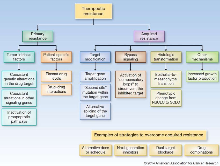
Dati Registro Tumori Modena- 2004
ADC
55-60%

                                             ° Classic
                                            Squamo Ca
                                              20-25%      SCLC
                                                         10-15%
          NSCLC nos        Non-smoker                               Other
            LCC            squamo Ca                              histotypes
             5%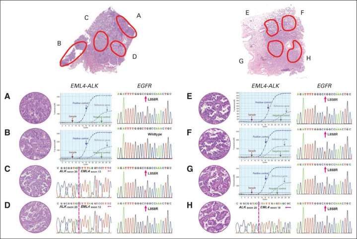
Clinico-radiologic presentation of lung
           cancer according to histotype
              Colby TV, Koss MN, Travis WD. AFIP, 1995

                                  SQC    ADC             SCLC LCC

•   Peripheral location           29% 70%                26% 65%
•   Central location              71% 5%                 > 70% 35%
•   Hilar/perihilar mass          40% 15%                80% 32%
•   Cavitation                    5-10% 1%               -     4%
•   5 yrs OS                      15.4% 16.6%            4.6% 11%
•   Distant mets                  50% 25%
•   Intrathoracic spread          25%  50% 15%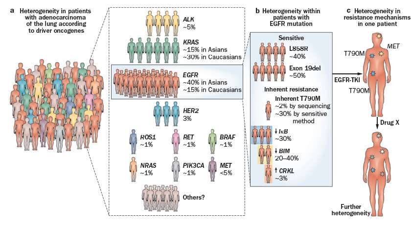
Adenocarcinoma histotype influences
       sampling procedures

Peripheral lesions & high frequency of
      lymph node involvement
1. Bronchoscopy over-rides radiology
  – More TBNA on lymph nodes or primary site
  – Cytology >>> Histology
2. Radiology over-rides bronchoscopy
  – More transthoracic biopsies
  – Histology >>> Cytology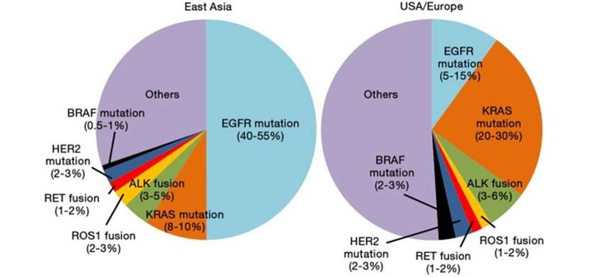
Type of material & analytical method
IHC FISH DNA RNA
(i.e.EGFR) (i.e. ALK) (i.e. EGFR/KRAS)
Cytology ++ ++ +++ +++
Cell block Stained slides do
no compromise
DNA quality
Biopsy +++ ++ ++ +
Surgery +++ +++ +++ +++
Lung Cancer alterations
                after EGFR & KRAS
                   -Small molecule     -Small molecule
 No drugs            (crizotinib)        (crizotinib)
   KRAS                  ALK                ROS1
- mutations         - FISH (IHC ?)          - FISH       2019
                                                          BRAF
 70’-80’                2007                 2012
                                                          HER2
            2004                     2011                PI3KCA
                                                           RET
          EGFR                      c-MET                C-MET
       - mutations         - Mutations / FISH / IHC       NRG1

   -Small molecules            - mAb (metmab)            PD-L1
                                                          TMB
  (gefitinib & erlotinib       -Small molecule
                                  (ARQ197)

to be continued ………………………………..

Evolution and importance of
            biomarkers in NSCLC
2013       Targets today                Targets in the future
           EGFR      ALK    ROS1
                                               KRAS                       BRAF HER2 RET          DDR2 MET        FGR1 P13K
                                               and others                                        and others
2008       Adenocarcinoma                                Large-cell carcinoma               Squamous cell carcinoma

2000       Non-small-cell lung cancer                                                       Small-cell lung cancer

1990       Lung cancer

 Adenocarcinoma                                             Large-cell carcinoma   Squamous cell carcinoma without oncogenic alteration
 Adenocarcinoma and treatable oncogenic alterations     Small-cell lung cancer     Squamous cell carcinoma with oncogenic alteration
 with approved drugs (EGFR mutation and ALK translocation)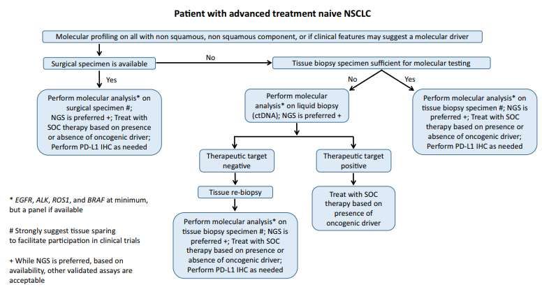
Geographical variability

                     Konho TLCR 2015 4:156-164
EGFR/KRAS: an ethnicity problem
Caucasian Asian
unselected people
EGFR mut 10% 30-60%
KRAS 25-35% 5%
Ciardiello F & Tortora G. NEJM 2008;358:1160-74Major NSCLC driver genes and
clinico-pathologic features
Driver gene EGFR KRAS ALK
• Gender F>M M>F M=F
• Age (years) >60 >60Awake VATS biopsy
1) Avoid side-effects of intubated general anesthesia
with single-lung mechanical ventilation.
2) Maintain more physiologic muscular, neurological,
and cardiopulmonary status.
Minimize the overall surgical
trauma assuring maximized
diagnostic accuracySurgical Approach Lung biopsy is performed by a single-incision VATS approach, under spontaneous ventilation, through local anesthesia with or without mild sedation.
Tipo
                                                              4
    Tipo
      4
               Tipo                             Tipo
                 3        Tipo                    1
                            2
                                  Tipo
                                    1                            Tipo
                                                                   5

   Biomarcatori predittivi in RER
Tipo 1: BiolMol estrattiva e BioLiq in U.O. Anatomia Patologica
Tipo 2: BiolMol estrattiva e BioLiq fuori dall’U.O. Anatomia Patologica
Tipo 3: BiolMol estrattiva all’interno dell’U.O. Anatomia Patologica; BioLiq/EGFR
in Oncologia
Tipo 4: BiolMol estrattiva in U.O. Anatomia Patologica; senza BioLiq
Tipo 5: BiolMol estrattiva e BioLiq fuori dall’U.O. Anatomia Patologica (fuori
Azienda)
1
2 Analisi polimorfismi
DPYD
Analisi polimorfismi
UGT
Analisi di mutazione
EGFR
su biopsia liquida
Analisi dell'instabilità dei Analisi dell'instabilità dei
microsatelliti microsatelliti Analisi di mutazione
MSI MSI Braf
con prelievo ematico con tessuto sano
Analisi di mutazione Analisi di mutazione Analisi di mutazione
Kras Nras Hras
Analisi di mutazione Analisi di mutazione Analisi di mutazione
PIK3CA KIT MET
Analisi di mutazione Analisi di mutazione Analisi di mutazione
EGFR ERBB2 IDH2
5 Analisi stato di
metilazione
Analisi di mutazione
IDH1
Analisi hotspot dell'esone
16
6 MGMT
Analisi di mutazione
Analisi presenza e
genotipizzazio
RET
Analisi di mutazione
PDGFRα GNAQ
ne HPV
Analisi di mutazione
Analisi di mutazione Analisi di mutazione
somatica BRCA1 e
RET GNA11
BRCA2
Analisi di espressione
Analisi di espressione
della proteina
della proteina
Profilo NGS PDL-1
ALK mediante
tessuto tumorale mediante
immunoistochi
immunoistochi
mica
mica
Analisi di espressione
della proteina Analisi traslocazione Analisi riarrangiamento
ROS1 EML4-ALK ROS1
mediante mediante mediante
immunoistochi FISH/ISH FISH/ISH
mica
Analisi di
Analisi traslocazione amplificazion
RET mediante e MET
FISH/ISH mediante
FISH/ISH
4 3Determinazioni EGFR eseguite da gennaio 2017 a Luglio 2018
presso il Laboratorio di Bioscienze IRST
Totale determinazioni: 933
Casi positivi per mutazione: 35 (26%)
135
tessuto
plasma
798
Casi positivi per
mutazione: 139 (17%)
Cortesia: Dott.ssa Paola Ulivi (IRST Meldola)Suggested diagnostic flow-chart May 2016
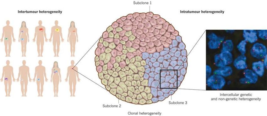
Courtesy: Prof. Fiorentino (S. Orsola – Bologna)NSCLS is a paradigm of
tumor heterogeneity
From Burrell RA Nature 2013Pathologic and genetic characteristics of two patients with ALK/EGFR
coaltered adenocarcinoma by reverse-transcriptase polymerase chain
reaction and amplification refractory mutation system assays.
Weijing Cai et al. JCO 2015;33:3701-
3709
©2015 by American Society of Clinical OncologyThe dogma of
mutual exclusivity of
mutationsSensitivity up to
Sensitivity 20-30% Sensitivity 1/10.000 1/1.000.000
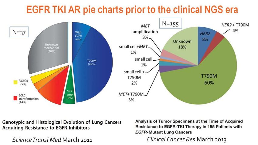
DOES IT MAKE CLINICAL SENSE TO ASSESS MUTATION IN 1/1.000.000 TUMOR CELLS?38 Paired samples (pre-post EGFR TKI): large panel NGS
reveals richer landscape of AR mechanisms
Pre-NGS pie chart Large panel NGS pie chart:
Acquired alterations
Helena Yu et al. CCR Mar 2018Tissue Sample DNA Library Prep Sequencing Data
Current:
Illumina HiSeq 2000 Illumina MiSeq Ion Torrent PGM
300 – 600 Gigabases 1.5 Gigabases 1 Gigabase
6 – 11 days 1 day 6 hoursWhich (New) Genes Should Be Tested for
Lung Cancer Patients?
3 categories
One set as an absolute minimum
- EGFR, ALK & ROS1, PD-L1
A second expanded panel:
- BRAF, MET, RET, HER2, and KRAS, if adequate
material is available
All other genes are investigationalWhat Is the Role of Testing for Circulating
cfDNA for Lung Cancer Patients?
There is currently insufficient evidence to support the use of
circulating plasma cfDNA molecular methods for establishing
a primary diagnosis of lung adenocarcinoma.
In some clinical settings in which tissue is limited and/or
insufficient for molecular testing, physicians may use a
cfDNA assay to identify EGFR mutations.
Physicians may use plasma cfDNA methods to identify EGFR
T790M mutations in lung adenocarcinoma patients with
progression or secondary clinical resistance to EGFR-
targeted TKIs;
testing of the tumor sample is recommended if the plasma result
is negative.EGFR Raccomandazioni AIOM-SIF- Rolfo C et al, J Thor SIAPEC 2018 Oncol 2018
The goal of ‘‘no mutation left behind’’ must be balanced against the
practical realities of resource utilization in the setting of a low
probability event. Furthermore, it should be noted that even in
resections, there is a possibility that a biologically combined carcinoma
may be vastly dominated by a single histology suggesting that even
resections showing squamous or small cell histology in younger or
never-/light-smokers should be considered for EGFR/ALK/ROS1 testing

Increasing use of multigene panels, especially NGS, encompassing a full
set of ‘‘pan-tumor’’ genes in a single assay, one can anticipate a
decreasing role of histology-based molecular test selection in the
future
Summary – Predictive
              Biomarkers NSCLC
             Tissue    Plasma      Method       Other      Approval
                                               techno

EGFR          +++     ++ (T790M)     PCR        NGS          Yes

ALK           +++         -        IHC>FISH     NGS          Yes
                                              Nanostring
ROS1          +++         -        FISH>IHC     NGS          Yes
                                              Nanostring
PD-L1         +++         -          IHC      TMB/NGS        Yes
BRAF          +++         -        PCR>IHC      NGS          Next
RET           +++         -          FISH       NGS          Next
                                              Nanostring
c-MET ex14    +++         -          PCR        NGS          Next
skip
Algorithm for Predictive Biomarkers in Advanced
(stage IIIB/IV) NSCLC Routine Practice
NSCLC
(+/- diagnostic immunostains)
Concurrently
TISSUE LIQUID
Mutations Gene fusions/amplifications TMB
NGSYou can also read