Segment-specific lymph node dissection and evaluation during anatomical pulmonary segmentectomy
←
→
Page content transcription
If your browser does not render page correctly, please read the page content below
Review Article
                                                                                                                                           Page 1 of 5

Segment-specific lymph node dissection and evaluation during
anatomical pulmonary segmentectomy
Ghulam Abbas1, Beebarg Raza2, Kamil Abbas3, Jason Lamb1, Alper Toker1
1
    Department of Cardiovascular & Thoracic Surgery, West Virginia University, Morgantown, WV, USA; 2St. George’s University, St. George’s,
Grenada; 3West Virginia University, Morgantown, WV, USA
Contributions: (I) Conception and design: G Abbas, J Lamb, A Toker; (II) Administrative support: B Raza; (III) Provision of study materials or patients:
All Authors; (IV) Collection and assembly of data: B Raza, K Abbas; (V) Data analysis and interpretation: All authors; (VI) Manuscript writing: All
authors; (VII) Final approval of manuscript: All authors.
Correspondence to: Ghulam Abbas, MD, MHCM, FACS. 1 Medical Center Dr., Morgantown, WV 26508, USA. Email: Ghulam.abbas@hsc.wvu.edu.

                  Abstract: Lung cancer continues to be the leading cause of cancer related deaths both in men and women.
                  Most patients present with locally advanced disease and are not candidates for resection. A recent surge of
                  lung cancer screening programs for high-risk patients across the western world has led to a rising number
                  of patients with early stage lung cancer. These patients with clinical stage I lung cancer and compromised
                  pulmonary reserves can be candidates for sub-lobar resection with curative intention and similar outcomes
                  as compare to lobectomy. Systemic or lobe-specific mediastinal lymph node dissection is an integral part
                  of lung cancer surgery, especially during lobectomy as nodal upstaging can occur up to 18% of clinical
                  stage I lung cancers and is associated with a worse prognosis. Nodal upstaging can occur in N1 lymph
                  nodes only or as a skip metastasis to the N2 lymph nodes or both. The characteristics and location of the
                  tumor plays an important role in lymph node metastasis. Recently, it has been suggested that a lobe-specific
                  mediastinal lymph node dissection is equivalent to multi-station aggressive nodal dissection for early stage
                  lung cancer detected during screening. Determining mediastinal and intersegmental lymph node metastasis
                  is important during segmentectomy as it is associated with an increase recurrence rate and poor survival.
                  These patients are perhaps better served with lobectomy rather than segmentectomy. The techniques and
                  method of standard mediastinal lymph node dissection are well described in literature but description of a
                  systematical approach for N1 lymph node dissection during a segmentectomy to efficiently identify the nodal
                  upstaging intra-operatively, is lacking. We describe a methodological evaluation of N1 lymph node during
                  segmentectomy in an effort to avoid failure to recognize nodal upstaging.

                  Keywords: Lymph node dissection; nodal upstaging; segmentectomy

                  Received: 23 April 2020; Accepted: 22 October 2020; Published: 15 June 2021.
                  doi: 10.21037/vats-2019-rcs-08
                  View this article at: http://dx.doi.org/10.21037/vats-2019-rcs-08

Lung cancer continues to be one of the most commonly                           resectable and may be candidate for segmentectomy (3).
diagnosed cancer and leading cause of cancer related deaths                    Lymph node metastasis in lung cancer is associated with
worldwide (1). Historically only one-third of the patients                     poor outcomes. Overall nodal upstaging can occur up to
with lung cancer presented with early stage disease. The                       18% of clinical stage 1 lung cancer (4-7). Skip lymph node
growing acceptance of the low dose computed tomography                         metastasis to N2 only nodes occurs in 4–7% of patients
(LDCT) for lung cancer screening is changing the paradigm                      and N2 upstaging can be found up to 8.8% of clinical stage
with the expectation that two-third of all lung cancers in                     1A non-small cell lung cancer (NSCLC) (4). Usual lymph
the screening population will be detected in their early                       node metastasis travels from intraparenchymal stations to
stages (2). These patients will be potentially surgically                      the interlobar stations and then to hilar and mediastinal

© Video-Assisted Thoracic Surgery. All rights reserved.               Video-assist Thorac Surg 2021;6:17 | http://dx.doi.org/10.21037/vats-2019-rcs-08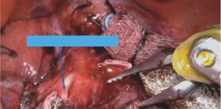
Page 2 of 5                                                                                     Video-Assisted Thoracic Surgery, 2021

lymph nodes with worsening outcomes. The higher                      nodal upstaging who had segmentectomy or lobectomy (18).
number of lymph node harvested and nodal upstaging are                  Lobectomy is perceived as a better surgical approach
considered a surrogate for good quality oncological surgical         by many authorities with the assumption that lobectomy
resection. This has led to recommendation of the systemic            allows for a more radical lymphadenectomy and better
mediastinal lymph node dissection during lung cancer                 margins thus decreasing the chances of local recurrence.
surgery. The technique and method of a methodological                Although no randomized study is available yet to prove
intraoperative evaluation and removal of mediastinal                 that segmentectomy and lobectomy have similar outcome
lymph nodes focusing on N2 lymph node dissection is                  for stage 1 lung cancer, but single institutional reports and
well described (8). Recently, many reports, especially from          meta-analyses have shown similar outcomes for at least T1a
Asia, has questioned the benefit of routine aggressive               and T1b tumors.
mediastinal lymph node dissection (9,10). The patterns of               In our experience at West Virginia University Medicine
nodal metastasis vary by the size and location of the tumor.         lobectomy was associated with 20% nodal upstaging as
For example, the incidence of level 7, 8 & 9 lymph node              compare to 10% with segmentectomy. Though the nodal
(lower mediastinal lymph nodes) metastasis is less than 1%           upstaging was higher in lobectomy group, the number of
for a
Video-Assisted Thoracic Surgery, 2021 Page 3 of 5
A1 - Apical segment artery Right upper lobe posterior segmentectomy (S2)
A3 - Anterior segment artery Dissection is done in the fissure. The pulmonary artery is
exposed. The lymph node over the pulmonary artery is sent
for frozen section. Subsequently, the posterior segment
vein (V2) arising from the central vein is dissected and
transected. Similarly, the pulmonary artery branch (A2)
is transected and the fissure is completed. This leads to
the exposure of the base of the right upper lobe bronchus,
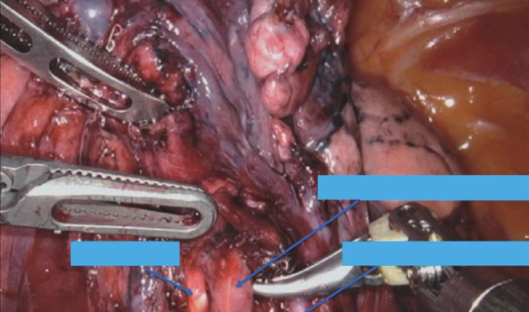
Figure 1 View of the first branch of right pulmonary artery origin of the right upper lobe posterior and anterior
dividing into anterior (A3) and apical branches (A1) with lung bronchi. All the lymph nodes in this area are removed and
retracted caudally. frozen section performed of a representative lymph node.
Then right upper lobe posterior segment bronchus (B2) is
transected.
Right upper lobe anterior segmentectomy (S3)
B1 - Apical segment bronchus
After the completion of the mediastinal and hilar lymph
node dissection, the space between the upper lobe and
middle lobe vein is dissected. Subsequently, the dissection
is done in fissure extending it anteriorly and exposing the
central vein of the right upper lobe. The anterior segment
Figure 2 Right upper lobe apical segment bronchus (B1) after the branch (V3) is transected leading to the exposure of the
transection of A1. bronchus and the artery along with small lymph nodes. It
will be appropriate to proceed with segmentectomy if the
frozen section is negative otherwise a lobectomy would be a
performed by skeletonizing the space over the right main better option (Figure 3)
stem bronchus till the inferior margin of the azygous vein.
Subsequently, lymph node between apical segment vein
and the anterior segmental pulmonary artery branch should Left upper lobe segmentectomies
be removed. If these lymph nodes are negative, then right Following left upper lobe segmentectomies are usually
upper lobe segmentectomy can be pursued with caution. performed.
Left upper lobe upper division segment (S1 + S2 + S3);
Right upper lobe apical segmentectomy (S1) Left upper lobe apicoposterior segment (S1 + S2);
Lingular segmentectomy (S4 + S5).
The mediastinal and hilar lymph node dissection is A standard left sided mediastinal lymph node dissection
completed as described. The apical vein is transected is performed. The pleura over the posterior surface of
leading to exposure of the lymph node at the origin of the the lung is opened and the level 10 lymph node over the
truncus over the pulmonary artery. If this lymph node is pulmonary artery is sent for frozen section. Subsequently,
not sent for frozen then it should be done at this point as the lymph node between the upper vein and the main
lobectomy would be a better approach if this lymph node pulmonary artery is evaluated.
is positive. Subsequently, the truncus branch is dissected
till its bifurcation into apical and anterior segment. The
Left upper lobe upper division segment (S1 + S2 + S3)
lymph node at bifurcation (level 12) is removed. The
apical branches are transected leading to the exposure of After the completion of the mediastinal and hilar lymph
B1. There are usually smaller lymph node exposed at the node dissection the fissure is completed posteriorly. The
meeting site of the three segmental bronchi. The bronchus posterior segmental arteries are transected leading to the
is cleared to the base and frozen section should be obtained exposure of the posterior surface of the bronchus and
of any suspicious lymph node (Figures 1,2). lymph node between lingular and upper division segmental
© Video-Assisted Thoracic Surgery. All rights reserved. Video-assist Thorac Surg 2021;6:17 | http://dx.doi.org/10.21037/vats-2019-rcs-08Page 4 of 5 Video-Assisted Thoracic Surgery, 2021
Transection of the targeted segmental artery leads to
the exposure of the lymph node over the bronchus which
should be sent to frozen.
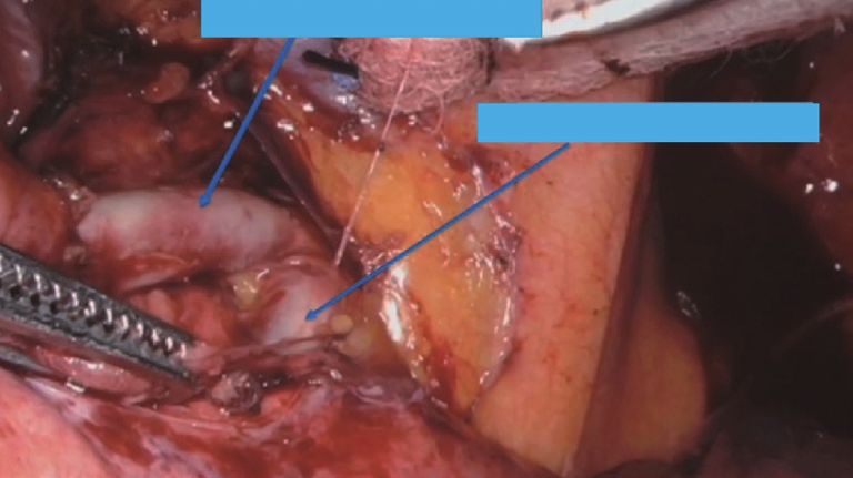
A3 - Anterior segment artery Lower lobe anteromedial (S7 + S8) and
posterolateral (S9 + S10) segmentectomy
Bronchus B3 A1 - Apical segment artery
Similar to common basilar segmentectomy, any suspicious
lymph node in the fissure is sent for frozen. Subsequently,
Figure 3 Exposure of right upper lobe anterior segment bronchus (B3). the targeted segmental branch is transected which exposes
the lymph node over the bronchus which is sent for frozen.
In conclusion, segmentectomy is an acceptable alternate
bronchi. The sample should be sent for frozen section. to lobectomy for tumors less than 2 cm.
Anteriorly the upper division vein is transected leading to In addition to the systemic or lobe-specific mediastinal
the exposure of the lymph node over the first branch of the lymph node dissection, a segment specific lymph node
pulmonary artery which also should be confirmed to be dissection should be performed before committing to the
negative before committing to segmentectomy. segmentectomy to avoid missing nodal upstaging.
Left upper lobe apicoposterior segment (S1 + S2) Acknowledgments
The steps are similar to upper division segmentectomy The authors acknowledge the editorial help of Syeda Sara
except that only the apicoposterior vein and bronchus rather Abbas.
than whole upper division and bronchus is transected. Funding: None.
Lingular segmentectomy (S4 + S5) Footnote
The mediastinal and hilar lymph node dissection is Provenance and Peer Review: This article was commissioned
completed. The dissection is done in the fissure and any by the editorial office, Video-Assisted Thoracic Surgery for
suspicious lymph node is sent for frozen. The lingular the series “Robotic Segmentectomies”. The article has
branches of the artery and vein are transected leading to undergone external peer review.
the exposure of lingular bronchus and lymph nodes in
secondary carina which should be sent for frozen before Conflicts of Interest: All authors have completed the ICMJE
committing to the lingular segmentectomy. uniform disclosure form (available at http://dx.doi.
org/10.21037/vats-2019-rcs-08). The series “Robotic
Segmentectomies” was commissioned by the editorial office
Right and left lower lobe superior and basilar
without any funding or sponsorship. Alper Toker served
segmentectomy [S6 + S7(8)–10]
as the unpaid Guest Editor of the series and serves as an
Lower lobe segment specific lymph node dissection depends unpaid editorial board member of Video-Assisted Thoracic
on the targeted segment. Segment 7 is missing on the left. Surgery from Jun 2019 to May 2021. The authors have no
The technique of lower lobe segmentectomy is mentioned other conflicts of interest to declare.
in a separate manuscript of this issue. Following is the
suggested technique for lymph node dissection. Ethical Statement: The authors are accountable for all
The pleura over the posterior surface of the lung is aspects of the work in ensuring that questions related
opened and the space between the vein and the bronchus is to the accuracy or integrity of any part of the work are
dissected to expose the level 11 lymph node which should appropriately investigated and resolved.
be confirmed to be negative.
Any suspicious lymph node over the pulmonary artery Open Access Statement: This is an Open Access article
and branches should be confirmed to be negative. distributed in accordance with the Creative Commons
© Video-Assisted Thoracic Surgery. All rights reserved. Video-assist Thorac Surg 2021;6:17 | http://dx.doi.org/10.21037/vats-2019-rcs-08Video-Assisted Thoracic Surgery, 2021 Page 5 of 5
Attribution-NonCommercial-NoDerivs 4.0 International 9. Deng HY, Zhou J, Wang RL, et al. Lobe-Specific Lymph
License (CC BY-NC-ND 4.0), which permits the non- Node Dissection for Clinical Early-Stage (cIA) Peripheral
commercial replication and distribution of the article with Non-small Cell Lung Cancer Patients: What and How?
the strict proviso that no changes or edits are made and the Ann Surg Oncol 2020;27:472-80.
original work is properly cited (including links to both the 10. Hattori A, Matsunaga T, Takamochi K, et al.
formal publication through the relevant DOI and the license). Significance of Lymphadenectomy in Part-Solid Lung
See: https://creativecommons.org/licenses/by-nc-nd/4.0/. Adenocarcinoma: Propensity Score Matched Analysis. Ann
Thorac Surg 2018;106:989-97.
11. Jensik RJ, Faber LP, Milloy FJ, et al. Segmental resection
References
for lung cancer. A fifteen-year experience. J Thorac
1. Bray F, Ferlay J, Soerjomataram I, et al. Global cancer Cardiovasc Surg 1973;66:563-72.
statistics 2018: GLOBOCAN estimates of incidence and 12. Ginsberg RJ, Rubinstein LV. Randomized trial of
mortality worldwide for 36 cancers in 185 countries. CA lobectomy versus limited resection for T1 N0 non-small
Cancer J Clin 2018;68:394-424. Erratum in: CA Cancer J cell lung cancer. Lung Cancer Study Group. Ann Thorac
Clin 2020;70:313. Surg 1995;60:615-22; discussion 622-3.
2. De Koning H, Van Der Aalst C, Ten Haaf K, et al. 13. Okada M, Nishio W, Sakamoto T, et al. Effect of tumor
PL02.05 Effects of Volume CT Lung Cancer Screening: size on prognosis in patients with non-small cell lung
Mortality Results of the NELSON Randomised- cancer: the role of segmentectomy as a type of lesser
Controlled Population Based Trial. J Thorac Oncol resection. J Thorac Cardiovasc Surg 2005;129:87-93.
2018;13:S185. 14. Bao F, Ye P, Yang Y, et al. Segmentectomy or lobectomy
3. Schuchert MJ, Abbas G, Awais O, et al. Anatomic for early stage lung cancer: a meta-analysis. Eur J
segmentectomy for the solitary pulmonary nodule and Cardiothorac Surg 2014;46:1-7.
early-stage lung cancer. Ann Thorac Surg 2012;93:1780-5; 15. Cao C, Chandrakumar D, Gupta S, et al. Could less be
discussion 1786-7. more?-A systematic review and meta-analysis of sublobar
4. Deng HY, Zhou J, Wang RL, et al. Surgical Choice for resections versus lobectomy for non-small cell lung
Clinical Stage IA Non-Small Cell Lung Cancer: View cancer according to patient selection. Lung Cancer
From Regional Lymph Node Metastasis. Ann Thorac Surg 2015;89:121-32.
2020;109:1079-85. 16. Zhang L, Li M, Yin R, et al. Comparison of the oncologic
5. Kneuertz PJ, Cheufou DH, D'Souza DM, et al. outcomes of anatomic segmentectomy and lobectomy for
Propensity-score adjusted comparison of pathologic nodal early-stage non-small cell lung cancer. Ann Thorac Surg
upstaging by robotic, video-assisted thoracoscopic, and 2015;99:728-37.
open lobectomy for non-small cell lung cancer. J Thorac 17. Bedetti B, Bertolaccini L, Rocco R, et al. Segmentectomy
Cardiovasc Surg 2019;158:1457-1466.e2. versus lobectomy for stage I non-small cell lung cancer:
6. Boffa DJ, Kosinski AS, Paul S, et al. Lymph node a systematic review and meta-analysis. J Thorac Dis
evaluation by open or video-assisted approaches in 11,500 2017;9:1615-23.
anatomic lung cancer resections. Ann Thorac Surg 18. Lutfi W, Schuchert MJ, Dhupar R, et al. Node-Positive
2012;94:347-53; discussion 353. Segmentectomy for Non-Small-Cell Lung Cancer: Risk
7. Wilson JL, Louie BE, Cerfolio RJ, et al. The prevalence Factors and Outcomes. Clin Lung Cancer 2019;20:e463-9.
of nodal upstaging during robotic lung resection in
early stage non-small cell lung cancer. Ann Thorac Surg
doi: 10.21037/vats-2019-rcs-08
2014;97:1901-6; discussion 1906-7.
Cite this article as: Abbas G, Raza B, Abbas K, Lamb J,
8. Lardinois D, De Leyn P, Van Schil P, et al. ESTS
Toker A. Segment-specific lymph node dissection and
guidelines for intraoperative lymph node staging in
evaluation during anatomical pulmonary segmentectomy.
non-small cell lung cancer. Eur J Cardiothorac Surg
Video-assist Thorac Surg 2021;6:17.
2006;30:787-92.
© Video-Assisted Thoracic Surgery. All rights reserved. Video-assist Thorac Surg 2021;6:17 | http://dx.doi.org/10.21037/vats-2019-rcs-08You can also read