Cancer Association of South Africa (CANSA)
←
→
Page content transcription
If your browser does not render page correctly, please read the page content below
Cancer Association of South Africa (CANSA)
Fact Sheet
on
Lung Cancer
Introduction
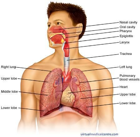
The chest contains two lungs, one lung on the
right side of the chest, and the other on the left
side of the chest. Each lung is made up of sections
called lobes – the right lung consists of three
lobes and the left lung consists of two lobes. The
lung is soft and protected by the ribcage. The
purposes of the lungs are to absorb oxygen (O2),
into the bloodstream for distribution throughout
the body and to remove carbon dioxide (CO2), a
waste product, from the body.
[Picture Credit – Anatomy of the Lungs]
Lung Cancer
Lung cancer is a disease characterised by uncontrolled cell growth in tissues of the lung. If left
untreated, this growth can spread beyond the lung in a process called metastasis into nearby tissue
and, eventually, into other parts of the body. Most cancers that start in lung, known as primary lung
cancers, are carcinomas that arise from epithelial cells.
Carter, B.W. & Erasmus, J.J. 2019. Current concepts in the diagnosis and staging of lung cancer. In:
Holder, J., Kubik-Huch, R./A. & von Schulthess, G.D. (Editors).
“Lung cancer is the leading cause of cancer-related death in the United States and accounts for more
deaths than colorectal, breast, prostate, and pancreatic cancers combined. The eighth edition of the
tumor-node-metastasis staging system (TNM-8), which has been accepted by the Union for
International Cancer Control and the American Joint Committee on Cancer based on proposals from
the International Association for the Study of Lung Cancer, includes key changes to T and M
descriptors and overall stage groups and introduces new recommendations for classifying patterns
of disease with multiple sites of pulmonary involvement. As accurate staging is necessary for the
formulation of appropriate treatment strategies, radiologists must be able to identify these features
on imaging studies.”
Researched and Authored by Prof Michael C Herbst
[D Litt et Phil (Health Studies); D N Ed; M Art et Scien; B A Cur; Dip Occupational Health; Dip Genetic Counselling; Dip Audiometry and
Noise Measurement; Diagnostic Radiographer; Medical Ethicist]
Approved by Ms Elize Joubert, Chief Executive Officer [BA Social Work (cum laude); MA Social Work]
March 2021 Page 1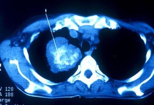
Incidence of Lung Cancer in South Africa
According to the outdated National Cancer Registry (2017), known for under reporting, the following
number of lung cancer cases was histologically diagnosed in South Africa during 2017:

Group - Males                        Actual                 Estimated            Percentage of
2017                               No of Cases             Lifetime Risk          All Cancers
All males                             1 880                     1:82                 4,70%
Asian males                             90                      1:65                 9,27%
Black males                            697                     1:147                 5,28%
Coloured males                         372                      1:45                 7,89%
White males                            721                      1:48                 3,40%

Group - Females                      Actual                 Estimated            Percentage of
2017                               No of Cases             Lifetime Risk          All Cancers
All females                           1 056                    1:191                 2,53%
Asian females                           32                     1:266                 2,41%
Black females                          260                     1:560                 1,35%
Coloured females                       219                      1:94                 4,81%
White females                          545                      1:69                 3,18%

The frequency of histologically diagnosed cases of lung cancer in South Africa for 2017 was as
follows (National Cancer Registry, 2017):

Group - Males                     0 – 19         20 – 29         30 – 39        40 – 49       50 – 59       60 – 69       70 – 79         80+
2017                              Years           Years           Years          Years         Years         Years         Years         Years
All males                           1               5              34             174           518           615           410           123
Asian males                         0               1               1              4             14            37            25            8
Black males                         1               2              22              91           240           231            90            20
Coloured males                      0               0               7              49           115           114            67            20
White males                         0               2               4              30           149           233           228            75

Group - Females                   0 – 19         20 – 29         30 – 39        40 – 49       50 – 59       60 – 69       70 – 79         80+
2017                              Years           Years           Years          Years         Years         Years         Years         Years
All females                         1               4              25             65            249           349           268            95
Asian females                       0               0               0              5             5             10             7            5
Black females                       0               4              13             28             85            73            42            15
Coloured females                    1               0               4             14             57            82            48            13
White females                       0               0               8             18            102           184           171            62
N.B. In the event that the totals in any of the above tables do not tally, this may be the result of uncertainties as to the age, race or sex of
the individual. The totals for ‘all males’ and ‘all females’, however, always reflect the correct totals.

Types of Lung Cancer
There are two main types of lung cancer, non-small cell lung cancer and small cell lung cancer. These
names refer to how the cancers look under a microscope to a pathologist (a person specifically
qualified to make a diagnosis by looking at items under a microscope. Most lung cancers are non-
small cell. There are also some subtypes of non-small cell lung cancer.

Researched and Authored by Prof Michael C Herbst
[D Litt et Phil (Health Studies); D N Ed; M Art et Scien; B A Cur; Dip Occupational Health; Dip Genetic Counselling; Dip Audiometry and
Noise Measurement; Diagnostic Radiographer; Medical Ethicist]
Approved by Ms Elize Joubert, Chief Executive Officer [BA Social Work (cum laude); MA Social Work]
March 2021                                                                                                                      Page 2
Causes of Lung Cancer
The main cause of lung cancer (internationally) is smoking of tobacco products. Lung cancer has
always been – and still is – more common among men. As more women have started smoking, the
number of women developing lung cancer has been on the increase.

People who do not smoke can also develop lung cancer. Approximately 10–15% of people who get
lung cancer have never smoked.

Other risk factors include the effects of past cancer treatment and exposure to asbestos, radon gas
and – in very rare cases – substances such as uranium, chromium and nickel. Lung cancer is not
infectious and can’t be passed on to other people.

Smoking - The more one smokes, the greater the risk of developing lung cancer. It is also more likely
to develop in people who start smoking at a young age. If someone stops smoking, their risk of
developing lung cancer reduces over time. After about 15 years, the chance of developing the
disease is similar to that of a non-smoker.

In a recent study by Alexandrov, et al., (2016) their analysis shows a direct link between the number
of cigarettes smoked in a lifetime and the number of mutations in tumour DNA.

The researchers found that, on average, smoking a packet of cigarettes a day led to:

     •    150 mutations in each lung cell every year
     •    97 in the larynx or voice box
     •    23 in the mouth
     •    18 in the bladder
     •    six in the liver

According to the researchers, the more mutations there are, the higher the chance that these will
occur in the key genes that are called cancer genes, which convert a normal cell into a cancer cell.

Passive smoking- breathing in other people’s cigarette smoke (passive smoking) increases the risk of
lung disease and cancer

Pipes and cigars - although many believe that pipe and cigar smokers have a lower risk of lung cancer
than cigarette smokers, there remains a risk of cancer.

Cannabis - Cannabis smoke contains a similar profile of carcinogenic (cancer causing) chemicals as
tobacco smoke and is usually inhaled more deeply. Although cannabis smoke is known to contain
similar harmful and carcinogenic substances to tobacco smoke, relatively little is understood
regarding the respiratory health effects from cannabis smoking (Gates, et al.).

Radon gas – radon is a colourless, odourless radioactive gas that forms from the decay of radioactive
elements such as uranium. Radon gas given off by soil and rock can enter homes and buildings
through cracks in floors and walls, pipes, wires and pumps. Radon concentrations are usually highest
in basements or in underground mining environments. Radon is the number one cause of lung
cancer among non-smokers, according to estimates from the Environmental Protection Agency
(EPA). Overall, radon is the second leading cause of lung cancer

Researched and Authored by Prof Michael C Herbst
[D Litt et Phil (Health Studies); D N Ed; M Art et Scien; B A Cur; Dip Occupational Health; Dip Genetic Counselling; Dip Audiometry and
Noise Measurement; Diagnostic Radiographer; Medical Ethicist]
Approved by Ms Elize Joubert, Chief Executive Officer [BA Social Work (cum laude); MA Social Work]
March 2021                                                                                                                      Page 3
Age - like most types of cancer, lung cancer is more common in older people. About 80% of lung
cancers are diagnosed in people over 60. Lung cancer rarely affects people under 40

Genetic risk - some people with a close relative who has had lung cancer may be at an increased risk
of it themselves, although the increase in risk is very small. The risk is slightly greater if a relative is a
non-smoker and developed lung cancer at an early age, or if more than one relative on the same
side of the family developed lung cancer

 Asbestos - people who have been in contact with asbestos have a higher risk of developing lung
cancer, especially smokers. Asbestos and tobacco smoke act together to increase the risk..

Industrial exposure - several industrial carcinogens, for example, arsenic and polycyclic hydrocarbons
as well as some occupations including non-ferrous metal production and painting, have been linked
to lung cancer

Exposure to Diesel Exhaust Fumes - diesel exhaust was classified as a cause of lung cancer by the
International Agency for Research on Cancer (IARC) in June 2012, following a review of evidence
mainly from highly-exposed workers. IARC cited a study of diesel exhaust exposure in miners, which
showed risk of lung cancer was increased approximately three times in those most heavily exposed

Occupational exposure to silica - silica exposure can result in silicosis with an increased risk for lung
cancer, but without silicosis there is no increased risk. The body of evidence supports an increased
risk of lung cancer with exposure to asbestos in non-smokers and that risks are especially high in
those who smoke, who also have past exposure to asbestos

Family History - a family history of lung cancer in a first-degree relative is associated with a two-fold
(double) increased risk, independent of smoking. If both cancers are diagnosed before the age of 60,
the risk ratio is almost five-fold. The association between family history and risk may be stronger in
black individuals than white

Screening for Lung Cancer
Three Common Lung Cancer Screening Methods
                                                                           Screening for lung cancer is usually
                                                                           accomplished using three methods.

                                                                           [Picture Credit: Medicinet]

                                                          Physical Examination - a physical exam will
                                                          look for signs of wheezing, shortness of
                                                          breath, cough, pain and other possible signs
                                                          of lung cancer. Depending on the
                                                          advancement of the cancer, other early
                                                          signs of lung cancer symptoms may include
                                                          a lack of sweating, dilated neck veins, face
                                                          swelling, excessively constricted pupils, and
other signs. The physical exam will also include the patient's history of smoking and a chest X-ray.

Researched and Authored by Prof Michael C Herbst
[D Litt et Phil (Health Studies); D N Ed; M Art et Scien; B A Cur; Dip Occupational Health; Dip Genetic Counselling; Dip Audiometry and
Noise Measurement; Diagnostic Radiographer; Medical Ethicist]
Approved by Ms Elize Joubert, Chief Executive Officer [BA Social Work (cum laude); MA Social Work]
March 2021                                                                                                                      Page 4
Sputum Cytology Examination - a sputum cytology exam involves a microscopic examination of a
patient's mucus (sputum).
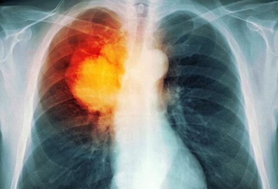
Spiral CT Scan Examination - this
method of CT scanning builds a detailed
image of the body's internal workings.
Inside a spiral CT machine, detailed
images are taken of the relevant parts
of the patient's body. Those images are
then linked to an X-ray machine to
create 3D images of the patient's
internal organs. These images may
reveal potentially cancerous tumours.
[Picture Credit: Medicinet]
A study by researchers suggested that
people aged 55 to 74 years old who had smoked at least one pack of cigarettes a day for 30 or more
years may benefit from a spiral CT study of the lungs.
Signs and Symptoms of Lung Cancer
Lung cancer typically doesn't cause signs and symptoms in its earliest stages. Signs and symptoms of
lung cancer typically occur only when the disease is advanced. Signs and symptoms of lung cancer
may include:
• a new cough that does not go away
• changes in a chronic cough or ‘smoker's cough’
• a cough that gets worse or does not go away
• coughing up blood, even a small amount
• shortness of breath or wheezing
• constant chest pain – especially when coughing
• frequent chest infections, such as pneumonia, or an infection that does not go away
• wheezing
• hoarseness
• swelling of the neck and face
• fatigue (feeling very tired all the time)
• loss of appetite
• losing weight without trying
Diagnosis of Lung Cancer
The following may be used to diagnose cancer of the lung:
Researched and Authored by Prof Michael C Herbst
[D Litt et Phil (Health Studies); D N Ed; M Art et Scien; B A Cur; Dip Occupational Health; Dip Genetic Counselling; Dip Audiometry and
Noise Measurement; Diagnostic Radiographer; Medical Ethicist]
Approved by Ms Elize Joubert, Chief Executive Officer [BA Social Work (cum laude); MA Social Work]
March 2021 Page 5Medical history - to find out if lung cancer may be present, the doctor evaluates a person's medical
history, smoking history, his/her exposure to environmental and occupational substances, and family
history of cancer.
Physical examination - the doctor also performs a physical exam and may order a test to take an
image of the chest or other tests. Seeing a spot on an image is usually how a doctor first suspects
that lung cancer may be present.
Sputum cytology - if lung cancer is suspected, the doctor may order a test called ‘sputum cytology’.
This is a simple test where a doctor examines a sample of mucous cells coughed up from the lungs
under a microscope to see if cancer is present.
Bronchoscopy – a procedure to observe the bronchi of the lungs. During a bronchoscopy the doctor
can collect cells or small samples of tissues from the airways and lungs.
Imaging tests - doctors may use imaging methods such as a spiral Computerised Tomography (CT)
scan or a Positron Emission Tomography (PET) scan to look for signs of cancer. A CT scan is a series of
detailed pictures of areas inside the body. A PET scan is a computerised image of the metabolic
activity of body tissues.
Staging of Lung Cancer
Staging is the process of finding out how far a cancer has spread. This is important because
treatment options and outlook for recovery and survival depend on the cancer's stage.
Staging of lung cancer can be done by means of:
• The TNM system for staging contains 3 key pieces of information:
▪ T describes the size of the primary tumour, measured in centimetres (cm), and
whether the cancer has spread to organs next to the tumour
▪ N describes the extent of spread to nearby (regional) lymph nodes
▪ M indicates whether the cancer has metastasised (spread) to other organs of the
body
Where Lung Cancer May Spread to in the Body
In the event of lung cancer spreading to other parts of the body, it may spread as indicated in the
bold section below:
Cancer Type: Main Sites of Metastasis (Spread)
Bladder Bone, liver, lung
Breast Bone, brain, liver, lung
Colon Liver, lung
Colorectal Liver, lung, peritoneum (lining of abdomen)
Kidney Adrenal gland, bone, brain, liver, lung
Lung Adrenal gland, bone, brain, liver, other lung
Melanoma Bone, brain, liver, lung, skin, muscle
Researched and Authored by Prof Michael C Herbst
[D Litt et Phil (Health Studies); D N Ed; M Art et Scien; B A Cur; Dip Occupational Health; Dip Genetic Counselling; Dip Audiometry and
Noise Measurement; Diagnostic Radiographer; Medical Ethicist]
Approved by Ms Elize Joubert, Chief Executive Officer [BA Social Work (cum laude); MA Social Work]
March 2021 Page 6Ovary Liver, lung, peritoneum (lining of abdomen)
Pancreas Liver lung, peritoneum (lining of abdomen)
Prostate Adrenal gland, bone, liver, lung
Stomach Liver, lung, peritoneum (lining of abdomen), ovaries
Thyroid Bone, liver, lung
Uterus Boner, liver, lung, peritoneum (lining of abdomen), vagina
Non-melanoma skin cancer Very rare: lymph nodes, lung, bone (if in head/neck region)
Treatment of Lung Cancer
The type of treatment a patient may receive for lung cancer depends on several factors, including:
• the type of lung cancer (non-small cell or small cell)
• the size and position of the cancer
• how far advanced the cancer is (the stage)
• patient’s overall health
Treatment options may include:
Surgery – surgical removal of cancerous tissue.
Radiation therapy - radiation therapy is a cancer treatment that uses high-energy x-rays or other
types of radiation to kill cancer cells or keep them from growing. There are two types of radiation
therapy. External radiation therapy uses a machine outside the body to send radiation toward the
cancer. Internal radiation therapy uses a radioactive substance sealed in needles, seeds, wires, or
catheters that are placed directly into or near the cancer.
Radiosurgery - is a method of delivering radiation directly to the tumour with little damage to
healthy tissue. It does not involve surgery and may be used to treat certain tumours in patients who
cannot have surgery.
Chemotherapy - chemotherapy is a cancer treatment that uses drugs to stop the growth of cancer
cells, either by killing the cells or by stopping them from dividing.
Targeted therapy - targeted therapy is a type of treatment that uses drugs or other substances to
identify and attack specific cancer cells without harming normal cells.
Immunotherapy – use is made of medicines to stimulate the immune system of the body to fight the
cancer.
Watchful waiting - watchful waiting is closely monitoring a patient’s condition without giving any
treatment until symptoms appear or change.
Chun, S.G., Liao, Z., Jeter, M.D., Chang, J.Y., Lin, S.H., Komaki, R.U., Guerrero, T.M., Mayo, R.C.,
Korah, B.M., Koshy, S.M., Heymach, J.V., Koong, A.C. & Skinner, H.D. 2020.
BACKGROUND: Metformin reduces glucose uptake in physiologic tissues and has been shown to
affect non-small cell lung cancer (NSCLC) metabolism. We hypothesized that positron emission
tomography (PET) scans could detect the impact of metformin on glucose uptake in NSCLC and we
sought to redundant test this hypothesis in a prospective clinical trial.
Researched and Authored by Prof Michael C Herbst
[D Litt et Phil (Health Studies); D N Ed; M Art et Scien; B A Cur; Dip Occupational Health; Dip Genetic Counselling; Dip Audiometry and
Noise Measurement; Diagnostic Radiographer; Medical Ethicist]
Approved by Ms Elize Joubert, Chief Executive Officer [BA Social Work (cum laude); MA Social Work]
March 2021 Page 7MATERIALS AND METHODS: A single-blinded phase II clinical trial was performed with subjects randomized 6:1 to 3 to 4 weeks of metformin versus placebo for inoperable early-stage NSCLC. PET scans were performed at baseline, mid-treatment (after 2 wk study medication), and 6 months postradiation. The primary endpoint of the trial was tumor metabolic response to metformin by PERCIST before definitive radiation. Stereotactic body radiotherapy to 50 Gy in 4 fractions was used for peripheral tumors and 70 Gy in 10 fractions for central tumors. RESULTS: There were 14 subjects randomized to the metformin and 1 to placebo. Histologies were 60% adenocarcinoma, 33.3% squamous cell carcinoma, and 6.7% poorly differentiated carcinoma. At mid-treatment PET scan, 57% of subjects randomized to metformin met PERCIST criteria for metabolic response, of which 75% had progressive metabolic disease and 25% had partial metabolic response, whereas the placebo subject had stable metabolic disease. At 6 months, the metformin arm had 69% complete metabolic response, 23% partial metabolic response and 1 progressive metabolic disease, and the subject treated with placebo had a complete metabolic response. There were no CTCAE grade ≥3 toxicities. CONCLUSIONS: Despite low accrual, majority of subjects treated with metformin had metabolic responses by PERCIST criteria on PET imaging. Contrary to the effect of metformin on most physiologic tissues, most tumors had increased metabolic activity in response to metformin. Hanna, N.H., Schneider, B.J., Temin, S., Baker, S. (Jr), Brahmer, Jm, Ellis, P.M., Gaspar, L.E., Haddad, R.Y., Hesketh, P.J., Jain, D., Jaiyesimi, I., Johnson, D.H., Leighl, N.B., Phillips, T., Riely, G.J., Robinson, A.G., Rosell, R., Schiller, J.H., Singh, N., Spigel, D.R., Stabler, J.O., Tashbar, J. & Masters, G. 2020. PURPOSE: The aim of this work is to provide evidence-based recommendations updating the 2017 ASCO guideline on systemic therapy for patients with stage IV non-small-cell lung cancer (NSCLC) without driver alterations. A guideline update for patients with stage IV NSCLC with driver alterations will be published separately. METHODS: The American Society of Clinical Oncology and Ontario Health (Cancer Care Ontario) NSCLC Expert Panel made updated recommendations based on a systematic review of randomized controlled trials from December 2015 to 2019. RESULTS: This guideline update reflects changes in evidence since the previous guideline update. Five randomized controlled trials provide the evidence base. Additional literature suggested by the Expert Panel is discussed. RECOMMENDATIONS: Recommendations apply to patients without driver alterations in epidermal growth factor receptor or ALK. For patients with high programmed death ligand 1 (PD-L1) expression (tumor proportion score [TPS] ≥ 50%) and non-squamous cell carcinoma (non-SCC), the Expert Panel recommends single-agent pembrolizumab. Additional treatment options include pembrolizumab/carboplatin/pemetrexed, atezolizumab/carboplatin/paclitaxel/bevacizumab, or atezolizumab/carboplatin/nab-paclitaxel. For most patients with non-SCC and either negative (0%) or low positive (1% to 49%) PD-L1, the Expert Panel recommends pembrolizumab/carboplatin/pemetrexed. Additional options are atezolizumab/carboplatin/nab- paclitaxel, atezolizumab/carboplatin/paclitaxel/bevacizumab, platinum-based two-drug combination chemotherapy, or non-platinum-based two-drug therapy. Single-agent pembrolizumab is an option for low positive PD-L1. For patients with high PD-L1 expression (TPS ≥ 50%) and SCC, the Expert Panel recommends single-agent pembrolizumab. An additional treatment option is pembrolizumab/carboplatin/(paclitaxel or nab-paclitaxel). For most patients with SCC and either negative (0%) or low positive PD-L1 (TPS 1% to 49%), the Expert Panel recommends pembrolizumab/carboplatin/(paclitaxel or nab-paclitaxel) or chemotherapy. Single-agent pembrolizumab is an option in select cases of low positive PD-L1. Recommendations are conditional Researched and Authored by Prof Michael C Herbst [D Litt et Phil (Health Studies); D N Ed; M Art et Scien; B A Cur; Dip Occupational Health; Dip Genetic Counselling; Dip Audiometry and Noise Measurement; Diagnostic Radiographer; Medical Ethicist] Approved by Ms Elize Joubert, Chief Executive Officer [BA Social Work (cum laude); MA Social Work] March 2021 Page 8
on the basis of histology, PD-L1 status, and/or the presence or absence of contraindications.
Additional information is available at www.asco.org/lung-cancer-guidelines.

Weiss, J.M., Csoszi, T., Maglakelidze, M., Hoyer, R.J., Beck, J.T., Domine Gomez, M., Lowczak, A.,
Aljumaily, R., Rocha Lima, C.M., Boccia, R.V., Hanna, W., Nikolinakos, P., Chiu, V.K., Owonikoko,
T.K., Schuster, S.R., Hussein, M.A., Richards, D.A., Sawrycki, P., Bulat, I., Hamm, J.T., Hart, L.L.,
Adler, S., Antal, J.M., Lai, A.Y., Sorrentino, J.A., Yang, Z., Malik, R.K., Morris, S.R., Roberts, P.J. &
Dragnev, K.H., & G1T28-02 Study Group. 2019.
BACKGROUND: Chemotherapy-induced damage of hematopoietic stem and progenitor cells (HSPC)
causes multi-lineage myelosuppression. Trilaciclib is an intravenous CDK4/6 inhibitor in
development to proactively preserve HSPC and immune system function during chemotherapy
(myelopreservation). Preclinically, trilaciclib transiently maintains HSPC in G1 arrest and protects
them from chemotherapy damage, leading to faster hematopoietic recovery and enhanced
antitumor immunity.
PATIENTS AND METHODS: This was a phase Ib (open-label, dose-finding) and phase II (randomized,
double-blind placebo-controlled) study of the safety, efficacy and PK of trilaciclib in combination
with etoposide/carboplatin (E/P) therapy for treatment-naive extensive-stage small-cell lung
cancer patients. Patients received trilaciclib or placebo before E/P on days 1-3 of each cycle. Select
end points were prespecified to assess the effect of trilaciclib on myelosuppression and antitumor
efficacy.
RESULTS: A total of 122 patients were enrolled, with 19 patients in part 1 and 75 patients in part 2
receiving study drug. Improvements were seen with trilaciclib in neutrophil, RBC (red blood cell) and
lymphocyte measures. Safety on trilaciclib+E/P was improved with fewer ≥G3 adverse events (AEs)
in trilaciclib (50%) versus placebo (83.8%), primarily due to less hematological toxicity. No trilaciclib-
related ≥G3 AEs occurred. Antitumor efficacy assessment for trilaciclib versus placebo, respectively,
showed: ORR (66.7% versus 56.8%, P = 0.3831); median PFS [6.2 versus 5.0 m; hazard ratio (HR)
0.71; P = 0.1695]; and OS (10.9 versus 10.6 m; HR 0.87; P = 0.6107).
CONCLUSION: Trilaciclib demonstrated an improvement in the patient's tolerability of
chemotherapy as shown by myelopreservation across multiple hematopoietic lineages resulting in
fewer supportive care interventions and dose reductions, improved safety profile, and no detriment
to antitumor efficacy. These data demonstrate strong proof-of-concept for trilaciclib's
myelopreservation benefits.
CLINICAL TRAIL NUMBER: NCT02499770.

Lowering the Risk for Lung Cancer
Reducing the risk for lung cancer can be achieved by not smoking. Other means include:

Smoking cessation - smoking is responsible for the majority of lung cancers. Quitting all forms of
smoking at any time can lower the risk of developing lung cancer, and appears to be beneficial after
a diagnosis of lung cancer as well.

Preventing exposure to Radon - exposure to radon in the home is the second leading cause of lung
cancer overall, and the number one cause in non-smokers. Radon is an invisible radioactive gas that
results from the normal decay of radium in the soil.

Researched and Authored by Prof Michael C Herbst
[D Litt et Phil (Health Studies); D N Ed; M Art et Scien; B A Cur; Dip Occupational Health; Dip Genetic Counselling; Dip Audiometry and
Noise Measurement; Diagnostic Radiographer; Medical Ethicist]
Approved by Ms Elize Joubert, Chief Executive Officer [BA Social Work (cum laude); MA Social Work]
March 2021                                                                                                                      Page 9
Not being exposed to secondhand smoke - exposure to second hand smoke increases the risk of lung
cancer in non-smokers two to three fold.

Asbestos - workplace exposure to asbestos increases the risk of lung cancer, and combined with
smoking the risk is exponential. Employers should have safety recommendations for those exposed.

About Clinical Trials
Clinical trials are research studies that involve people. They are conducted under controlled
conditions. Only about 10% of all drugs started in human clinical trials become an approved drug.

Clinical trials include:
     • Trials to test effectiveness of new treatments
     • Trials to test new ways of using current treatments
     • Tests new interventions that may lower the risk of developing certain types of cancers
     • Tests to find new ways of screening for cancer

The South African National Clinical Trials Register provides the public with updated information on
clinical trials on human participants being conducted in South Africa. The Register provides
information on the purpose of the clinical trial; who can participate, where the trial is located, and
contact details.

For additional information, please visit: www.sanctr.gov.za/

Medical Disclaimer
This Fact Sheet is intended to provide general information only and, as such, should not be
considered as a substitute for advice, medically or otherwise, covering any specific situation. Users
should seek appropriate advice before taking or refraining from taking any action in reliance on any
information contained in this Fact Sheet. So far as permissible by law, the Cancer Association of
South Africa (CANSA) does not accept any liability to any person (or his/her dependants/
estate/heirs) relating to the use of any information contained in this Fact Sheet.

Whilst the Cancer Association of South Africa (CANSA) has taken every precaution in compiling this
Fact Sheet, neither it, nor any contributor(s) to this Fact Sheet can be held responsible for any action
(or the lack thereof) taken by any person or organisation wherever they shall be based, as a result,
direct or otherwise, of information contained in, or accessed through, this Fact Sheet.

References and Sources Consulted or Utilised

Alexandrov, L.B., Ju, Y.S., Haase, K., Van Loo, P., Martincorena, I., Nik-Zainal, S., Totoki, Y., Fujimoto, A., Nakagawa, H.,
Shibata, T., Campbell, P.J., Vineis, P., Phillips, D.H. & Stratton, M.R. 2016. Mutational signatures associated with tobacco
smoking in human cancer. Science 04 Nov 2016: Vol. 354, Issue 6312, pp. 618-622. DOI: 10.1126/science.aag0299.

Researched and Authored by Prof Michael C Herbst
[D Litt et Phil (Health Studies); D N Ed; M Art et Scien; B A Cur; Dip Occupational Health; Dip Genetic Counselling; Dip Audiometry and
Noise Measurement; Diagnostic Radiographer; Medical Ethicist]
Approved by Ms Elize Joubert, Chief Executive Officer [BA Social Work (cum laude); MA Social Work]
March 2021                                                                                                                     Page 10
Anatomy of Lungs http://www.google.co.za/imgres?hl=en&sa=X&rlz=1G1LENN_ENZA491&biw=1366&bih=613&tbm=isch&prmd=imvns&tbni d=X5h4_-6dVZSdbM:&imgrefurl=http://www.virtualmedicalcentre.com/diseases/asthma/12&docid=AZ8_VrQ5- bVyRM&imgurl=http://www.virtualmedicalcentre.com/uploads/VMC/DiseaseImages/2218_respiratory_anatomy_450.jpg &w=449&h=439&ei=AP9WUOzyBq6W0QXSxoCYBg&zoom=1&iact=hc&vpx=799&vpy=278&dur=441&hovh=222&hovw=22 7&tx=119&ty=133&sig=107310304455409594391&page=2&tbnh=149&tbnw=152&start=21&ndsp=28&ved=1t:429,r:25,s: 21,i:219]. Bruni, L., Albero, G., Serrano, B., Mena, M., Gómez, D., Muñoz, J., Bosch, F.X.& de Sanjosé, S. 2019. ICO/IARC Information Centre on HPV and Cancer (HPV Information Centre). Human Papillomavirus and Related Diseases in South Africa. Summary Report 17 June 2019. [Date Accessed] Carter, B.W. & Erasmus, J.J. 2019. Current concepts in the diagnosis and staging of lung cancer. In: Holder, J., Kubik-Huch, R./A. & von Schulthess, G.D. (Editors). Chun, S.G., Liao, Z., Jeter, M.D., Chang, J.Y., Lin, S.H., Komaki, R.U., Guerrero, T.M., Mayo, R.C., Korah, B.M., Koshy, S.M., Heymach, J.V., Koong, A.C. & Skinner, H.D. 2020 Therapy for stage IV non-small-cell lung cancer without driver alterations: ASCO and OH (CCO) Joint Guideline Update. Am J Clin Oncol. 2020 Jan 27. doi: 10.1097/COC.0000000000000632. [Epub ahead of print] Computed Tomography https://www.cancer.gov/news-events/cancer-currents-blog/2015/medicare-lung-cancer-screening Gates, P., Jaffe, A. & Copeland, J. 2014. Cannabis smoking and respiratory health: consideration of the literature. Respirology, doi: 10.1111/resp. 12298. [Epub ahead of print] Hanna, N.H., Schneider, B.J., Temin, S., Baker, S. (Jr), Brahmer, Jm, Ellis, P.M., Gaspar, L.E., Haddad, R.Y., Hesketh, P.J., Jain, D., Jaiyesimi, I., Johnson, D.H., Leighl, N.B., Phillips, T., Riely, G.J., Robinson, A.G., Rosell, R., Schiller, J.H., Singh, N., Spigel, D.R., Stabler, J.O., Tashbar, J. & Masters, G. 2020. J Clin Oncol. 2020 Jan 28:JCO1903022. doi: 10.1200/JCO.19.03022. [Epub ahead of print] LungCancer.Org http://www.lungcancer.org/reading/types.php Medicinet https://www.medicinenet.com/lung_cancer_pictures_slideshow/article.htm?ecd=mnl_spc_031521 Schar, B. 2015. Screening for lung cancer: a review. South African Respiratory Journal, Vol 21 (4):101-107. United States Environmental Protection Agency http://www.epa.gov/radon/healthrisks.html Weiss, J.M., Csoszi, T., Maglakelidze, M., Hoyer, R.J., Beck, J.T., Domine Gomez, M., Lowczak, A., Aljumaily, R., Rocha Lima, C.M., Boccia, R.V., Hanna, W., Nikolinakos, P., Chiu, V.K., Owonikoko, T.K., Schuster, S.R., Hussein, M.A., Richards, D.A., Sawrycki, P., Bulat, I., Hamm, J.T., Hart, L.L., Adler, S., Antal, J.M., Lai, A.Y., Sorrentino, J.A., Yang, Z., Malik, R.K., Morris, S.R., Roberts, P.J. & Dragnev, K.H., & G1T28-02 Study Group. 2019. Myelopreservation with the CDK4/6 inhibitor trilaciclib in patients with small-cell lung cancer receiving first-line chemotherapy: a phase Ib/randomized phase II trial. Ann Oncol. 2019 Oct;30(10):1613-1621. doi: 10.1093/annonc/mdz278. Epub 2019 Dec 27. Researched and Authored by Prof Michael C Herbst [D Litt et Phil (Health Studies); D N Ed; M Art et Scien; B A Cur; Dip Occupational Health; Dip Genetic Counselling; Dip Audiometry and Noise Measurement; Diagnostic Radiographer; Medical Ethicist] Approved by Ms Elize Joubert, Chief Executive Officer [BA Social Work (cum laude); MA Social Work] March 2021 Page 11
You can also read