Cancer Association of South Africa (CANSA)
←
→
Page content transcription
If your browser does not render page correctly, please read the page content below
Cancer Association of South Africa (CANSA)
Fact Sheet
on
Cancer of the Gums
Introduction
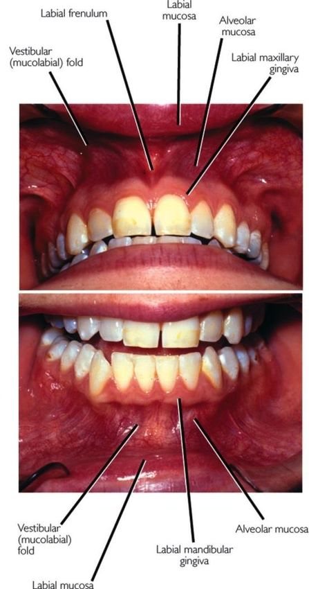
The gums are part of the soft tissue lining of the mouth. The
gums surround the teeth and provide a seal around them.
[Picture Credit: Gums]
Compared with the soft tissue linings of the lips and cheeks,
most of the gingivae are tightly bound to the underlying
bone which helps resist the friction of food passing over
them. Thus when healthy, it presents an effective barrier to
the barrage of periodontal insults to deeper tissue. Healthy
gums are usually coral pink, but may contain melanin
pigmentation.
The gums comprises connective tissue that consists of two
layers. The outermost gingival layer is a continuation of the
mucous membranes of the oral cavity. The layer beneath
that is composed of fibrous tissue. The gingiva fills the
spaces between the teeth and also surrounds the roots of
every tooth. The gingiva is attached to the cementum layer
of the root as well as to the jaw bone.
Cancer of the Gums
Gum cancer is a type of malignancy that occurs when there
is an uncontrolled growth of cancer cells in the gums. Gum
cancer is a type of oral cancer and a relatively rare form of cancer in general.
Gum cancer is most treatable and curable if caught in the earliest stage of the disease. Gum cancer
grows relatively slowly, but untreated and/or advanced gum cancer can spread into the deeper tissues
of the mouth and neck. In advanced stages, gum cancer can spread through the lymph nodes and
blood to other parts of the body where the cancer cells can form another cancerous tumour. This is
called metastasis. Gum cancer and other forms of oral cancer have a high risk of recurring after
treatment.
Researched and Authored by Prof Michael C Herbst
[D Litt et Phil (Health Studies); D N Ed; M Art et Scien; B A Cur; Dip Occupational Health; Dip Genetic Counselling; Dip
Audiometry and Noise Measurement; Diagnostic Radiographer; Medical Ethicist]
Approved by Ms Elize Joubert, Chief Executive Officer [BA Social Work (cum laude); MA Social Work]
January 2021 Page 1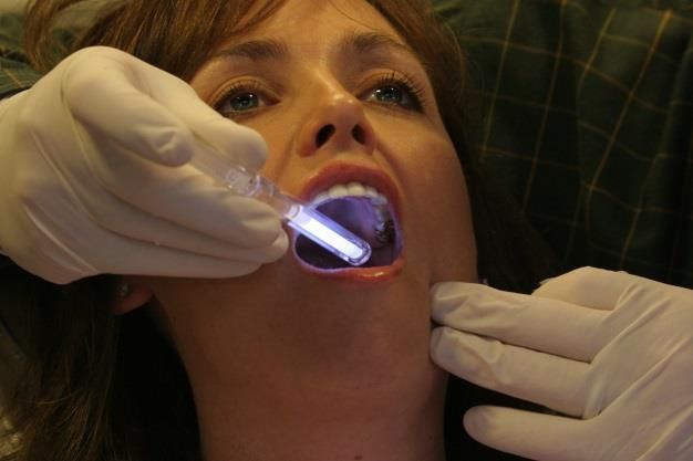
Naik, R., Chatura, K.R., Mujib, B.R.A., Verrappa, S. & Gopal, S. 2019.
“Metastatic dissemination to the oral cavity is extremely rare and constitutes about 1%-2.1% of all
oral malignancies. The very first sign of the metastasis into the oral cavity indicates an occult
malignancy in the distant site. It gives the evidence of widespread disease with an average survival
rate of less than 7 months. Adenocarcinomas account for up to 60% of all metastatic neoplasms of
unknown primary origin. Here, we report a case of metastatic adenocarcinoma of unknown primary
origin in a 60-year-old male patient in the upper and lower gingiva without involvement of the
underlying bone which is a very rare case reported in the literature till date.”

Malinoski, H., Reddy, R., Cohen, D.M., Bhattacharyya, I., Islam, M.N. & Brwn, T.L.4 th . 2019.
PURPOSE: To describe and discuss the demographic and clinical features of oral melanomas, which
are relatively rare but deadly neoplasms, and list the criteria for their diagnosis to increase early
detection.
MATERIALS AND METHODS: An institutional review board-approved retrospective search of oral
melanomas was performed in the archives of the University of Florida Oral and Maxillofacial
Pathology Biopsy Service (Gainesville, FL) from 2015 through 2018. Exclusion criteria included cases
with inconclusive diagnosis, skin involvement, and missing clinical data or slide material. Of 7 patients
with a diagnosis of melanoma of the head and neck region, 6 (87.5%) were found to be diagnosed
with oral melanomas and met the inclusion criteria.
RESULTS: All 6 patients were at least 45 years (range, 45 to 87 yr). The male-to-female ratio was 4:2.
Three patients were asymptomatic and 3 experienced symptoms, including pain, swelling, and
tingling. Seven lesions were detected in these 6 patients. Three of these lesions were located on the
maxillary gingiva, 2 were on the mandibular gingiva, and 2 involved the palate. Two lesions were
diagnosed as spindle cell melanoma, 4 were diagnosed as melanoma, and 1 was diagnosed as a
mucosal lentiginous melanoma.
CONCLUSION: Oral melanomas should be considered in the differential diagnosis of oral pigmented
lesions, especially on the gingiva or palate, in middle-age and elderly patients. Oral melanomas have
a male bias. In addition, supportive criteria enabling early diagnosis of oral melanomas is addressed.

Incidence of Gum Cancer in South Africa
According to the outdated National Cancer Registry (2017) the following number of Cancer of the
Gums cases was histologically diagnosed in South Africa during 2017:

 Group - Males                    Actual              Estimated        Percentage of
 2017                           No of Cases         Lifetime Risk       All Cancers
 All males                          24                 1:5 572             0,06%
 Asian males                         0                     -                  -
 Black males                         9                 1:9 303             0,06%
 Coloured males                      3                 1:3 135             0,06%
 White males                        12                 1:2 552             0,06%

 Group - Females                  Actual              Estimated        Percentage of
 2017                           No of Cases         Lifetime Risk       All Cancers
 All females                        15                 1:11 905            0,04%
 Asian females                       2                  1:4 609            0,16%
 Black females                       7                 1:20 834            0,04%
 Coloured females                    0                     -                  -
 White females                       6                  1:5 051            0,04%

Researched and Authored by Prof Michael C Herbst
[D Litt et Phil (Health Studies); D N Ed; M Art et Scien; B A Cur; Dip Occupational Health; Dip Genetic Counselling; Dip
Audiometry and Noise Measurement; Diagnostic Radiographer; Medical Ethicist]
Approved by Ms Elize Joubert, Chief Executive Officer [BA Social Work (cum laude); MA Social Work]
January 2021                                                                                                             Page 2

The frequency of histologically diagnosed cases of Cancer of the Gums in South Africa for 2017 was as
follows (National Cancer Registry, 2017):

 Group - Males                    0 – 19         20 – 29         30 – 39       40 – 49       50 – 59      60 – 69       70 – 79         80+
 2017                             Years           Years           Years         Years         Years        Years         Years         Years
 All males                           0              1               0             2             7            3             8             3
 Asian males                         0              0               0             0             0            0             0             0
 Black males                         0              0               0             2             3            1             2             1
 Coloured males                      0              0               0             0             0            0             2             1
 White males                         0              1               0             0             4            2             4             1

 Group - Females                  0 – 19         20 – 29         30 – 39       40 – 49       50 – 59      60 – 69       70 – 79         80+
 2017                             Years           Years           Years         Years         Years        Years         Years         Years
 All females                         0              0               0             1             5            3             6             0
 Asian females                       0              0               0             1             1            0             0             0
 Black females                       0              0               0             0             4            1             2             0
 Coloured females                    0              0               0             0             0            0             0             0
 White females                       0              0               0             0             0            2             4             0
N.B. In the event that the totals in any of the above tables do not tally, this may be the result of uncertainties as to the age, race or sex of
the individual. The totals for ‘all males’ and ‘all females’, however, always reflect the correct totals.

Causes and Risk Factors for Gum Cancer
Men get gum cancer more often than women and people over the age of 40 are affected more often
than younger people. However, more recently, gum cancer is occurring in greater numbers in younger
people.

The possibility of getting gum cancer is increased dramatically in those who smoke, those who chew
tobacco and those who don't take care of their oral health with regular brushing, flossing and dental
checkups.
People at risk for developing gum cancer include:
• smokers (cigarettes, cigars and pipes)
• using smokeless tobacco (chewing tobacco, snus)
• people who drink alcohol excessively
• being infected with the human papillomavirus (HPV)
• a diet that is low in fruits and vegetables

Suprewicz, Ł., Tokajuk, G., Cieśluk, M., Deptuła, P., Sierpińska, T., Wolak, P., Wollny, T., Tokajuk, J.,
Głuszek, S., Piktel, E. & Bucki, R. 2020.
“Understanding the importance of oral microbiota in human health and disease also leads to an
expansion of the knowledge on functional, metabolic, and molecular alterations directly contributing
to oral and systemic pathologies. To date, a compelling number of studies have documented the
crucial role of some oral cavity-occurring microbes in the initiation and progression of cancers.
Although this effect was noted primarily for Fusobacterium spp., the potential impact of other oral
microbes is also worthy of investigation. In this study, we aimed to assess the effect of Enterococcus
faecalis, Actinomyces odontolyticus, and Propionibacterium acnes on the proliferation capability and
mechanical features of gingival cells and cell lines derived from lung, breast, and ovarian cancers. For
this purpose, we incubated selected cell lines with heat-inactivated bacteria and supernatants
collected from biofilms, cultured in both anaerobic and aerobic conditions, in the presence of
surgically removed teeth and human saliva. The effect of oral bacteria on cell population growth is
Researched and Authored by Prof Michael C Herbst
[D Litt et Phil (Health Studies); D N Ed; M Art et Scien; B A Cur; Dip Occupational Health; Dip Genetic Counselling; Dip
Audiometry and Noise Measurement; Diagnostic Radiographer; Medical Ethicist]
Approved by Ms Elize Joubert, Chief Executive Officer [BA Social Work (cum laude); MA Social Work]
January 2021                                                                                                             Page 3
variable, with the highest growth-promoting abilities observed for E. faecalis in relation to human
primary gingival fibroblasts (HGF) and lung cancer A549 cells, and P. acnes in relation to breast cancer
MCF-7 and ovarian cancer SKOV-3 cells. Notably, this effect seems to depend on a delicate balance
between the pro-stimulatory and toxic effects of bacterial-derived products. Regardless of the diverse
effect of bacterial products on cellular proliferation capability, we observed significant alterations in
stiffness of gingival and lung cancer cells stimulated with E. faecalis bacteria and corresponding
biofilm supernatants, suggesting a novel molecular mechanism involved in the pathogenesis of
diseases in oral cavities and tooth tissues. Accordingly, it is proposed that analysis of cancerogenic
features of oral cavity bacteria should be multivariable and should include investigation of potential
alterations in cell mechanical properties. These findings corroborate the important role of oral hygiene
and root canal treatment to assure the healthy stage of oral microbiota.”

Irani, S., Barati, I. & Badiei, M. 2020.
“Gingival tissues are attacked by oral pathogens which can induce inflammatory reactions. The
immune-inflammatory responses play essential roles in the patient susceptibility to periodontal
diseases. There is a wealth of evidence indicating a link between chronic inflammation and risk of
malignant transformation of the affected oral epithelium. Periodontitis is associated with an increased
risk of developing chronic systemic conditions including autoimmune diseases and different types of
cancers. Besides, some risk factors such as smoking, alcohol consumption and human papilloma virus
have been found to be associated with both periodontitis and oral cancer. This review article aimed
to study the current concepts in pathogenesis of chronic periodontitis and oral cancer by reviewing
the related articles.”

Tuominen, H. & Rautava, J. 2020.
“Oral microbiota are among the most diverse in the human body. More than 700 species have been
identified in the mouth, and new sequencing methods are allowing us to discover even more species.
The anatomy of the oral cavity is different from that of other body sites. The oral cavity has mucosal
surfaces (the tongue, the buccal mucosa, the gingiva, and the palate), hard tissues (the teeth), and
exocrine gland tissue (major and minor salivary glands), all of which present unique features for
microbiota composition. The connection between oral microbiota and diseases of the human body
has been under intensive research in the past years. Furthermore, oral microbiota have been
associated with cancer development. Patients suffering from periodontitis, a common advanced
gingival disease caused by bacterial dysbiosis, have a 2-5 times higher risk of acquiring any cancer
compared to healthy individuals. Some oral taxa, especially Porphyromonas gingivalis and
Fusobacterium nucleatum, have been shown to have carcinogenic potential by several different
mechanisms. They can inhibit apoptosis, activate cell proliferation, promote cellular invasion, induce
chronic inflammation, and directly produce carcinogens. These microbiota changes can already be
seen with potentially malignant lesions of the oral cavity. The causal relationship between microbiota
and cancer is complex. It is difficult to accurately study the impact of specific bacteria on carcinoma
development in humans. This review focuses on the elucidating the interactions between oral cavity
bacterial microbiota and cancer. We gather literature on the current knowledge of the bacterial
contribution to cancer development and the mechanisms behind it.”

Bagan, J., Murillo-Cortes, J., Leopoldo-Rodado, M., Sanchis-Bielsa, J.M., Bagan, L. 2019.
BACKGROUND: Proliferative verrucous leukoplakia, recently coined as proliferative leukoplakia (PL),
is associated with a strong tendency to recur after treatment and an elevated rate of malignant
transformation. We compared the clinical characteristics of patients with gingival PL with and without
progression to oral squamous cell carcinoma.

Researched and Authored by Prof Michael C Herbst
[D Litt et Phil (Health Studies); D N Ed; M Art et Scien; B A Cur; Dip Occupational Health; Dip Genetic Counselling; Dip
Audiometry and Noise Measurement; Diagnostic Radiographer; Medical Ethicist]
Approved by Ms Elize Joubert, Chief Executive Officer [BA Social Work (cum laude); MA Social Work]
January 2021                                                                                                             Page 4
METHODS: The patients were divided into two groups: group 1 included 33 patients with gingival PL
that did not progress to cancer, and group 2 included 30 patients with PL who developed malignant
transformation during follow-up. We compared age, sex, tobacco habit, clinical characteristics of
gingival PL lesions, and location, tumor-node-metastasis (TNM) stage, and clinical characteristics of
gingival malignancy between groups.
RESULTS: Female sex was predominant in the group with gingival cancer, and simultaneous
involvement of the buccal mucosa, tongue, and palate was more common in this group than in the
group without cancer. PL lesions were also largest in the group of patients with cancer. Most
gingival cancer occurred in areas with teeth and took the form of oral ulceration. TNM stage I was
most common.
CONCLUSIONS: The simultaneous presence of lesions on the buccal mucosa, grade of lesion
extension, and presence of ulcerative lesion were significantly associated with gingival cancer in
patients with PL.

Signs and Symptoms of Gum Cancer
Regular medical or dental check-ups can detect the early stages of oral cancer or conditions that may
lead to oral cancer.

Common symptoms of oral cancer include:
• patches inside the mouth or on the lips that
   are white, a mixture of red and white, or red
   ▪ white patches (leukoplakia) are the most
       common - white patches sometimes
       become malignant

                               [Picture Credit: Leukoplakia]

    ▪    mixed red and white patches
         (erythroleukoplakia) are more likely than
         white patches to become malignant
    ▪    red patches (erythroplakia) are brightly coloured, smooth areas that often become malignant

                                                                                                [Picture Credit: Erythroplakia]

•    a sore on the lip or in the mouth that won't
     heal
•    bleeding in the mouth
•    loose teeth
•    difficulty or pain when swallowing
•    difficulty wearing dentures
•    a lump in the neck
•    earache

Anyone with these symptoms should see a
doctor or dentist so that any problem can be
diagnosed and treated as early as possible. Most

Researched and Authored by Prof Michael C Herbst
[D Litt et Phil (Health Studies); D N Ed; M Art et Scien; B A Cur; Dip Occupational Health; Dip Genetic Counselling; Dip
Audiometry and Noise Measurement; Diagnostic Radiographer; Medical Ethicist]
Approved by Ms Elize Joubert, Chief Executive Officer [BA Social Work (cum laude); MA Social Work]
January 2021                                                                                                             Page 5
often, these symptoms do not mean cancer. An infection or another problem can cause the same
symptoms.
Diagnosis of Gum Cancer
If the doctor suspects a patient may have oral cancer, one or more of the following tests may be used
to find out if he/she has cancer and whether it has spread:
Biopsy - if any abnormalities are discovered during the exam, a small tissue sample, or biopsy, usually
is taken. This biopsy is important, as it is the only sure way to know if the abnormal area is cancer.
A biopsy may be obtained by:
• Brush biopsy or exfoliative cytology - this relatively new type of biopsy is painless and does not
require anesthetic. The dentist or doctor rotates a small stiff-bristled brush on the area, causing
abrasion or pinpoint bleeding. Cells from the area are collected and examined under a microscope
by a pathologist. If results are inconclusive or show cancer, an incisional biopsy will be completed.
• Incisional biopsy - this is the traditional, most frequently used type of biopsy. The doctor or dentist
surgically removes part or all of the tissue where cancer is suspected. Usually, this procedure is
completed in the doctor's office or clinic under local anesthesia. But if the tumour is inside the
throat, the biopsy may be done in an operating room under general anesthesia.
• Fine-needle-aspiration biopsy (FNA) - this type of biopsy often is used if a patient has a lump in
the neck that can be felt. In this procedure, a thin needle is inserted into the area. Then cells are
withdrawn and examined under a microscope.
Mucosal staining - a blue dye called toluidine blue O is applied to the area where cancer is suspected.
If any blue areas remain after rinsing, they probably will be investigated with a biopsy.
Chemiluminescent light (the emission of light or
luminescence as the result of a chemical reaction) -
after having rinsed the mouth with a mild acid
solution, the mouth is examined with a special light.
Healthy cells do not reflect the light, whereas
cancerous cells will reflect the light.
[Picture Credit: Chemiluminescence]
Imaging tests, which may include:
• Computed axial tomography (CAT) scans
• Positron emission tomography (PET) scans
• Magnetic resonance imaging (MRI) scans
• Chest and dental X-rays
• Barium swallow - also called an upper GI (gastrointestinal) series, this set of X-rays of the
esophagus and stomach may be used to look for other cancers and determine how well the patient
swallows
• Endoscopy
Liu, G-M., Lu, T-C., Liu, Y. & Luo, Y-G. 2020.
Objectives: Early diagnosis of and markers for gingival oral squamous cell carcinoma (OSCC) is
important for effective treatment.
Researched and Authored by Prof Michael C Herbst
[D Litt et Phil (Health Studies); D N Ed; M Art et Scien; B A Cur; Dip Occupational Health; Dip Genetic Counselling; Dip
Audiometry and Noise Measurement; Diagnostic Radiographer; Medical Ethicist]
Approved by Ms Elize Joubert, Chief Executive Officer [BA Social Work (cum laude); MA Social Work]
January 2021 Page 6Methods: The current study performed a whole exome sequencing of gingival OSCC tissues in thirteen Chinese patients to explore exonic mutants. Results: Eighty-five genes emerged as mutants in patients with primary gingival OSCC. CCL4L1 presented a G>A transversion at chr17 17q12, position 36212480, exon 3. KDM5B presented a T>TA insertion at chr1 1q32.1, position 202766506, exon 6. ANKRD36C presented a C>G transition at chr2 2q11.1, position 95945175, exon 18. Conclusion: These three mutants might be new markers of gingival OSCC. The finding may provide new targets to diagnose and treat gingival OSCC. Treatment of Gum Cancer The type of treatment your doctor will recommend depends on the tumour site and how far the cancer has spread. Treatment may include: • Surgery (which may include surgery to the lymph glands draining the area • Chemotherapy • Radiation Therapy • A combination of two or more of the above Recurrent Oral Cavity or Oropharyngeal Cancer When cancer come backs after treatment, it is called recurrent cancer. Recurrence can be local (in or near the same place it started), regional (in nearby lymph nodes), or distant (spread to bone or organs such as the lungs). Treatment options for recurrent cancers depend on the location and size of the cancer, what treatments have already been used and on the person’s general health. If the cancer comes back in the same area and radiation therapy was used as the first treatment, surgery is often the next treatment if possible. Usually, external beam radiation therapy cannot be repeated in the same site except in selected cases. However, brachytherapy can often be used to control the cancer if it has recurred in the place of origin. If the cancer comes back in the lymph nodes in the neck, these are often removed with surgery. This may be followed by radiation. Watters, C., Brar, S. & Pepper, T. 2020. “Oral mucosal cancer is cancer that arises from the lining (mucosa) of the oral cavity. The oral cavity is compromised of the mucosa lining of the lips and cheeks, the teeth, gingiva (gums), anterior two- thirds of the tongue, the floor of the mouth, hard palate, and the retromolar trigone posterior to the wisdom teeth. It has a close anatomical relationship with the oropharynx, the boundary of which is the border between the hard and soft palate, the border between the anterior 2/3rds and posterior 1/3rd of the tongue, and the anterior pillars of the tonsils. The main risk factors are smoking and alcohol consumption. The mainstay of treatment is surgery, often with adjuvant radiotherapy.” Nassiri, A.M., Campbell, B.R., Mannion, K., Sinard, R.J., Nettervil;e. J.L. & Rhode, S.L. 2019. OBJECTIVES: To measure disease-free, disease-specific, and overall survival among patients with T4aN0M0 mandibular gingival squamous cell carcinoma who were treated with surgery alone. STUDY DESIGN: Case series with chart review. SETTING: Tertiary care center. Researched and Authored by Prof Michael C Herbst [D Litt et Phil (Health Studies); D N Ed; M Art et Scien; B A Cur; Dip Occupational Health; Dip Genetic Counselling; Dip Audiometry and Noise Measurement; Diagnostic Radiographer; Medical Ethicist] Approved by Ms Elize Joubert, Chief Executive Officer [BA Social Work (cum laude); MA Social Work] January 2021 Page 7
SUBJECTS AND METHODS: A retrospective chart review was performed of all adult patients treated
surgically with an oral cavity composite resection between January 2005 and March 2017. Among
other data, patient preoperative characteristics were recorded (eg, age, sex, smoking history, alcohol
use, and clinical stage); operative notes were reviewed to determine tumor subsite involvement,
reconstruction method, and intraoperative surgical complications; and pathology reports were
evaluated for various pathologic findings. Survival outcomes were determined with Kaplan-Meier
analysis.
RESULTS: The mean follow-up was 18.5 months (range, 0.1-100). The 1- and 5-year disease-free
survival rates were 90.5% and 84.5%, respectively, while the 1- and 5-year disease-specific survival
rates were 87.8% and 81.9%. The 1- and 5-year overall survival rates were 86.4% and 80.6%.
CONCLUSIONS: Patients with T4aN0M0 squamous cell carcinoma of the mandibular gingiva treated
with surgery alone have a 5-year overall survival of 80.6%. Treatment with surgery alone obviates
morbidities associated with adjuvant therapy while upholding survival outcomes.

About Clinical Trials
Clinical trials are research studies that involve people. They are conducted under controlled
conditions. Only about 10% of all drugs started in human clinical trials become an approved drug.

Clinical trials include:
     • Trials to test effectiveness of new treatments
     • Trials to test new ways of using current treatments
     • Tests new interventions that may lower the risk of developing certain types of cancers
     • Tests to find new ways of screening for cancer

The South African National Clinical Trials Register provides the public with updated information on
clinical trials on human participants being conducted in South Africa. The Register provides
information on the purpose of the clinical trial; who can participate, where the trial is located, and
contact details.

Medical Disclaimer
This Fact Sheet is intended to provide general information only and, as such, should not be considered
as a substitute for advice, medically or otherwise, covering any specific situation. Users should seek
appropriate advice before taking or refraining from taking any action in reliance on any information
contained in this Fact Sheet. So far as permissible by law, the Cancer Association of South Africa
(CANSA) does not accept any liability to any person (or his/her dependants/estate/heirs) relating to
the use of any information contained in this Fact Sheet.

Whilst the Cancer Association of South Africa (CANSA) has taken every precaution in compiling this
Fact Sheet, neither it, nor any contributor(s) to this Fact Sheet can be held responsible for any action
(or the lack thereof) taken by any person or organisation wherever they shall be based, as a result,
direct or otherwise, of information contained in, or accessed through, this Fact Sheet.

Researched and Authored by Prof Michael C Herbst
[D Litt et Phil (Health Studies); D N Ed; M Art et Scien; B A Cur; Dip Occupational Health; Dip Genetic Counselling; Dip
Audiometry and Noise Measurement; Diagnostic Radiographer; Medical Ethicist]
Approved by Ms Elize Joubert, Chief Executive Officer [BA Social Work (cum laude); MA Social Work]
January 2021                                                                                                             Page 8
Sources and References Consulted or Utilised American Cancer Society http://www.cancer.org/cancer/oralcavityandoropharyngealcancer/detailedguide/oral-cavity-and-oropharyngeal-cancer- treating-by-stage Bagan, J., Murillo-Cortes, J., Leopoldo-Rodado, M., Sanchis-Bielsa, J.M., Bagan, L. 2019. Oral cancer on the gingiva in patients with proliferative leukoplakia: a study of 30 cases. J Periodontol. 2019 May 9. doi: 10.1002/JPER.18-0620. [Epub ahead of print] Cancer Research UK http://www.cancerresearchuk.org/about-cancer/type/mouth-cancer/treatment/surgery/types-of-mouth-cancer- operations Chemiluminescence https://www.google.co.za/search?q=chemiluminescent+light&source=lnms&tbm=isch&sa=X&ei=LpX7UajlLcOZhQfh_YCICg &sqi=2&ved=0CAcQ_AUoAQ&biw=1366&bih=614#facrc=_&imgdii=_&imgrc=tDeI2gOrXaVj4M%3A%3BEmNr4YZekGhutM %3Bhttp%253A%252F%252Fc1- preview.prosites.com%252F67026%252Fwy%252Fimages%252F0106zila%252520062.jpg%3Bhttp%253A%252F%252Fwww .twincitiesdentalstudio.com%252Fvizilite%2525C2%2525AE-plus%252F%3B3504%3B2336 E-How http://www.ehow.com/about_4706811_beginning-stages-gum-cancer.html Endo, H., Takayama, K., Mitsudo, K., Nakamura, T., Seto, I., Yamaguchi, H., Ono, T., Suzuki, M., Azami, Y., Wada, H., Murakami, M. & Tohnai, I. 2018. Proton beam therapy in combination with intra arterial infusion chemotherapy for T4 squamous cell carcinoma of the maxillary gingiva. Cancers (Basel). 2018 Sep 15;10(9). pii: E333. doi: 10.3390/cancers10090333. Erythroplakia https://www.google.co.za/search?q=erythroplakia+pictures&source=lnms&tbm=isch&sa=X&ei=tWb7UcLzCsK4hAffq4DAD Q&ved=0CAcQ_AUoAQ&biw=1366&bih=614#facrc=_&imgdii=_&imgrc=QbRcn1gqr0XrRM%3A%3Bi86yGdMlesmROM%3Bh ttp%253A%252F%252Fwww.rborl.org.br%252Fconteudo%252Facervo%252Fimages%252F75-02-21- fig01.jpg%3Bhttp%253A%252F%252Fwww.rborl.org.br%252Fconteudo%252Facervo%252Fprint_acervo_english.asp%253Fi d%253D3823%3B350%3B271 Gums http://medical-dictionary.thefreedictionary.com/Gum+(anatomy) Irani, S., Barati, I. & Badiei, M. 2020. Periodontitis and oral cancer – current concepts of the etiopathogenesis. Oncol Rev. 2020 Mar 18;14(1):465. Leukoplakia https://www.google.co.za/search?q=leukoplakia+pictures&source=lnms&tbm=isch&sa=X&ei=gmf7UYTmA86zhAeg7IGQBw &sqi=2&ved=0CAcQ_AUoAQ&biw=1366&bih=614#facrc=_&imgdii=_&imgrc=9Yivy52EL3rpvM%3A%3BvzRYyOn17bTivM%3 Bhttp%253A%252F%252F1.bp.blogspot.com%252F-iC-sxcZFeBg%252FThsNMSFsqXI%252FAAAAAAAAAHg%252FHWthu- zCg1A%252Fs320%252Fleukoplakia%252Bof%252Bgingiva.jpg%3Bhttp%253A%252F%252Fdentallecnotes.blogspot.com%2 52F2011%252F07%252Fleukoplakia-is-most-common-premalignant.html%3B780%3B503 Liu, G-M., Lu, T-C., Liu, Y. & Luo, Y-G. 2020. Gene expression analysis of primaty gingival cancer by whole exome sequencing in thirteen Chinese patients. Int J Clin Exp Pathol. 2020 Jul 1;13(7):1909-1914. Malinoski, H., Reddy, R., Cohen, D.M., Bhattacharyya, I., Islam, M.N. & Brwn, T.L.4th . 2019. Oral melanomas: a case series of a deadly neoplasm. J Oral Maxillofac Surg. 2019 Mar 26. pii: S0278-2391(19)30343-X. doi: 10.1016/j.joms.2019.03.018. [Epub ahead of print] MD Anderson Cancer Center http://www.mdanderson.org/patient-and-cancer-information/cancer-information/cancer-types/oral- cancer/diagnosis/index.html Researched and Authored by Prof Michael C Herbst [D Litt et Phil (Health Studies); D N Ed; M Art et Scien; B A Cur; Dip Occupational Health; Dip Genetic Counselling; Dip Audiometry and Noise Measurement; Diagnostic Radiographer; Medical Ethicist] Approved by Ms Elize Joubert, Chief Executive Officer [BA Social Work (cum laude); MA Social Work] January 2021 Page 9
MedicineNet.Com
http://www.medicinenet.com/oral_cancer/page3.htm#oral_cancer_whos_at_risk

Naik, R., Chatura, K.R., Mujib, B.R.A., Verrappa, S. & Gopal, S. 2019. Gingival metastasis of adenocarcinoma with an
unknown occult primary. J Family Med Prim Care. 2019 Mar;8(3):1246-1249. doi: 10.4103/jfmpc.jfmpc_132_19.

Nassiri, A.M., Campbell, B.R., Mannion, K., Sinard, R.J., Nettervil;e. J.L. & Rhode, S.L. 2019. Survival outcomes in T4N0M0
mandibular gingival squamous cell carcinoma treated with surgery alone. Otolaryngol Head neck Surg. 2019 May;160(5):870-
875. doi: 10.1177/0194599818821892. Epub 2019 Jan 1.

National Cancer Institute
http://www.cancer.gov/clinicaltrials/learningabout/what-are-clinical-trials
Oral Cancer Foundation
http://www.oralcancerfoundation.org/treatment/

Prenhall
http://wps.prenhall.com/wps/media/objects/2976/3047536/MC20.pdf

Right Diagnosis
http://www.rightdiagnosis.com/g/gum_cancer/

Suprewicz, Ł., Tokajuk, G., Cieśluk, M., Deptuła, P., Sierpińska, T., Wolak, P., Wollny, T., Tokajuk, J., Głuszek, S., Piktel, E.
& Bucki, R. 2020. Bacteria Residing at Root Canals Can Induce Cell Proliferation and Alter the Mechanical Properties of
Gingival and Cancer Cells. Int J Mol Sci. 2020 Oct 24;21(21):7914.

Tuominen, H. & Rautava, J. 2020. Oral microbiota and cancer development. Pathobiology. 2020 Nov 11;1-11.

Watters, C., Brar, S. & Pepper, T. 2020. Oral mucosa cancer. In: StatPearls [Internet]. Treasure Island (FL): StatPearls
Publishing; 2020 Jan. 2020 Dec 5.

Researched and Authored by Prof Michael C Herbst
[D Litt et Phil (Health Studies); D N Ed; M Art et Scien; B A Cur; Dip Occupational Health; Dip Genetic Counselling; Dip
Audiometry and Noise Measurement; Diagnostic Radiographer; Medical Ethicist]
Approved by Ms Elize Joubert, Chief Executive Officer [BA Social Work (cum laude); MA Social Work]
January 2021                                                                                                           Page 10
You can also read