Oral-3D: Reconstructing the 3D Structure of Oral Cavity from Panoramic X-ray
←
→
Page content transcription
If your browser does not render page correctly, please read the page content below
Oral-3D: Reconstructing the 3D Structure of Oral Cavity from Panoramic X-ray
                                                                       Weinan Song * , Yuan Liang * , Jiawei Yang, Kun Wang, Lei He
                                                                                         University of California, Los Angeles, USA

                                                                        Abstract
arXiv:2003.08413v4 [eess.IV] 9 Jan 2021

                                            Panoramic X-ray (PX) provides a 2D picture of the patient’s
                                            mouth in a panoramic view to help dentists observe the in-
                                            visible disease inside the gum. However, it provides limited
                                            2D information compared with cone-beam computed tomog-
                                            raphy (CBCT), another dental imaging method that gener-
                                            ates a 3D picture of the oral cavity but with more radiation
                                            dose and a higher price. Consequently, it is of great interest
                                            to reconstruct the 3D structure from a 2D X-ray image, which
                                            can greatly explore the application of X-ray imaging in den-
                                            tal surgeries. In this paper, we propose a framework, named
                                            Oral-3D, to reconstruct the 3D oral cavity from a single PX
                                            image and prior information of the dental arch. Specifically,
                                            we first train a generative model to learn the cross-dimension       Figure 1: An overview of Oral-3D. We first back-project the
                                            transformation from 2D to 3D. Then we restore the shape of           panoramic image into a flattened 3D image of the oral cavity
                                            the oral cavity with a deformation module with the dental arch
                                                                                                                 with a generative network, then we utilize the dental arch
                                            curve, which can be obtained simply by taking a photo of
                                            the patient’s mouth. To be noted, Oral-3D can restore both           information to map the generation result into a curved plane
                                            the density of bony tissues and the curved mandible surface.         to reconstruct the final 3D structure.
                                            Experimental results show that Oral-3D can efficiently and
                                            effectively reconstruct the 3D oral structure and show criti-
                                            cal information in clinical applications, e.g., tooth pulling and    39.4× radiation (Brooks 2009) and takes 3.7× of the price
                                            dental implants. To the best of our knowledge, we are the first
                                            to explore this domain transformation problem between these
                                                                                                                 (Petersen et al. 2014) on average than PX. This problem is
                                            two imaging methods.                                                 especially evident for those sensitive to the radiation dose
                                                                                                                 and the developing countries where people are unwilling to
                                                                                                                 invest much in dental healthcare. Therefore, it is of great
                                                                   Introduction                                  interest to directly reconstruct the 3D structure of the oral
                                          Extra-oral imaging techniques such as PX and CBCT are                  cavity from a PX image.
                                          widely used in dental offices as examination methods before               However, it is of great challenge to reconstruct a 3D object
                                          the treatment. Both methods can show detailed bone infor-              from a single 2D image due to the lack of spatial information
                                          mation, including the tooth, mandible, and maxilla, of the             in the rendering direction. Most works rely on additional in-
                                          entire oral cavity. However, during the imaging process of             formation, such as shadow or prior shape of the object, to
                                          PX, the X-ray tube moves around the patient’s head and can             regularize the reconstruction result. Furthermore, this prob-
                                          only take a 2D panoramic picture. This has limited its appli-          lem is more difficult for the oral cavity due to the compli-
                                          cation in the cases when the disease needs to be located. In           cated shape of the mandible and detailed density informa-
                                          comparison, CBCT can reconstruct the whole 3D structure                tion of the teeth. To overcome such challenges, we propose
                                          of the lower head with divergent X-rays and provide abun-              a two-stage framework, named Oral-3D, to generate a high-
                                          dant information about the health condition of oral cavity.            resolution 3D structure of the oral cavity by decoupling the
                                          Nevertheless, the patient needs to take more radiation dose            reconstruction process of shape and density. We first train
                                          and pay a higher price during a CBCT scan. We summarize                a generation model to extract detailed density information
                                          the characteristics of these two imaging methods in Table 1.           from the 2D space, then restore the mandible shape with the
                                          We can see that although CBCT can provide more informa-                prior knowledge of the dental arch. Although our method
                                          tion in clinical applications (Momin et al. 2009), it generates        can not totally replace CBCT in the dental examination, we
                                                                                                                 provide a compromise solution to obtain the 3D oral struc-
                                             * Equal   Contributions                                             ture when only the PX is available.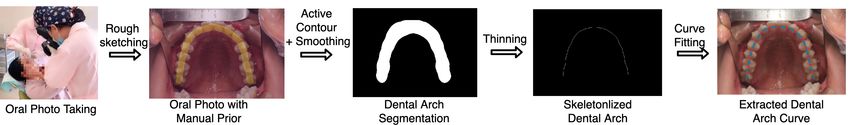
Table 1: A comparison of CBCT and panoramic X-ray on common dental disease.

     Imaging    Dimension       Imaging Cost         Radiation Dose    Diagnostic Accuracy   Wisdom   Tooth   Implant Orthodontics
     Method                 (Petersen et al. 2014)   (Brooks 2009)     (Momin et al. 2009)    Tooth   Decay   Planning
      CBCT         3D             e 184.44           58.9-1025.4 µSv         94.8%             3        3        3          3
       PX          2D             e 49.29             5.5-22.0 µSv           83.3%             3        3        7          7

   Our work can be summarized as a combination of a                    method for instance-level segmentation and identification of
single-view reconstruction problem and a cross-modality                teeth in the CBCT image. (Lee et al. 2018) trains a deep
transformation problem, where the model should recover                 neural network to detect and diagnose dental caries from pe-
both the shape and density information of the target object            riapical radiographic images. (Imangaliyev et al. 2016) de-
from a single image. We show an overview of Oral-3D in                 signs a classification model for red auto-fluorescence plaque
Figure 1. At first, we train a generation network to learn the         images to assist in detecting dental caries and gum diseases.
cross-dimension transformation that can back-project the 2D            (Prajapati, Nagaraj, and Mitra 2017) uses transfer learning
PX image into 3D space, where the depth information of                 to classify three different oral diseases for X-ray images. Al-
teeth and mandible can be learned automatically from the               though these methods have improved oral healthcare service
paired 2D and 3D images. In the second step, we register               by providing intelligent assistance, the model needs to be
this generated 3D image, a flattened oral structure, into a            trained with annotations on large datasets, which requires
curved plane to restore the original shape according to the            both professional knowledge and tedious labour. Compare
dental arch. This prior knowledge effectively restricts the            with these works, our model helps dental healthcare without
shape and location of the 3D structure and can be obtained in          the supervision of labelled data, where the 3D reconstruc-
many ways, such as by fitting the β function with the width            tion is learned from the latent relationship between 2D and
and depth of the mouth (Braun et al. 1998). To show the                3D images.
effectiveness of our framework, we first compare Oral-3D
with other methods on synthesized images generated from a
CBCT dataset. Then we evaluate the reconstruction results              Cross-Modality Transfer in Medical Imaging
for some clinical cases to prove the feasibility of our method.
Experimental results show that Oral-3D can reconstruct the             The target of cross-modality transfer is to find a non-linear
3D oral structure with high quality from a single panoramic            relationship between medical images in different modali-
X-ray image and keep the density information simultane-                ties. It can help reduce the extra acquisition time and ad-
ously. In conclusion, we make the following contributions:             ditional radiation in a medical examination or provide ad-
• We are the first to explore the cross-modality transfer of           ditional training samples without repetitive annotation work
  images in different dimensions for dental imaging by deep            to augment the dataset. Most works take this as a pix-to-pix
  learning. In addition to restoring the 3D shape and surface          problem, where the layout and the structure are consistent,
  of the bone structure, our model can restore the density in-         but the colour distribution is changed after the transforma-
  formation simultaneously, which is of great help for den-            tion between images in different modalities. For example,
  tal diagnosis.                                                       as shown in Figure 2, (Costa et al. 2017) takes the vessel
                                                                       tree of eyes as a condition to synthesis new images for fun-
• We decouple the reconstruction process for density and               dus photography. (Choi and Lee 2018) proposes a generation
  shape recovery by proposing a deformation module that                network to produce realistic structural MR images from flor-
  embeds a flattened 3D image into a curved plane. This                betapir PET images. However, few studies have discussed
  has not been addressed in previous research and can sig-             the cross-modality transfer problem from a lower-dimension
  nificantly improve the reconstruction performance.                   image to a higher-dimension one, which is more challeng-
• We propose an automatic method to generate paired 2D                 ing as the model needs to infer high-dimension information
  and 3D images to train and evaluate the reconstruction               from the lower-dimension image. We only find two works
  models, where Oral-3D achieves relatively high perfor-               that achieve a similar target to ours. Specifically, (Henzler
  mance and can show key features of some typical cases.               et al. 2018) uses an encoder-decoder network to reconstruct
  Meanwhile, we propose a workflow to evaluate our model               3D skull volumes of 175 mammalian species from 2D cra-
  on a real-world dataset, which indicates the feasibility of          nial X-rays, but the result is subject to too much ambiguity.
  clinical applications.                                               To improve the visual quality, (Ying et al. 2019) utilizes bi-
                                                                       planar X-rays to extract 3D anatomical structures of body
                      Related Work                                     CT with adversarial training and reconstruction constraints.
                                                                       However, our problem is quite different from theirs as the
Deep Learning for Oral Health                                          PX image can not be synthesized only from the orthogonal
Deep learning has dramatically promoted the computer as-               projection over the corresponding CT. Besides, our task is
sistance system for dental healthcare by automatically learn-          more challenging due to the complicated structure of the oral
ing feature representations from large amounts of data. For            cavity, where the model is required to restore more details of
example, (Cui, Li, and Wang 2019) proposes an automatic                the teeth and mandible.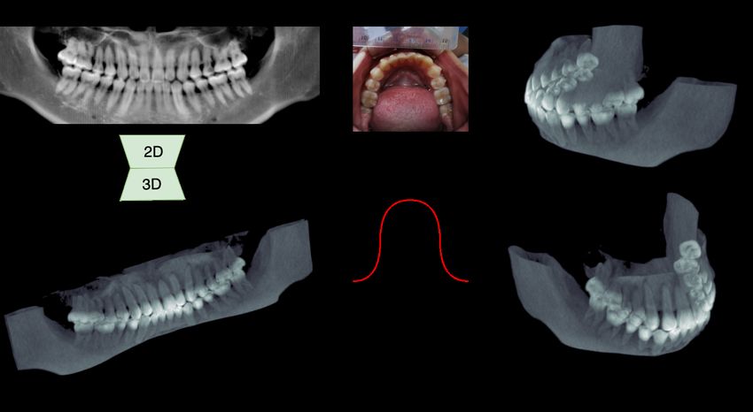
inator D in an adversarial way. The generator learns to out-
                                                                 put a fake image from a random vector to deceive the dis-
                                                                 criminator, while the discriminator tries to distinguish sam-
                                                                 pling data between real and fake images. As we aim to gen-
                                                                 erate the consistent 3D content from the semantic informa-
                                                                 tion of the panoramic X-ray image, we utilize conditional
                                                                 GANs (Mirza and Osindero 2014) as the generative model
Figure 2: We show some examples of cross-modality trans-         to learn the back-projection transformation.
fer for Retina → Fundus (Costa et al. 2017) and PET →
MRI (Choi and Lee 2018), where the source image and the
target image usually contains consistent physiological struc-    Objective Function To improve the generation quality
tures although in different modalities.                          and guarantee the stable training process, we use LSGAN
                                                                 (Mao et al. 2017) as the keystone to train the generator and
                                                                 discriminator. The adversarial loss can be summarized as:
3D Reconstruction from 2D Image
                                                                       LossD =Ey (D(y) − 1)2 + Ex D(G(x))2

Recent work of 3D reconstruction from 2D images can be
concluded as two categories: multi-view reconstruction and                                                                 (1)
                                                                        LossG =Ex D(G(x)) − 1)2 ,

single-view reconstruction. For the first one, the method
generally requires little prior knowledge about the 3D ob-       where x is the PX image and y is the flattened oral structure.
ject as the images taken from multiple angles can restrict the
                                                                    To maintain the structural consistency of the input and
reconstruction shape. For example, (Kolev, Brox, and Cre-
                                                                 generation result, we also introduce the reconstruction loss
mers 2012) computes the most probable 3D shape that gives
                                                                 and projection loss to improve the generation quality. These
rise to the observed colour information from a series of cal-
                                                                 proposed loss functions can bring voxel-wise and plane-wise
ibrated 2D images. (Choy et al. 2016) learns the mapping
                                                                 regularization to the generation network, which can be de-
function from arbitrary viewpoints to a 3D occupancy grid
                                                                 fined as:
with a 3D recurrent neural network. As a comparison, re-
construction from a single-view image usually requires ad-                  LossR =Ex,y (y − G(x))2

ditional information, e.g., prior shape, to inference the ob-                                                               (2)
                                                                            LossP =Ex,y (P (y) − P (G(x)))2 ,

ject shape. As such, (Yang et al. 2018) proposes a unified
framework trained with a small amount of pose-annotated
images to reconstruct a 3D object. (Wu et al. 2018) takes the    where the function P () is achieved by orthogonal projec-
adversarially learned shape priors as a regularizer to penal-    tions along each dimension of the generated 3D image. In
ize the reconstruction model. However, the PX image can be       summary, the total optimization problem can be concluded
either seen as a single-view image taken by a moving cam-        as:
era or a concatenate image blended with multiple views. In         D∗ = arg min LossD
this paper, we take our problem as the first kind to decou-                    D
ple the reconstruction process for the bone density and the        G∗ = arg min λ1 · LossG + λ2 · LossR + λ3 · LossP .
mandible shape. In the experiment, we also show that this                      G
                                                                                                                            (3)
can significantly promote performance over the multi-view
reconstruction model both in quality and quantity.
                                                                 Generator During the X-ray imaging, the depth informa-
                         Method                                  tion can be reflected in the absorption of radiation through
In this section, we introduce our framework that reconstructs    the bone. Therefore it is reasonable to extract the thickness
a high-resolution 3D oral cavity from a 2D PX image. We          of the tooth and the mandible from a PX image. Then the
choose to break this problem into two stages to recover more     objective for the generator is to find a cross-dimension trans-
details of the bone density. We show the structure of Oral-      formation G from 2D to 3D, which can be denoted as:
3D, which consists of a back-projection module and a defor-
mation module in Figure 3. The back-projection module de-                               2D     3D
                                                                                   G : IH×W → IH×W ×D ,                     (4)
velops from generative adversarial networks (GAN (Good-
fellow et al. 2014)), where the generator is trained to learn    where I 2D is the PX image with a size of H × W and I 3D is
a back-projection transformation by exploring the depth in-      the flattened 3D structure with a size of H × W × D. In this
formation contained in the X-ray image. The deformation          paper, we utilize 2D convolution to retrieve the latent depth
module takes in the generated 3D image (Image c) from the        information. The 3D information is embedded into differ-
back-projection module and the dental arch (Image e) to re-      ent channels of feature maps. As shown in Fig. 3, the en-
store the curved shape of the mandible.                          coding network decreases the resolution of feature maps but
                                                                 increases the number of feature channels, while the decod-
Back-Projection Module                                           ing network increases the resolution to generate a 3D object.
GANs have proved to be an effective model to learn latent        The output voxel value is restricted to (−1, 1) with a tanh
data distribution by training the generator G and the discrim-   layer at the end.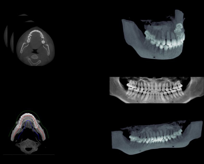
Figure 3: Our framework consists of two modules to decouple the recovery of bone density and the mandible shape. The back-
projection module utilizes a generation network to restore the 3D density information from the 2D space, and the deformation
module transforms the flattened 3D image into a curve plane according to the prior knowledge in the dental arch.

Dense Block Dense connections (Huang et al. 2017) have           paired of 3D images when training the discriminator.
shown compelling advantages for feature extraction in deep
neural networks. This architecture is especially efficient in    Deformation Module
forwarding 3D information as each channel of the output          With the generation of a 3D image from the back-projection
has a direct connection with intermediate feature maps. In       module, the deformation model maps the flattened 3D struc-
the projection module, we utilize two kinds of dense blocks,     ture into the curved space according to the arch curve to
noted as A and B, to extract depth information from the X-       output the final reconstruction object. As shown in the right
ray image. As shown at the bottom of Figure 3, the dense         part of Figure 3, we propose a registration algorithm that
block A explores the depth information by increasing the         can best restore the shape of the oral cavity and keep the
channel number of feature maps. In contrast, the dense block     recovered density information. We first sample the gener-
B fuses feature maps from the up − samplinglayer and             ated 3D image (Image c) into slices (Image f ) in the sagit-
the skip-connections but maintain the number of channels         tal plane, then interpolate these slices along the dental arch
to forward the depth information. In the end, the number of      curve (Image e). To achieve this, we sample a number of
stacked features in the output is equal to the depth of the      points from the curve with equal distance and embed the
generated 3D image.                                              slices into the curve. In the end, we interpolate the voxels
                                                                 between the neighbouring slices to output a smooth 3D im-
Discriminator The discriminator has been frequently              age (Image g). For computation convenience, we combine
used in many generative models to improve the generation         these steps together and conclude it in Algorithm 1, where
quality by introducing an instance-level loss. In the back-      we assume that the generated 3D image and the bone model
projection module, we adopt a patch discriminator intro-         has the same height of H.
duced by (Isola et al. 2017) to improve the generation quality
of tooth edges by learning high-frequency structures in the                             Experiment
flattened 3D structure. We set the patch size as 70 × 70 × 70
and follow a similar structure in (Isola et al. 2017) but re-    Dataset
place 2D convolution with 3D. The discrimination network         As grouped data of PX image, dental arch shape, and 3D
ends with a Sigmoid layer to predict the probability of the      oral structure of the same patient, especially in the same pe-
samples belonging to the real image. To be noted, we sample      riod, is hard to find, we first use synthesized data to evaluate
the same number of 3D patches at the same position from the      the performance. We collect 100 CBCT scans from a major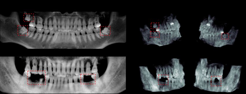
Algorithm 1 Embedding and Interpolation
 1: function REGISTER(Slices, W3D , D3D , curve)
 2:    W, H, D ← SHAPE(Slices)
 3:    OralImage ← ZEROS(W3D , H, D3D )
 4:    SampleP oints ← SAMPLE(curve)
 5:    for i = 0; i < W3D ; i + + do
 6:        for j = 0; j < D3D ; j + + do
 7:            id, dist ← DIST((i, j), SampleP oints)
 8:            Slice ← Slices[id, :, :]
 9:            Slice ← INTERPOLATE(Slice, dist)
10:            OralImage[i, :, j] ← Slice
11:        end for
12:    end for
13:    return OralImage
14: end function

stomatological hospital in China and re-sample these 3D im-      Figure 4: An overview of generating paired data for 3D oral
ages into a size of 288 × 256 × 160. The dataset is finally      structure and 2D panoramic X-ray is shown in this picture.
normalized into a range of (−1, 1) and split into a ratio of     We first get the MIP image from the CBCT scan to obtain
3 : 1 : 1 for training, validation, and testing.                 the dental arch curve (red), and boundaries of the dental area
   An overview of preparing the synthesized data can be seen     (blue and green). Then we obtain the flattened oral structure,
in Figure 4. We first obtain a 2D image in the axial plane       PX image, and the 3D oral structure by re-sampling, projec-
by maximum intensity projection (MIP) (Image b) over the         tion, and extraction, respectively.
CBCT slices (Image a). Then we obtain the dental curve
with a similar method as in (Yun et al. 2019) to estimate the
curve function and boundaries of the dental arch. To gener-      • Overall: To combine these three metrics together, we also
ate the PX image (Image d), we simulate projection with the        define a score S = (P SN R/20 + Dice + SSIM )/3 to
Beer-Lambert absorption-only model along the arch curve.           compare the overall performance of the 3D reconstruc-
This imaging process is similar to the way for a real PX ma-       tion.
chine, where the manufacturer usually improves the imag-
ing quality by designing a trajectory of the camera to fit       Comparison Models
the mandible shape. Finally, we extract the 3D oral struc-
ture (Image e) by removing the unrelated tissues with the        To show the effectiveness and efficiency of Oral-3D, we also
boundaries and generate the flattened 3D structure (Image        compare our framework with other models that work on a
c) by re-sampling along the arch curve.                          similar problem:
                                                                 • Residual CNN: An encoder-decoder network that has
Evaluation Metrics                                                 been introduced in (Henzler et al. 2018) to reconstruct the
• PSNR: Peak signal-to-noise ratio (PSNR) is often used            3D model with a single X-ray.
  to measure the difference between two signals. Compared
                                                                 • GAN: A generative model based on (Goodfellow et al.
  with mean squared error, PSNR can be normalized by the
                                                                   2014) that takes the Res-CNN as the backbone for gener-
  signal range and expressed in terms of the logarithmic
                                                                   ator with reconstruction loss and the same discriminator
  decibel scale. We take this to measure the density recov-
                                                                   as Oral-3D.
  ery of our models.
• Dice: In order to reflect the deformation of the reconstruc-   • R2N2: We transform our task into a multi-view recon-
  tion, we use dice coefficient between our reconstruction         struction problem to train R2N2 (Choy et al. 2016) by tak-
  results and the groundtruth in a volume level of the oral        ing the PX image as a composition of X-ray image taken
  cavity. The 3D volume of the oral cavity is obtained by          from three different views.
  setting a threshold (e.g.,−0.8 over the reconstruction re-     • Auto Encoder: We remove the discriminative network in
  sult.                                                            Oral-3D and keep the encoder-decoder network only in
• SSIM: We use the structure similarity index (SSIM)               the back-projection module.
  (Wang et al. 2004) as the key criterion to quantify the per-
  formance of density recovery.SSIM considers the bright-        Training
  ness, contrast and structure information at the same time      All the experiment are trained by Adam optimizer (Kingma
  and can match better the subjective evaluation of humans.      and Ba 2014) with a batch size of 1 for 300 epochs. The
  It can effectively indicate the reconstruction quality and     learning rate starts at 1 × 10⁻³ and decreases 10 times every
  is widely used in other similar works, such as (Ying et al.    50 epochs. We use the validation data as the stop criterion,
  2019).                                                         and all models converge after 300 epochs. For adversarial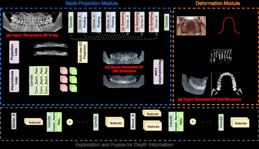
Figure 5: We show the qualitative comparison from different views and rendering ways in this picture. We can see that our
method generates the best results with more detailed density and a more sharp surface.
Comparison with Other Methods
We first show the 3D bone structure in two rendering ways as
in Figure 5, where the volume rendering can show the recon-
structed surface and the maximum projection can indicate
the restored density information. Then we summarize the
evaluation metrics in Table 2 to compare with other meth-
ods. We can see that Oral-3D has the best performance over
other models. Comparing Oral-3D and Auto Encoder with
the Residual CNN and GAN, we can see the importance of
Figure 6: We show reconstruction results for patients with decoupling the back-projection and deformation process. To
wisdom/missing teeth and mark the key features with red be noted, R2N2 achieves the worst performance, where the
bounding boxes. We can see that our method can accurately model only learns the shape of the oral cavity but loses de-
locate these positions, which can be an important reference tails of teeth. This has indicated the defect when converting
during the surgery. the PX image as a collection of multi-view images. Addi-
tionally, we see that Auto Encoder has the closest perfor-
mance to Oral, although the latter has a more clear surface.
networks, i.e. Oral-3D and GAN, we introduce the discrim- This has proved the promotion brought by the adversarial
inative network after 100 epochs to alleviate the influence of loss.
discrimination loss at the beginning.
Identification of Wisdom/Missing Teeth
In this paragraph, we show two of the most common cases
Results in dental healthcare, e.g., dental implants and tooth pulling,
to see if Oral-3D can provide dentist useful reference. Both
In this section, we evaluate the reconstruction performance cases require to locate the operation location before the
of Oral-3D from different different perspectives. We first surgery. In the first row of Figure 6, three wisdom teeth
compare Oral-3D with other methods qualitatively and can be seen clearly on both sides in PX. These features also
quantitatively. Then we show the results of special cases for present in the two sides of the reconstruction results. In the
some common dental applications. In the end, we do clinical second row, the patient misses two teeth on both sides of
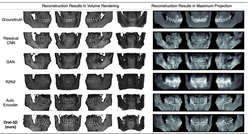
trails by evaluating our method on real-word images. the mandible. While the missing place can also be locatedFigure 7: We show a workflow to apply Oral-3D to obtain the dental arch curve in real-world applications in this picture. We
first take a picture of the patient’s mouth and segment then dental area semi-automatically. Then we use a cubic function to the
fit points sampled from the skeletonized image of the binary mask.
Table 2: Quantitative Evaluation of 3D Reconstruction
Method View Prior D-Net PSNR (dB) SSIM (%) Dice (%) Overall
Residual CNN 1 No No 17.46±9.58 72.90±2.09 57.95±7.43 73.54
GAN 1 No Yes 17.71±1.04 69.96±1.91 57.80±7.76 73.78
R2N2 3 No No 18.06±0.94 71.94±1.36 57.71±6.52 73.32
Oral-3D (Auto-Encoder) 1 Yes No 19.04±0.85 76.78±1.65 69.68±4.98 80.56
Oral-3D (GAN) 1 Yes Yes 19.22±0.83 78.27±1.74 71.28±4.69 81.89
Table 3: Evaluation results of different combination of dis-
crimination loss (DL), reconstruction loss (RL), and projec-
tion loss (PL).
DL only DL+PL DL+RL DL+RL+PL
PSNR 8.06 18.06(+10.00) 19.14(+11.08) 19.22(+11.16) Figure 8: Although the quality decreases in density details
SSIM 46.61 73.02(+26.41) 78.41(+31.80) 78.27(+31.66) for real-word PX, we can still identify each tooth in the re-
Dice 35.50 64.53(+29.03) 70.89(+35.39) 71.28(+35.78) construction result.
Overall 40.79 75.95(+35.16) 81.66(+40.87) 81.89(+41.10)
Table 4: Evaluation results on real-world images is shown in Figure 7. We use cycleGAN (Zhu et al. 2017) to
alleviate the colour variance between the training and test-
Dataset PSNR SSIM Dice
ing PX images. As shown in Table 4, the drop mainly comes
from the PSNR and SSIM, which is because the colour vari-
Real 17.36±0.70 69.30±2.03 71.44±3.66
ance in different CBCT machines. From Figure 8 we can
Synthesized 19.22±0.83 78.27±1.74 71.28±4.69 that although the quality decreases in density details, we can
still identify each tooth in the reconstruction result.
accurately in the reconstruction image. Conclusion
Ablation Study In this paper, we propose a two-stage framework to recon-
struct the 3D structure of the oral cavity from a single 2D
To reveal the factors that influence the reconstruction quality PX image, where individual shape information of the dental
of the generation network, we also do an ablation by chang- arch is provided as prior knowledge. We first utilize a gen-
ing the combination of the loss functions. As shown in Ta- erative model to back-project the 2D image into 3D space,
ble 3, we see that the model shows the worst performance if then deform the generated 3D image into a curved plane to
trained only with the adversarial loss. This is mainly because restore the oral shape. We first use synthesized data to com-
the adversarial loss can not bring voxel-wise optimization. pare with different methods, then evaluate the model with
We can also see that the major improvement comes from the real-world data to see the feasibility in clinical applications.
reconstruction loss, while the projection loss brings much Experimental results show that our model can recover both
less promotion, especially when trained with the reconstruc- the shape and the density information in high resolution. We
tion loss together. This is also reasonable as the reconstruc- hope this work can help improve dental healthcare from a
tion loss can supervise the generation network to learn more novel attitude.
detailed information.
Clinical Trials Acknowledgement
In the end, we evaluate Oral-3D on real-world data from 6 We thank Dr. Liang Chengwen, Dr. Wangbin, and Dr. Wang
patients. The workflow of collecting dental arch information Weiqian for collecting data.References Mao, X.; Li, Q.; Xie, H.; Lau, R. Y.; Wang, Z.; and
Braun, S.; Hnat, W. P.; Fender, D. E.; and Legan, H. L. 1998. Paul Smolley, S. 2017. Least squares generative adversarial
The form of the human dental arch. The Angle Orthodontist networks. In Proceedings of the IEEE International Confer-
68(1): 29–36. ence on Computer Vision, 2794–2802.
Brooks, S. L. 2009. CBCT dosimetry: orthodontic consid- Mirza, M.; and Osindero, S. 2014. Conditional generative
erations. In Seminars in Orthodontics, volume 15, 14–18. adversarial nets. arXiv preprint arXiv:1411.1784 .
Elsevier. Momin, M. A.; Okochi, K.; Watanabe, H.; Imaizumi, A.;
Omura, K.; Amagasa, T.; Okada, N.; Ohbayashi, N.; and
Choi, H.; and Lee, D. S. 2018. Generation of structural MR
Kurabayashi, T. 2009. Diagnostic accuracy of cone-beam
images from amyloid PET: application to MR-less quantifi-
CT in the assessment of mandibular invasion of lower gin-
cation. Journal of Nuclear Medicine 59(7): 1111–1117.
gival carcinoma: comparison with conventional panoramic
Choy, C. B.; Xu, D.; Gwak, J.; Chen, K.; and Savarese, S. radiography. European journal of radiology 72(1): 75–81.
2016. 3d-r2n2: A unified approach for single and multi- Petersen, L. B.; Olsen, K. R.; Christensen, J.; ; and Wen-
view 3d object reconstruction. In European conference on zel, A. 2014. Image and surgery-related costs comparing
computer vision, 628–644. Springer. cone beam CT and panoramic imaging before removal of
Costa, P.; Galdran, A.; Meyer, M. I.; Abràmoff, M. D.; impacted mandibular third molars. Dentomaxillofacial Ra-
Niemeijer, M.; Mendonça, A. M.; and Campilho, A. 2017. diology 43(6): 20140001.
Towards adversarial retinal image synthesis. arXiv preprint Prajapati, S. A.; Nagaraj, R.; and Mitra, S. 2017. Classi-
arXiv:1701.08974 . fication of dental diseases using CNN and transfer learning.
Cui, Z.; Li, C.; and Wang, W. 2019. ToothNet: automatic In 2017 5th International Symposium on Computational and
tooth instance segmentation and identification from cone Business Intelligence (ISCBI), 70–74. IEEE.
beam CT images. In Proceedings of the IEEE Conference Wang, Z.; Bovik, A. C.; Sheikh, H. R.; and Simoncelli, E. P.
on Computer Vision and Pattern Recognition, 6368–6377. 2004. Image quality assessment: from error visibility to
Goodfellow, I.; Pouget-Abadie, J.; Mirza, M.; Xu, B.; structural similarity. IEEE transactions on image process-
Warde-Farley, D.; Ozair, S.; Courville, A.; and Bengio, Y. ing 13(4): 600–612.
2014. Generative adversarial nets. In Advances in neural Wu, J.; Zhang, C.; Zhang, X.; Zhang, Z.; Freeman, W. T.;
information processing systems, 2672–2680. and Tenenbaum, J. B. 2018. Learning shape priors for
Henzler, P.; Rasche, V.; Ropinski, T.; and Ritschel, T. 2018. single-view 3d completion and reconstruction. In Pro-
Single-image Tomography: 3D Volumes from 2D Cranial X- ceedings of the European Conference on Computer Vision
Rays. In Computer Graphics Forum, volume 37, 377–388. (ECCV), 646–662.
Wiley Online Library. Yang, G.; Cui, Y.; Belongie, S.; and Hariharan, B. 2018.
Huang, G.; Liu, Z.; Van Der Maaten, L.; and Weinberger, Learning single-view 3d reconstruction with limited pose
K. Q. 2017. Densely connected convolutional networks. In supervision. In Proceedings of the European Conference on
Proceedings of the IEEE conference on computer vision and Computer Vision (ECCV), 86–101.
pattern recognition, 4700–4708. Ying, X.; Guo, H.; Ma, K.; Wu, J.; Weng, Z.; and Zheng, Y.
2019. X2CT-GAN: reconstructing CT from biplanar X-rays
Imangaliyev, S.; van der Veen, M. H.; Volgenant, C. M.; Kei-
with generative adversarial networks. In Proceedings of the
jser, B. J.; Crielaard, W.; and Levin, E. 2016. Deep learn-
IEEE conference on computer vision and pattern recogni-
ing for classification of dental plaque images. In Interna-
tion, 10619–10628.
tional Workshop on Machine Learning, Optimization, and
Big Data, 407–410. Springer. Yun, Z.; Yang, S.; Huang, E.; Zhao, L.; Yang, W.; and Feng,
Q. 2019. Automatic reconstruction method for high-contrast
Isola, P.; Zhu, J.-Y.; Zhou, T.; and Efros, A. A. 2017. Image- panoramic image from dental cone-beam CT data. Com-
to-image translation with conditional adversarial networks. puter methods and programs in biomedicine 175: 205–214.
In Proceedings of the IEEE conference on computer vision
and pattern recognition, 1125–1134. Zhu, J.-Y.; Park, T.; Isola, P.; and Efros, A. A. 2017. Un-
paired image-to-image translation using cycle-consistent ad-
Kingma, D. P.; and Ba, J. 2014. Adam: A method for versarial networks. In Proceedings of the IEEE international
stochastic optimization. arXiv preprint arXiv:1412.6980 . conference on computer vision, 2223–2232.
Kolev, K.; Brox, T.; and Cremers, D. 2012. Fast joint esti-
mation of silhouettes and dense 3d geometry from multiple
images. IEEE Transactions on Pattern Analysis and Ma-
chine Intelligence 34(3): 493–505.
Lee, J.-H.; Kim, D.-H.; Jeong, S.-N.; and Choi, S.-H.
2018. Detection and diagnosis of dental caries using
a deep learning-based convolutional neural network algo-
rithm. Journal of dentistry 77: 106–111.You can also read