Accuracy of Dental Monitoring 3D digital dental models using photograph and video mode
←
→
Page content transcription
If your browser does not render page correctly, please read the page content below
TECHNO BYTES

Accuracy of Dental Monitoring 3D digital
dental models using photograph
and video mode
Ryan S. Morris,a Lauren N. Hoye,a Mohammed H. Elnagar,b Phimon Atsawasuwan,b
Maria Therese Galang-Boquiren,b Jennifer Caplin,b Grace Costa Viana,b Ales Obrez,b and Budi Kusnotob
Chicago, Ill

      Introduction: This study aimed to test the accuracy of the 3-dimensional (3D) digital dental models generated by
      the Dental Monitoring (DM) smartphone application in both photograph and video modes over successive DM
      examinations in comparison with 3D digital dental models generated by the iTero Element intraoral scanner.
      Methods: Ten typodonts with setups of class I malocclusion and comparable severity of anterior crowding
      were used in the study. iTero Element scans along with DM examination in photograph and video modes
      were performed before tooth movement and after each set of 10 Invisalign aligners for each typodont. Stereo-
      lithography (STL) files generated from the DM examinations in photograph and video modes were superimposed
      with the STL files from the iTero scans using GOM Inspect software to determine the accuracy of both photo-
      graph and video modes of DM technology. Results: No clinically significant differences, according to the Amer-
      ican Board of Orthodontics–determined standards, were found. Mean global deviations for the maxillary arch
      ranged from 0.00149 to 0.02756 mm in photograph mode and from 0.0148 to 0.0256 mm in video mode.
      Mean global deviations for the mandibular arch ranged from 0.0164 to 0.0275 mm in photograph mode and
      from 0.0150 to 0.0264 mm in video mode. Statistically significant differences were found between the 3D
      models generated by the iTero and the DM application in photograph and video modes over successive DM
      examinations. Conclusions: 3D digital dental models generated by the DM smartphone application in photo-
      graph and video modes are accurate enough to be used for clinical applications. (Am J Orthod Dentofacial
      Orthop 2019;156:420-8)

T
      eledentistry is one segment of telemedicine. Tele-                              provider. Teledentistry can be used for remote dental
     dentistry is the combination of telecommunica-                                   consultation, treatment planning and monitoring,
     tions and dentistry to provide more access to                                    appliance fabrication, or on-site job training.1-5
dental care, it involves the digital exchange of clinical in-                             Specific to orthodontics, teledentistry has been
formation between a patient and a health care center or                               shown to be an effective method for positively identi-
                                                                                      fying appropriate referrals, increasing treatment effec-
                                                                                      tiveness, and saving time for both patients and
a
                                                                                      orthodontists.4 The main goal of teledentistry is to allow
 Private practice, Chicago, Ill.
b
 Department of Orthodontics, College of Dentistry, University of Illinois at Chi-     the orthodontist to maintain treatment control of pa-
cago, Chicago, Ill.                                                                   tients who are unable to come into the office for regular
Ryan S. Morris is the principal investigator, collected the data, and wrote the       visits. This is especially important in remote areas or
article. Lauren N. Hoye is the co-principal investigator and collected the data
and wrote the article. Mohammed H. Elnagar is co-investigator, collected the          where there is a shortage of providers and long waits
data, wrote the article, and designed the study. Phimon Atsawasuwan, Maria            for patients who have been appropriately referred.5,6
Therese Galang-Boquiren, Jennifer Caplin, and Ales Obrez are committee mem-               With the creation of smartphones, 2-way, real-time pa-
bers and edited the manuscript. Grace Costa Viana is a committee member and
edited the manuscript and statistics. Budi Kusnoto is a committee adviser, edited     tient-doctor interaction is easier and more feasible than
the manuscript, and designed the study.                                               ever.7 In the orthodontic field, text message applications,
All authors have completed and submitted the ICMJE Form for Disclosure of Po-         chat rooms, and e-mail reminders have been shown to
tential Conflicts of Interest, and none were reported.
Address correspondence to: Mohammed H. Elnagar, Department of Orthodontics            improve oral hygiene as well as reduce treatment duration,
(M/C 841), College of Dentistry, University of Illinois at Chicago, RM 237D, 801 S.   bracket bond failure, and failed or late attendance.4-8
Paulina Street, Chicago, IL 60612-7211; e-mail, melnagar@uic.edu.                         A recently invented technology (Dental Monitoring
Submitted, July 2018; revised and accepted, February 2019.
0889-5406/$36.00                                                                      [DM]) allows orthodontists to monitor their patients
https://doi.org/10.1016/j.ajodo.2019.02.014                                           remotely using a smartphone, using photographs or
420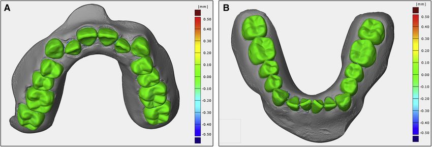
Morris et al                                                                                                        421

video scans taken by patients. Moreover, it can construct    pretreatment or posttreatment changes. To use the Doc-
3-dimensional (3D) digital models from these scans.5,9       tor Dashboard, the orthodontist first uploads an initial
    With continuing advances in 3D digital imaging           3D digital model in stereolithography (STL) file format.
technology, conventional elastomeric impressions and         The patient then takes a pretreatment video or photo-
dental stone casts are being replaced by digital impres-     graph examination with a DM-patented cheek retractor,
sions captured with intraoral scanners.10,11 Digital         using the DM application on their own smartphone (Fig
models provide the advantage of not requiring                1, A). DM uses the initial scan and pretreatment video or
disinfection, but also can be nondestructively modified       photograph examination to establish a baseline of tooth
for better examination of individual teeth. 3D mapping       position and occlusion from which to calculate future
of tooth movement is achievable by superimposing             movements. Successive DM examinations then use the
digital models on stable structures with 3D software         immediately preceding 3D model to perform calcula-
systems.12-14                                                tions to produce the next 3D model.5
    With the trend toward digital impressions, recent            Ten typodonts were set up in class I anterior crowd-
studies have focused on evaluating the accuracy of dig-      ing wax forms. All occlusion screws on the typodont oc-
ital scanners.15-18 Digital impressions captured by          cluders were adjusted to stabilize the occlusion of the
intraoral scanners have been shown to be as accurate         maxillary and mandibular arches and the associated
in both trueness and precision as conventional               forces that could result in unwanted tooth movements
elastomeric impressions, and thus are acceptable for         within the wax.
clinical use.16-18 Numerous studies have tested the              Each of the 10 models was scanned with the iTero
accuracy of full-arch iTero (Align Technology, San           Element intraoral scanner according to the manufac-
Jose, Calif)-generated digital models and demonstrated       turer's instructions. The scans were exported from the
good results.19-23                                           MyAligntech online account as separate open source
    Over the past decade, increasing numbers of adults       STL files for the maxillary and mandibular arches. The
aged 18 years or older have sought orthodontic treat-        STL files were then uploaded to the DM platform as
ment24; this has resulted in a correspondingly greater       the initial 3D reference model for each typodont from
demand for more esthetic and invisible orthodontic ap-       which the next 3D model would be generated. The
pliances.25 Analyses of hundreds of thousands of Invis-      same STL file was also sent to Align Technology through
align cases show that the iTero scanner works better         the Invisalign Doctor site, with instructions to align teeth
with the Invisalign workflow than with traditional poly-      and resolve crowding as much as possible in 10 sets of
vinyl siloxane impressions.26                                maxillary and mandibular aligner trays without using at-
    The objective of this study was to compare the accu-     tachments or moving posterior teeth. After scanning,
racy of 3D digital dental models generated from the          initial photograph and video examinations for each ty-
smartphone application in photograph and video modes         podont were done using the DM mobile application
with 3D digital dental models generated from the iTero       (version 2.38) on an iPhone 7, model A1660 (Apple, Cu-
Element intraoral scanner. The null hypothesis was           pertino, Calif), running iOS 11.2. The initial iTero scans
that there would be no statistically significant mean dif-    were compared with the DM photograph and video re-
ferences in global deviations when comparing 3D digital      cords to ensure that there were no STL file errors from
dental models generated by the smartphone application        the iTero scan or inadvertent tooth movements from
in photograph and video modes with the 3D digital            handling the typodonts during the scanning and
dental models generated by the iTero Element intraoral       capturing examination.
scanner over successive smartphone photograph and                The first of 10 maxillary and mandibular Invisalign
video examinations.                                          trays were then seated onto each typodont and the typo-
                                                             donts were placed in a Polystat Immersion Circulator hot
                                                             water bath (Fisherbrand; Fisher Scientific, Waltham,
MATERIAL AND METHODS
                                                             Mass) at 45 C for 5 minutes. On removal from the water
    DM (Paris, France) is a digital technology software.27   bath, light pressure was applied manually to ensure that
DM technology allows orthodontists to monitor their          the maxillary and mandibular trays were fully seated to
patients remotely using a smartphone, using photo-           achieve the planned tooth movement. The typodonts
graphs or video scans taken by patients. DM comprises        were cooled for 15 minutes at room temperature to re-
3 interconnected platforms: a smartphone application         harden the wax and stabilize the teeth in their new po-
for patients, a patented tooth movement tracking algo-       sitions before removing the Invisalign trays. Each
rithm, and an online Doctor Dashboard on which ortho-        typodont was then scanned using the iTero Element
dontists can view patient treatment progress and             intraoral scanner as tray 1. Photograph and video

American Journal of Orthodontics and Dentofacial Orthopedics                    September 2019  Vol 156  Issue 3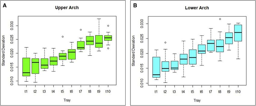
422                                                                                                         Morris et al

         Fig 1. A, Dental Monitoring smartphone application. B, Operator scanning the model with the smart-
         phone. C, Photograph scans.

examinations using the DM application were also                modes and from the iTero Element scans for each typo-
captured as described for each typodont (Fig 1).               dont after each tray. Separate files were generated for
    This process was repeated for all 10 sets of maxillary     the maxillary and mandibular arches. Three sets of
and mandibular Invisalign trays for each of the 10 typo-       maxillary and mandibular arch trays were excluded
donts and labeled as Invisalign trays 1-10.                    because of quality control errors. Thus, 97 maxillary
    Each photograph examination consisted of 7 photo-          arch and 97 mandibular arch files from the DM photo-
graphs: right posterior, right canine, center, left canine,    graph and video examinations and the iTero Element
left posterior, maxillary occlusal, and mandibular             scans were generated for this study.
occlusal. Each video examination consisted of 3 videos            Data were collected from the Dental Monitoring
(6 seconds long): right posterior to the left canine, right    Research and Development headquarters in Montreuil,
canine to left posterior, and maxillary occlusal to            France, but processed and analyzed at the University
mandibular occlusal. All examinations were taken               of Illinois, College of Dentistry, in Chicago, Ill. The 3D
against a white background under controlled lighting           image STL files generated from the DM scans in photo-
conditions for standardization. All examinations were          graph and video modes were superimposed on the STL
captured by the same operator utilizing 2 iPhone 7 mo-         files generated from the iTero Element scans for each
bile devices, 1 with the DM mobile application locked in       tray of every typodont to compare the differences in
photograph mode and the other locked in video mode.            geometric shape. The simulated gingiva in the model
    Two operators performed the typodont scanning us-          was removed from the DM STL files so that only the
ing 2 iTero Element intraoral scanners that were cali-         maxillary and mandibular teeth remained. This allowed
brated to the same settings. 3D image STL files were            more accurate global deviation calculation by removing
generated from the DM scans in photograph and video            any error introduced by the unwanted movement of the

September 2019  Vol 156  Issue 3                     American Journal of Orthodontics and Dentofacial Orthopedics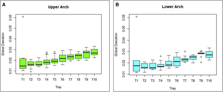
Morris et al 423
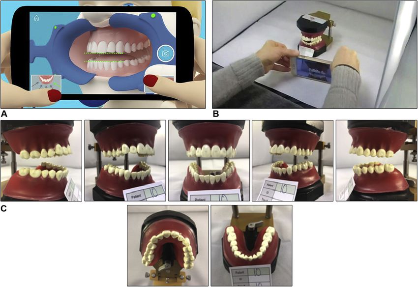
Fig 2. A, Maxillary and B, mandibular examples of heat maps created from the superimposition of the
3D digital models generated from the DM photographs/video scans on the 3D digital models generated
from the iTero Element scans.
to show discrepancies ranging from 0.5 to 0.5 mm in
Table I. Average global deviation values in photo- areas of expansion and contraction of the DM 3D digital
graph mode model compared with the iTero model. Deviations
Maxillary Mandibular Combined of .0.5 mm or \ 0.5 mm appear gray in the heat
arch (mm) arch (mm) (mm) map (Fig 2).
0.0212 0.022 0.0216 Exploratory data analysis was performed using SPSS
(IBM SPPS for Windows, version 22.0; IBM Corp, Ar-
monk, NY) to investigate whether there were any statis-
simulated gingiva during handling of the typodonts. The tically significant mean differences in global deviations
differences between the DM- and iTero Element- between 3D digital dental models generated by the
generated 3D models were measured using GOM Inspect DM application in photograph and video modes and
(GOM, Braunschweig, Germany) 3D evaluation software the 3D digital dental models generated by the iTero
to obtain a global deviation, measured in millimeters, Element over successive DM photograph and video ex-
from over 200,000 surface data points. The global devi- aminations. Descriptive statistics (mean, SD, 95% confi-
ation is the overall best-fit alignment of the individual dence interval [CI] of the mean) along with graphic
tooth positions between the 2 digital models. The soft- displays were used for mean deviation comparisons.
ware calculates global deviation using a prealignment The Shapiro-Wilk test was carried out to determine the
calculation. According to the GOM manual, “Pre-align- data distribution.
ment aligns the actual data (measurement, meshes. . .)
automatically to the nominal data, independently of RESULTS
start positions. A pre-alignment can be carried out The assumption of normality of the data using the
with subsequent automatic best-fit alignment.”28 Shapiro-Wilk test showed that all variables are normally
Professional standards established by the American distributed.
Board of Orthodontics (ABO) objective grading system In photograph mode, global deviations in the maxil-
consider deviations of \0.5 mm for alignment and mar- lary arch averaged 0.0212 mm, the mandibular arch
ginal ridge categories to be clinically acceptable,29 and, averaged 0.022 mm, and that of the maxillary and
according to the previous study, 0.5 mm is of minimal mandibular arches combined was 0.0216 mm (Table I).
clinical significance when investigating the accuracy of The mean deviations were all \0.5 mm and thus were
Invisalign-predicted tooth movements utilizing software not clinically significant. Differences in global deviations
similar in function to GOM Inspect.30 Because the soft- throughout successive DM photograph examinations
ware is capable of detecting deviations that are less than were averaged for all 10 typodonts at each tray. Maxil-
what is clinically relevant, maximum and minimum de- lary arch 95% CI comprised values of 0.0119-
viation threshold values of 0.5 and 0.5 mm were cho- 0.0293 mm; mandibular arch 95% CI comprised values
sen. A heat map was generated for each superimposition of 0.0084-0.0299 mm (Tables II and III). The 95% CI
American Journal of Orthodontics and Dentofacial Orthopedics September 2019 Vol 156 Issue 3424 Morris et al
Table II. Maxillary arch mean, SD, and 95% CI over successive DM photograph examinations
Invisalign maxillary arch (mm)
Trays Tray 1 Tray 2 Tray 3 Tray 4 Tray 5 Tray 6 Tray 7 Tray 8 Tray 9 Tray 10
Descriptive 0.0149 0.0163 0.0174 0.0198 0.0208 0.0222 0.0239 0.0238 0.0249 0.0258
mean
SD 0.0036 0.0033 0.0027 0.0046 0.0034 0.0032 0.0032 0.00263 0.0041 0.0024
95% CI 0.0119- 0.0014- 0.0146- 0.0162- 0.0184- 0.0183- 0.0216- 0.0219- 0.0218- 0.0258-
0.0179 0.0187 0.0183 0.0233 0.0233 0.0247 0.0262 0.0256 0.0281 0.0293
Table III. Mandibular arch mean, SD, and 95% CI over successive DM photograph examinations
Invisalign mandibular arch (mm)
Trays Tray 1 Tray 2 Tray 3 Tray 4 Tray 5 Tray 6 Tray 7 Tray 8 Tray 9 Tray 10
Descriptive 0.0221 0.0164 0.0170 0.0194 0.0195 0.0215 0.0234 0.0244 0.0276 0.0275
mean
SD 0.0164 0.0031 0.0032 0.0032 0.0037 0.0044 0.0028 0.0024 0.0030 0.0033
95% CI 0.0085- 0.0142- 0.0148- 0.0167- 0.0168- 0.0183- 0.0214- 0.0227- 0.0253- 0.0252-
0.0358 0.0187 0.0193 0.0221 0.0221 0.0246 0.0255 0.0261 0.0299 0.0299
deviations for each tray indicated that these differences
Table IV. Range of mean, SD, and 95% CI over succes-
were statistically significant (P \0.05). However, the
sive DM photograph examinations
global deviations were not clinically significant
Maxillary Mandibular because all values were \0.5 mm. Graphic displays
Arch arch (mm) arch (mm) showed a positive trend for both maxillary and
Descriptive mean 0.0149-0.0276 0.0164-0.0275 mandibular arches over successive DM video examina-
SD 0.0023-0.0044 0.0116-0.0044
95% CI 0.0119-0.0293 0.0084-0.0358
tions (Fig 4). No statistically significant differences ex-
isted between the maxillary and mandibular arches.
The increased mean global deviation for both arches
of the mean differences in global deviations for each tray was approximately the same over successive DM exam-
indicated that these differences were statistically signif- inations.
icant (P \0.05) (Table IV). However, because all global Trend lines of the mean global deviation values of
deviations were \0.5 mm, this finding was not clinically maxillary and mandibular arches were generated for
significant. Graphic displays show a positive trend for photograph and video modes by plotting the mean
both maxillary and mandibular arches over successive global deviation values for all 10 typodonts at each
DM photograph examinations (Fig 3). The preliminary step of Invisalign tray. The maxillary arch trend line
global deviation of 0.0609 mm in the first tray of patient equation in photograph mode was y 5 0.0013x 1
2 skews this trend initially. No statistically significant 0.0138, and the mandibular arch equation was
differences existed between the maxillary and mandib- y 5 0.0012x 1 0.0153. The maxillary arch trend line
ular arches. equation in video mode was y 5 0.0013x 1 0.0128,
In video mode, the global deviations for the maxil- and the mandibular arch equation was y 5 0.0013x 1
lary arch averaged 0.0202 mm; the global deviations 0.0131. To determine when clinical significance may
for the mandibular arch averaged 0.0205 mm. The be reached throughout successive DM photograph and
mean global deviation for maxillary and mandibular video examinations, 0.5 mm was substituted for “y” in
arches combined was 0.0204 mm (Table V). Differences the preceding equations. This resulted in a projected
in global deviations throughout successive DM video clinical significance in photograph mode after 374 suc-
examinations were averaged for all 10 typodonts at cessive photograph examinations in the maxillary arch
each tray. For the maxillary arch, the 95% CI included and 403 photograph examinations in the mandibular
values ranging from 0.0112 to 0.0273 mm. The arch. For video mode, clinical significance may be
mandibular arch 95% CI included values ranging reached after 375 successive upper arch and lower arch
from 0.0115 to 0.0290 mm (Tables VI, VII and VIII; DM video examinations have been captured for each
Fig 4). The 95% CI of the mean differences in global typodont.
September 2019 Vol 156 Issue 3 American Journal of Orthodontics and Dentofacial OrthopedicsMorris et al 425
Fig 3. A, Maxillary and B, mandibular arch means, SD, and 95% CI over successive DM photograph
examinations.
Table V. Average global deviation values in video mode a second time after the final set of trays, before and after
the photograph and video examinations were taken, to
Maxillary Mandibular Combined account for errors introduced while handling the
arch (mm) arch (mm) mean (mm)
typodonts. The deviation between these 2 scans for
0.0202 0.0205 0.0204
both arches in all 10 typodonts had an overall mean of
0.0330 mm. These deviations were within the range of
precision, 0.02984-0.1039 mm, of the iTero scanner
DISCUSSION for full-arch scanning found in previous
This study aimed to determine the accuracy of the 3D studies.10,15,22,23,31 Therefore, any errors that may have
digital dental models generated by the DM application in been introduced while scanning the typodonts with
photograph and video modes compared with the 3D the iTero and capturing the DM examinations were
models generated by the iTero. There were no statisti- insignificant.
cally significant mean differences in global deviations The clinical significance of remote 3D monitoring is
among the studied groups. This finding indicates that that it could be especially useful to increase access to
3D digital dental models generated by the DM applica- care for patients who live in remote areas or commute
tion in photograph and video modes were comparable long distances to receive orthodontic treatment. The
with the 3D digital dental models generated by the iTero average orthodontic patient appointment interval is 4
Element intraoral scanner. Moreover, the results showed to 6 weeks.32 With technology such as the DM applica-
that average differences between DM- and iTero tion, orthodontists can remotely monitor patients for
Element-generated 3D models were 0.021 mm for pho- scheduling optimization and possibly treat patients
tographs and 0.020 mm for video mode, suggesting with higher efficiency. The default DM examination is
clinical insignificance. Professional standards estab- scheduled for every 2 weeks, but the intervals can be
lished by the ABO objective grading system consider de- customized for each patient to accommodate the need
viations of \0.5 mm to be clinically acceptable.29 of oral hygiene or monitoring of elastic wear. More
The results also showed that the mean global devia- frequent monitoring and communication with these pa-
tion gradually increased from tray 1 to tray 10 in tients may enhance motivation and cooperation.
both arches of all studied typodonts using DM in both Because most clear aligners, customized fixed appli-
photograph and video modes. The 95% CI of the ances, and customized orthodontic archwires depend on
mean global deviation differences indicated statistical 3D digital models, intraoral self-scanning by smart-
significance for all trays (P \0.05). However, these dif- phones may reduce the cost and chairtime needed for
ferences were not clinically significant because all values the scanning process. If there is a discrepancy between
were \0.027 mm.29,30 Each typodont was also scanned actual and planned tooth movement, the refinement
American Journal of Orthodontics and Dentofacial Orthopedics September 2019 Vol 156 Issue 3426 Morris et al
Table VI. Maxillary arch mean, SD, and 95% CI over successive DM examinations in video mode
Invisalign maxillary arch (mm)
Trays Tray 1 Tray 2 Tray 3 Tray 4 Tray 5 Tray 6 Tray 7 Tray 8 Tray 9 Tray 10
Descriptive 0.0148 0.0156 0.0165 0.0172 0.0192 0.0201 0.0231 0.0241 0.0248 0.0256
mean
SD 0.0043 0.0033 0.0026 0.0028 0.0029 0.0024 0.0024 0.0026 0.0035 0.0023
95% CI 0.0112- 0.0133- 0.0146- 0.015- 0.017- 0.0184- 0.0214- 0.0222- 0.022- 0.0239-
0.0184 0.0180 0.0183 0.0193 0.0212 0.0218 0.0248 0.0260 0.0273 0.0272
Table VII. Mandibular arch mean, SD, and 95% CI over successive DM examinations in video mode
Invisalign mandibular arch (mm)
Trays Tray 1 Tray 2 Tray 3 Tray 4 Tray 5 Tray 6 Tray 7 Tray 8 Tray 9 Tray 10
Descriptive 0.0150 0.0167 0.0162 0.0194 0.0195 0.0215 0.0241 0.0240 0.0256 0.0264
mean
SD 0.0041 0.0038 0.0023 0.0032 0.0036 0.0026 0.0030 0.0039 0.0025 0.0037
95% CI 0.0115- 0.0135- 0.0136- 0.0167- 0.0165- 0.0193- 0.0216- 0.0206- 0.0235- 0.023-
0.0184 0.0198 0.0180 0.0221 0.0224 0.0236 0.0266 0.0272 0.0277 0.0291
Table VIII. Range of mean, SD, and 95% CI over suc- optimal time interval between appointments during
cessive DM examinations in video mode each stage of treatment. It also may be possible to eval-
uate the efficiency and effectiveness of new and existing
Maxillary Mandibular appliances and products in ways that could not be
Arch arch (mm) arch (mm)
achieved in the past.
Descriptive mean 0.0148-0.0256 0.0150-0.0264
SD 0.0023-0.0043 0.0021-0.0041
Live patients are instructed to take the first photo-
95% CI 0.0112-0.0272 0.0115-0.0290 graph and video examination with teeth in occlusion.
However, to avoid inadvertent movement of the typo-
dont teeth from forces of occlusion, in this study no
visit could be scheduled for optimal clinical outcome. photograph or video examinations were taken with the
Oral hygiene and compliance also could be improved typodonts in occlusion. Therefore, this study cannot
by these applications. In the future, intraoral scans could assess the ability of DM to accurately track interocclusal
theoretically be achieved remotely with this technology relationships.
by having DM generate a 3D STL file from patient- In addition, because this study was performed using
administered photograph or video examinations rather only class I malocclusion with mild crowding, and all
than requiring an additional office visit. teeth fully erupted and visible in the arches, the accuracy
In this study, we demonstrated the accuracy of DM of DM tooth tracking cannot be assumed for all types of
technology to be as accurate as the iTero Element, malocclusions and dental problems.
although more studies in vivo are warranted to confirm Because this study captured DM examinations after
the results. There may be potential for fabrication of ap- each Invisalign tray, it only investigated DM's accuracy
pliances, such as Essix retainers, from patient- in tracking small tooth movements.34 In vivo studies
administered DM photograph or video examination are warranted to test the accuracy of DM when other fac-
alone. This would eliminate time and travel for in- tors come into play, such as saliva, plaque and calculus,
office appointments for impressions, as well as reducing teeth with various restorations, and limited mouth open-
office chairtime and material expense.33 Remote moni- ing. Studies verifying the ability of DM to accurately re-
toring can enhance practice efficiency because the cord patient occlusion, including functional shifts, are
orthodontist can notify patients about treatment prog- also recommended. In our study, scans were carried
ress and problems before in-person appointments. out by professionals on typodonts without lips or cheeks.
Finally, DM has the potential to be a source of data The ability of patients to accurately capture images of
for research in the orthodontic field. Since this technol- their own teeth with standard smartphones needs to
ogy quantifies and tracks tooth movements at specific be examined. It is claimed that DM, using a cone-
intervals of time, it could be used to determine the beam computed tomography scan for the initial scan,
September 2019 Vol 156 Issue 3 American Journal of Orthodontics and Dentofacial OrthopedicsMorris et al 427
Fig 4. A, Maxillary and B, mandibular arch means, SD, and 95% CI over successive DM video exam-
inations.
allows for the tracking of root movements. If accurate, Telecommunications in Health Care. Washington, DC: National
root tracking could possibly allow orthodontists to pre- Academies Press (US); 1996: Available at: https://www.ncbi.nlm.
nih.gov/books/NBK45445/. Accessed January 27, 2018.
plan their bracket positioning. However, future studies
2. Chen JW, Hobdell MH, Dunn K, Johnson KA, Zhang J. Teledentistry
that test the validity of this claim need to be performed and its use in dental education. J Am Dent Assoc 2003;134:342-6.
before orthodontists rely on this functionality. 3. Jampani ND, Nutalapati R, Dontula BSK, Boyapati R. Applications
of teledentistry: a literature review and update. J Int Soc Prev Com-
CONCLUSIONS mun Dent 2011;1:37-44.
4. Mandall NA, O’Brien KD, Brady J, Worthington HV, Harvey L. Tele-
dentistry for screening new patient orthodontic referrals. part 1: a
1. 3D digital dental models generated by the DM smart-
randomised controlled trial. Br Dent J 2005;199:659-62: discus-
phone's application scans from photograph and sion 653.
video modes are comparable to iTero scans and are 5. Kravitz ND, Burris B, Butler D, Dabney CW. Teledentistry, do-
accurate enough to be used in clinical applications. it-yourself orthodontics, and remote treatment monitoring. J
2. Based on the trends displayed in this study, it was Clin Orthod 2016;50:718-26.
6. Eppright M, Shroff B, Best AM, Barcoma E, Lindauer SJ. Influence
predicted that deviations would not reach clinical
of active reminders on oral hygiene compliance in orthodontic
significance of 0.5 mm according to ABO- patients. Angle Orthod 2014;84:208-13.
determined standards until 374 successive DM 7. Bowen TB, Rinchuse DJ, Zullo T, DeMaria ME. The influence of
photograph examinations in the upper arch and text messaging on oral hygiene effectiveness. Angle Orthod
403 examinations in the lower arch have been 2015;85:543-8.
8. Li X, Xu Z-R, Tang N, Ye C, Zhu XL, Zhou T, et al. Effect of inter-
captured. Clinical significance was predicted to be
vention using a messaging app on compliance and duration of
reached after 375 successive extraoral DM video ex- treatment in orthodontic patients. Clin Oral Investig 2016;20:
aminations have been captured for both upper and 1849-59.
lower arches. 9. How Dental Monitoring works. Dental Monitoring: Available at:
3. Intraoral examination and other factors in vivo may https://dental-monitoring.com/how-dental-monitoring-works/.
Accessed January 31, 2018.
lead to larger deviations that cause DM examination
10. Patzelt SBM, Emmanouilidi A, Stampf S, Strub JR, Att W. Accuracy
to reach clinical significance earlier than the results of full-arch scans using intraoral scanners. Clin Oral Investig 2014;
shown in this study. 18:1687-94.
11. Burhardt L, Livas C, Kerdijk W, van der Meer WJ, Ren Y. Treatment
REFERENCES comfort, time perception, and preference for conventional and
digital impression techniques: a comparative study in young
1. Institute of Medicine (US) Committee on Evaluating Clinical Appli- patients. Am J Orthod Dentofacial Orthop 2016;150:261-7.
cations of TelemedicineField MJ. Evolution and current applica- 12. Thiruvenkatachari B, Al-Abdallah M, Akram NC, Sandler J,
tions of telemedicine. In: Telemedicine: A Guide to Assessing O’Brien K. Measuring 3-dimensional tooth movement with a
American Journal of Orthodontics and Dentofacial Orthopedics September 2019 Vol 156 Issue 3428 Morris et al
3-dimensional surface laser scanner. Am J Orthod Dentofacial 23. Hack GD, Patzelt SBM. Evaluation of the accuracy of six intraoral
Orthop 2009;135:480-5. scanning devices: an in-vitro investigation. ADA Prof Prod Rev
13. Fleming PS, Marinho V, Johal A. Orthodontic measurements on 2015;10:1-5.
digital study models compared with plaster models: a systematic 24. A study to smile about: new study shows record number of adults
review. Orthod Craniofac Res 2011;14:1-16. are seeking orthodontic treatment. Am Assoc Orthod 2016: Avail-
14. Elnagar MH, Elshourbagy E, Ghobashy S, Khedr M, Kusnoto B, able at: https://res.cloudinary.com/dorhu9mrb/image/upload/
Evans CA. Dentoalveolar and arch dimension changes in patients q_57/v1454081658/AAO_Press_Release_Increase_in_Adult_Patients_
treated with miniplatesanchored maxillary protraction. Am J Or- 1-28-16.pdf.
thod Dentofacial Orthop 2017;151:1092-106. 25. Wheeler TT. Orthodontic clear aligner treatment. Semin Orthod
15. Renne W, Ludlow M, Fryml J, Schurch Z, Mennito A, Kessler R, 2017;23:83-9.
et al. Evaluation of the accuracy of 7 digital scanners: an in vitro 26. iTero intraoral scanner. Align Technology, Inc: Available at: http://
analysis based on 3-dimensional comparisons. J Prosthet Dent www.itero.com/en-us; 2016. Accessed January 31, 2018.
2017;118:36-42. 27. Welcome. Dental monitoring: Available at: https://dental-
16. Zhang F, Suh KJ, Lee KM. Validity of intraoral scans compared with monitoring.com. Accessed January 23, 2018.
plaster models: an in-vivo comparison of dental measurements 28. GOM Inspect - 2016 Training Outline; 2016. Available at: https://
and 3D surface analysis. PLoS One 2016;11:e0157713. www.gom.com. Accessed July 18, 2019.
17. Gr€unheid T, McCarthy SD, Larson BE. Clinical use of a direct 29. Casko JS, Vaden JL, Kokich VG, Damone J, James RD,
chairside oral scanner: an assessment of accuracy, time, and pa- Cangialosi TJ, et al. Objective grading system for dental casts
tient acceptance. Am J Orthod Dentofacial Orthop 2014;146: and panoramic radiographs. American Board of Orthodontics.
673-82. Am J Orthod Dentofacial Orthop 1998;114:589-99.
18. Arag on MLC, Pontes LF, Bichara LM, Flores-Mir C, Normando D. 30. Gr€unheid T, Loh C, Larson BE. How accurate is Invisalign in non-
Validity and reliability of intraoral scanners compared to conven- extraction cases? Are predicted tooth positions achieved? Angle
tional gypsum models measurements: a systematic review. Eur J Orthod 2017;87:809-15.
Orthod 2016;38:429-34. 31. Anh JW, Park JM, Chun YS, Kim M, Kim M. A comparison of the
19. Logozzo S, Franceschini G, Kilpela A, Caponi M, Governi L, Blois L. precision of three-dimensional images acquired by 2 digital intrao-
A comparative analysis of intraoral 3d digital scanners for restor- ral scanners: effects of tooth irregularity and scanning direction.
ative dentistry. Internet J Med Technol 2008;5:1-18. Korean J Orthod 2016;46:3-12.
20. Naidu D, Freer TJ. Validity, reliability, and reproducibility of the 32. What to expect from your braces follow-up visits. Oral-B: Available
iOC intraoral scanner: a comparison of tooth widths and Bolton ra- at: https://oralb.com/en-us/oral-health/life-stages/braces/follow-
tios. Am J Orthod Dentofacial Orthop 2013;144:304-10. up-visits-what-to-expect. Accessed January 31, 2018.
21. Fl€
ugge TV, Schlager S, Nelson K, Nahles S, Metzger MC. Precision 33. The Dental Monitoring range. Dental Monitoring: Available at:
of intraoral digital dental impressions with iTero and extraoral https://dental-monitoring.com/doctor/resources/range. Accessed
digitization with the iTero and a model scanner. Am J Orthod Den- February 1, 2018.
tofacial Orthop 2013;144:471-8. 34. Kravitz ND, Kusnoto B, BeGole E, Obrez A, Agran B. How well does
22. Ender A, Attin T, Mehl A. In vivo precision of conventional and dig- Invisalign work? A prospective clinical study evaluating the effi-
ital methods of obtaining complete-arch dental impressions. J cacy of tooth movement with Invisalign. Am J Orthod Dentofacial
Prosthet Dent 2016;115:313-20. Orthop 2009;135:27-35.
September 2019 Vol 156 Issue 3 American Journal of Orthodontics and Dentofacial OrthopedicsYou can also read